DOI:
10.1039/C5RA19199H
(Paper)
RSC Adv., 2015,
5, 100912-100922
Kaempferide, the most active among the four flavonoids isolated and characterized from Chromolaena odorata, induces apoptosis in cervical cancer cells while being pharmacologically safe†
Received
17th September 2015
, Accepted 26th October 2015
First published on 26th October 2015
Abstract
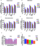
Chromolaena odorata, commonly known as Siam weed, is popular as a traditional medicine. We report the isolation and characterization of four compounds from a cytotoxic fraction, F-17, isolated from the dichloromethane (DCM) extract of C. odorata by bioactivity-guided fractionation. The organic extracts were screened in five cancer cell lines of various origins for their cytotoxic effect. The DCM extract exhibited maximum cytotoxicity and was purified by silica gel column chromatography to obtain four major compounds. The compounds were characterized by 1H-NMR, 13C-NMR, and HR-MS methods and were found to be acacetin (1), dihydrokaempferide (2), isosakuranetin (3), and kaempferide (4). MTT assay was used for preliminary evaluation of the cytotoxicity of these compounds. Among the cancer cell lines that were screened, HeLa was the most sensitive to kaempferide (IC50: 16 μM) followed by acacetin (174 μM), dihydrokaempferide (>200 μM) and isosakuranetin (>200 μM). Kaempferide (4) induced morphological characteristics of apoptosis in HeLa cells and was non-toxic to rapidly dividing normal human fibroblasts up to 100μM. Annexin V staining, characteristic of early stage of apoptosis was further confirmed by FACS analysis. Induction of apoptosis was illustrated by its potential to induce the cleavage of caspases and PARP. FACS analysis demonstrated that kaempferide (4)-induced cytotoxicity is independent of cell cycle arrest. Acute and chronic toxicity studies conducted in vivo proved that the compound is pharmacologically safe. To the best of our knowledge, this is the first study reporting the anticancer potential and pharmacological safety of kaempferide (4).
1. Introduction
Cervical cancer, the most common female cancer in developing countries, contributes considerably to cancer-related mortality among women in Africa, Central America and South-Central Asia and has the highest disease frequency in India.1 The causative agent of cervical cancer is human papilloma virus (HPV) which is implicated in sexually transmitted diseases and most of the victims of this cancer possess a poor socio-economic status and cannot afford costly drugs. Chromolaena odorata, formerly known as Eupatorium odoratum, is native to North America and has been introduced to tropical Asia, West Africa, and parts of Australia.2 Although regarded as a serious weed, C. odorata has potential medicinal uses. Traditionally, fresh leaf juice or a decoction of C. odorata is used to cure amenorrhea, skin diseases, poisonous bites, wounds, amygdalitis, fever, inflammatory diseases, malaria, jaundice and rheumatism.3–5 In St. Lucia, C. odorata is used for the treatment of the early stages of cancer as a decoction in which this plant is boiled with four other plants.6 Also, people in Machang, Kelantan, and Malaysia use this plant to treat wounds, uterus-related problems, and to arrest bleeding.7 This plant is reported to have anti-inflammatory, antioxidant, wound healing, antimicrobial and haemostatic properties.3,8–10 Previous phytochemical studies on C. odorata have led to the isolation of flavonoids, anthraquinones, alkaloids, triterpenoids, and steroids.11–14 Preliminary studies have reported on the cytotoxic effects of the ethanolic extract of the whole plant on various cancer types, including cancers of the blood, breast, lungs, liver, cervix, and prostate.7,15–17 Recently, chromomoric acid C–I, an activator of Nrf2 has been reported from the methanolic extract of C. odorata.18 Even though some of the compounds isolated from this plant have been shown to have anti-cancer potential, studies pertaining to the anticancer potential of kaempferide (4) are not reported. In the present study, we report for the first time the presence of a potent anticancer principle kaempferide (4) along with three other less cytotoxic compounds from the DCM extract of the leaves of C. odorata. We have also investigated the mode of cell death induced by this compound in cervical cancer cells. In this study, we report some promising pro-apoptotic activity of kaempferide (4) against cervical cancer cells, as evidenced by morphological features such as membrane blebbing, nuclear dye uptake of acridine orange/ethidium bromide, Annexin V-PI stain and the activation of the caspase cascade, which is a classical marker of apoptotic cell death. We have also conducted in vivo studies to ensure the biological safety of the compound.
2. Materials and methods
2.1 Reagents and antibodies
Annexin V apoptosis detection kit and antibody against PARP (poly(ADP-ribose)polymerase) were obtained from Santa Cruz Biotechnology (Santa Cruz, CA, USA). Antibodies against caspases 3, 7, 8 and 9 were purchased from Cell Signaling Technology (Beverly, MA, USA). Silica gel for column chromatography, and silica gel F254 for thin layer chromatography were obtained from Sigma Chemicals (St. Louis, MO, USA). Solvents were purchased from Merck (Germany). Curcumin and MTT (3-(4,5-dimethyl-2-thiazolyl)-2,5-diphenyl-2H-tetrazolium bromide) were purchased from Sigma Chemicals (St. Louis, MO, USA). Immobilon Western blotting reagent was purchased from Millipore Corporation, Billerica, USA.
2.2 Methods
2.2.1 Extraction from the leaves of C. odorata. Fresh plant was collected in August 2009 from local areas in Thiruvanathapuram and was identified by Dr G. Valsaladevi, Curator, Dept of Botany, University of Kerala, and a voucher specimen has been deposited at the author’s laboratory (voucher no: CRP04). Leaves of the plants were washed thoroughly and shade-dried at room temperature (25–30 °C) for 7 days. The well dried sample was powdered and a defined quantity (100 g) was serially extracted using solvents with increasing polarity viz. hexane, dichloromethane, ethyl acetate and methanol at 37 °C, 120 rpm, 24 h in a shaker incubator. The individual solvent extracts were concentrated separately under vacuum using a rotary evaporator; the resulting crude extracts were dissolved in minimal volume of DMSO and were stored in −20 °C freezer as stock solutions (100 mg mL−1 or 50 mg mL−1).
2.2.2 Isolation of bioactive fractions using bioassay-guided fractionation. The crude DCM extract (3 g) was subjected to silica gel column chromatography (60–120 mesh size, column size 40 cm × 20 cm) using hexane/dichloromethane (100/0, 80/20, 60/40, 50/50, 25/75, 0/100), dichloromethane/chloroform (100/0, 80/20, 60/40, 25/75, 0/100), and chloroform/methanol (100/0, 80/20, 60/40, 0/100). A total of 50 fractions were collected, visualized with a UV chamber (256 nm and 365 nm) and then the fractions possessing a similar TLC profile were pooled. Each pooled fraction (F-1 to F-20) was subjected to cytotoxicity testing and F-17 which was eluted by a dichloromethane/chloroform 60/40 ratio was found to be the most cytotoxic fraction. Hence, F-17 was subjected to silica gel column chromatography, eluted with EtOAc/hexane (20/80 to 50/50), to yield a mixture of two compounds. The mixture of compounds in EtOAc/hexane along with a few drops of methanol in a 50 mL rough surfaced glass vial was kept aside for slow evaporation of the solvents at room temperature. After complete evaporation of the solvents, a mixture of pale yellow and white solids was observed which was carefully separated with the help of a sharp steel syringe needle. The pale yellow colored solid acacetin (1) and the white solid dihydrokaempferide (2) (Fig. 2) were isolated as 15 and 10 mg, respectively. Further elution of the column with EtOAc/hexane (50/50) to 100% EtOAc afforded pure isosakuranetin (3) (Fig. 2) as a white solid (60 mg). Some fractions of compound 3 were isolated as a mixture (80 mg) with a yellow colored compound. The mixture was then subjected to purification by column chromatography using the mobile phase gradient system hexane/DCM (30/70, 40/60, 20/80) to acetone/DCM (1/99) affording compound 3 (40 mg) as a white solid, and kaempferide (4) (Fig. 2) as yellow crystals (25 mg). All four compounds were previously found to be derivable from C. odorata as well as from other plants as reported in the literature.9,12,19–23
2.2.3 Cell culture and cell viability. The cancer cell lines viz. HeLa (cervical), MDA-MB-231 (breast), HCT-116 (colon) and HL60 (leukemia) were procured from the National Centre for Cell Sciences, Pune and are maintained under standard conditions in our laboratory. The lung cancer cell line, H1299 and normal fibroblasts were gifted by Dr Bharat Aggarwal, MD Anderson Cancer center, Houston, USA.
MTT assay. Briefly, the cells were seeded in 96-well plates (2000 cells per well). After overnight incubation, cells were treated with different concentrations of organic extracts (25–250 μg mL−1) and isolated compounds (5–200 μM) along with the positive control curcumin (5–30 μM) for 72 h and cytotoxicity was measured. Fresh media containing 25 μL of MTT solution (5 mg mL−1 in PBS) and 75 μL of complete medium was added to the wells and incubated for 2 h. At the end of the incubation, MTT lysis buffer (20% sodium dodecyl sulphate in 50% dimethyl formamide) was added to the wells (0.1 mL per well) and incubated for another 1 h at 37 °C. At the end of the incubation, the optical density was measured at 570 nm using a plate reader (Bio-Rad). The relative cell viability as a percentage was calculated as (A570 of treated samples/A570 of untreated samples) × 100.24 The IC50 values were extrapolated from a polynomial regression analysis of the experimental data.
2.2.4 Acridine orange/ethidium bromide staining. Morphological changes characteristic of apoptosis were assessed by fluorescent microscopy using an acridine orange/ethidium bromide staining method. Briefly, cells were seeded in 96-well plates and treated with kaempferide (4) as in the MTT assay for 24 h. After washing once with PBS, the cells were stained with 100 μL of a 1
:
1 mixture of acridine orange/ethidium bromide 4 μg mL−1 solutions. The cells were immediately washed with PBS, viewed under a Nikon inverted fluorescent microscope (TE-Eclipse 300) and photographs were taken.25
2.2.5 Detection of apoptosis by Annexin V staining. As apoptosis causes changes in membrane permeability, there is a transient leakage of phosphatidylserine to the membrane, which is considered to be an early marker of apoptosis. Annexin V preferentially binds to phosphatidylserine as it is a negatively charged phospholipid. Hence, using FITC (fluorescein isothiocyanate) conjugated Annexin V, apoptotic cells were detected with the help of a fluorescence microscope using the manufacturer’s protocol (Santa Cruz, CA, USA). Briefly, the cells were seeded in 96-well plates and treated with kaempferide (4) as in the MTT assay for 16 h. The cells were first washed with PBS and then with 1× assay buffer after which, 0.5–5 μL (0.1–1 μg) of Annexin V FITC per 100 μL assay buffer was added. After incubating for 15 min at room temperature in the dark, the cells were washed with PBS and immediately photographed using a fluorescence microscope.26
2.2.6 Estimation of apoptosis by FACS. The extent of apoptosis induced by kaempferide was estimated by FACS using an Annexin V apoptosis kit (Santa Cruz, CA, USA). Briefly, cells were seeded in 60 mm culture plates, and incubated with different concentrations of kaempferide. After 16 h, cells were trypsinized and pelleted down by low speed centrifugation, washed with PBS and were suspended in 1× assay buffer. To the buffer 5 μL of FITC conjugated Annexin V and 10 μL of propidium iodide were added and incubated for 15 min in the dark at room temperature. The cells were then analyzed immediately by flow cytometry to get the % of apoptotic cells (FACS Aria™, BD Bioscience).27
2.2.7 Western blot analysis. For the detection of apoptotic proteins, HeLa cells (106 cells/60 mm culture dish) were treated with the kaempferide (4) for 24 h. The cells were then washed with PBS and lysed by keeping them on ice for 30 min with whole cell lysis buffer containing 20 mM Tris of pH 7.4, 250 mM NaCl, 2 mM EDTA, 0.1% Triton, 1 mM DTT (1,4-dithiothreitol), 0.5 mM PMSF, 4 mM sodium orthovanadate, aprotinin (5 μg mL−1) and leupeptin (5 μg mL−1). The supernatants were collected by centrifugation at 13
000 rpm for 10 min at 4 °C and boiled in 5× loading dye before separating the proteins by SDS-polyacrylamide gel electrophoresis (SDS-PAGE) and Western blotting with antibodies against caspase proteins (pro-caspase 3, pro-caspase 7, pro-caspase 8 and pro-caspase 9), and poly(ADP-ribose)polymerase (PARP). Immunoreactive proteins were detected with horseradish peroxidase-coupled secondary antibodies and visualized by an enhanced chemiluminescence detection kit (Millipore Corporation, Billerica, MA, USA).28
2.2.8 Flow cytometry and cell cycle analysis. Cell cycle analysis helps in distinguishing the distribution of population of cells at different stages of the cell cycle. Briefly, cells were seeded in 60 mm plates and subjected to kaempferide (4) treatment for 24 h and 48 h followed by trypsinization. Curcumin (25 μM, 24 h) was used as a positive control. The cell pellets were fixed in 70% ice-cold ethanol, treated with 100 mg mL−1 RNase A and 50 mg mL−1 propidium iodide, and subjected to flow cytometry (BD Biosciences).27
2.2.9 Toxicological evaluation.
Acute toxicity study. Animals: Six to eight-week-old female Swiss albino mice (18–22 g) were obtained from the RGCB Animal Research Facility and the experiment was performed under protocols approved by the RGCB Institutional animal ethical committee (IAEC no: 189(a)/RUBY/2012).The experimental design: Swiss albino mice were randomly divided into 3 groups of 6 animals each and were allowed to acclimatize for a week. Group I was taken as the control, which received only cremophor vehicle, while Groups II and III received a single dose of kaempferide (4) (50 and 200 mg kg−1 body weight respectively) dissolved in cremophor as an intraperitoneal injection. The mice were observed continuously for 1 h, for any gross behavioral changes and death, and then intermittently for the next 6 h and 24 h. The behavioral parameters monitored were convulsion, hyperactivity, sedation, grooming, food and water intake, etc. The animals were observed frequently for the next 7 days from the day of treatment after which, the animals were euthanized in a CO2 chamber. The blood was collected for analysis of the biochemical parameters of liver function, abnormal values of which are indicative of toxicity. The liver was fixed in 10% buffered formalin and thin cryostat sections (LEICA CM 1850UV Cryostat) were stained with haematoxylin and eosin for histopathological evaluation. The weight of the animals as well as those of the individual vital organs were also recorded.29
Chronic toxicity. Swiss albino mice were randomly divided into 2 groups of 6 animals each and were allowed to acclimatize for a week. Group I was taken as the control, which received only cremophor vehicle, while Group II received 75 mg kg−1 body weight of kaempferide (4) dissolved in cremophor as an intraperitoneal injection on alternate days, thrice in a week, for 3 months. The animals were observed frequently during this period after which, the animals were euthanized in a CO2 chamber. The blood was collected for analyzing biochemical parameters of liver function, the abnormal values of which are indicative of hepatotoxicity. The liver was fixed in 10% buffered formalin and the thin cryostat sections were stained with haematoxylin and eosin for histopathological evaluation. The weight of the animals as well as that of the individual vital organs were also recorded.29
2.2.10 Statistical analysis. For the flow cytometry, data analysis was performed using the BD FACS Diva software version 5.0.2. The statistical analysis was performed using Graph Pad Prism software (Graph Pad software Inc., San Diego, CA, USA). Statistical significance was defined as p < 0.05. The error bars represent ±SD, taken from three independent experiments.
3. Results
3.1 DCM extract of C. odorata induces maximum cytotoxicity in cervical cancer cells
The cytotoxic effect of organic extracts of C. odorata was evaluated in cancer cells of different origin. The cells were treated with different concentrations of the extracts for 72 h, and cell viability was determined by MTT assay. Among the four extracts screened, the DCM extract is active against four cancer cells of different origin except the leukemia cell line (Fig. 1A–D). We selected HeLa for further studies since it turned out to be the most sensitive cell line in which the DCM extract induced a dose-dependent cytotoxicity (IC50 37.5 μg mL−1) (data not shown) and showed an array of spots in TLC in the solvent system, chloroform/methanol (96/4) (data not shown).
 |
| Fig. 1 17th fraction eluted by 60 : 40 (DCM : chloroform) is responsible for the cytotoxic effect of the DCM extract of C. odorata. (A–D) The effect of C. odorata organic extracts on cancer cells of various origin. A total of 2000 cells in triplicate were exposed to the indicated concentration of four extracts (25–250 μg mL−1) for 72 h and subjected to MTT assay. Relative cell viability was determined as % absorbance over untreated control. Data represent three independent sets of experiments and results are shown as the mean ± SD. (E) Cytotoxic effect of different column fractions eluted by column chromatography. The most active DCM extract was subjected to column chromatography using combinations of different solvent systems (hexane/DCM/chloroform/methanol). The most active fraction, F-17 was identified by MTT assay. (F) Dose dependent effect of F-17 on HeLa cells. A total of 2000 cells in triplicate were exposed to the indicated concentrations of F-17 (5–25 μg mL−1) for 72 h and subjected to MTT assay. Relative cell viability was determined as % absorbance over untreated control. The data represent three independent sets of experiments and results are shown as the mean ± SD. | |
3.2 Kaempferide (4) was found to be most cytotoxic among the four compounds isolated from the DCM extract of C. odorata
The active DCM extract was fractionated as discussed in Section 2.2.2. F-17 was found to be cytotoxic against the cervical cancer cell line (Fig. 1E and F). Purification of F-17 resulted in four compounds which were characterized (Fig. 2, Table 1). Kaempferide (4) (IC50 16 μM) is the most cytotoxic followed by acacetin (1) (IC50 178 μM), dihydrokaempferide (2) (IC50 277 μM), and isosakuranetin (3) (IC50 312 μM) (Fig. 3A). Curcumin was used as a positive control for this study. Kaempferide (4) brought down the IC50 to 16 μM in HeLa cells, while being non-toxic to normal human fibroblasts up to 100 μM (Fig. 3B), while curcumin induced the same effect at 16.67 μM demonstrating that both the compounds have almost the same cytotoxic effect in HeLa cells.
 |
| Fig. 2 Structures of acacetin (1); dihydrokaempferide (2); isosakuranetin (3); kaempferide (4). | |
Table 1 NMR data for acacetin (1); dihydrokaempferide (2); isosakuranetin (3); kaempferide (4)
Position |
1 |
2 |
3 |
4 |
δ 13Ca |
δ 1H mult [Ja (Hz)] |
δ 13Cb |
δ 1H mult [Jb (Hz)] |
δ 13Cb |
δ 1H mult [Jb (Hz)] |
δ 13Ca |
δ 1H mult [Ja (Hz)] |
Measured in DMSO-d6. Measured in methanol-d. |
2 |
163.7 |
|
84.8 |
5.03 d (12) |
78.8 |
5.34 d (13) |
146.7 |
|
3 |
103.9 |
6.79 s |
73.6 |
4.54 d (12) |
42.6 |
2.69, 3.08 dd, dd (3, 17) |
136.5 |
|
4 |
182.2 |
|
198.4 |
|
196.2 |
|
176.4 |
|
5 |
161.9 |
|
165.3 |
|
164.0 |
|
161.1 |
|
6 |
99.4 |
6.15 d (2) |
97.3 |
5.93 d (2) |
95.7 |
5.88 s |
98.7 |
6.10 s |
7 |
164.8 |
|
168.7 |
|
166.9 |
|
164.4 |
|
8 |
94.5 |
6.45 d (2) |
96.3 |
5.89 d (2) |
94.8 |
5.89 s |
94.0 |
6.40 s |
9 |
157.8 |
|
164.5 |
|
163.3 |
|
156.7 |
|
10 |
104.1 |
|
101.8 |
|
101.9 |
|
103.5 |
|
11 |
123.3 |
|
130.5 |
|
130.8 |
|
123.7 |
|
12 |
128.7 |
7.98 d (9) |
130.3 |
7.45 d (9) |
127.5 |
7.39 d (9) |
129.8 |
8.07 d (9) |
13 |
115.0 |
7.05 d (9) |
114.7 |
6.97 d (9) |
113.6 |
6.94 d (9) |
114.5 |
7.05 d (9) |
14 |
162.7 |
|
161.6 |
|
160.0 |
|
160.9 |
|
15 |
56.0 |
3.82 s |
55.7 |
3.82 s |
54.3 |
3.79 s |
55.8 |
3.70 s |
 |
| Fig. 3 Kaempferide (4) was found to be most cytotoxic among the four compounds, as assessed by a cell viability assay and analysis of morphological features and nuclear membrane damage, while being non-toxic to normal fibroblasts. (A) Effect of flavonoids of F-17, isolated from the DCM extract of C. odorata on HeLa cells. A total of 2000 cells in triplicate were exposed to the indicated concentration of test sample (2.5–200 μM) for 72 h and subjected to MTT assay. Relative cell viability was determined as % absorbance over untreated control. Data represent three independent sets of experiments and results are shown as the mean ± SD. Curcumin was used as the positive control (5–30 μM). (B) Effect of kaempferide (4) on normal fibroblast cells, FS as well as on cervical cancer cells, HeLa, SiHa and Caski. A total of 2000 cells in triplicate were exposed to the indicated concentrations of kaempferide for 72 h and subjected to MTT assay. Relative cell viability was determined as % absorbance over untreated control. Data represent three independent sets of experiments and results are shown as the mean ± SD. (C) Morphological changes induced by kaempferide (4) in fibroblasts, FS and HeLa by phase contrast microscopy. (D) The early stage and late stage of apoptosis were evaluated by acridine orange/ethidium bromide staining. AO/EB positive cells in various fields were counted, and the average was taken and plotted. | |
3.3 Kaempferide (4) is non-toxic to normal fibroblasts while inducing morphological changes, membrane flip-flop and nuclear membrane damage, characteristic of apoptosis in HeLa cells
We compared the morphological effect of kaempferide (4) in HeLa cells and normal fibroblasts. While phenotypic changes characteristic of apoptosis were induced in HeLa cells from 10 μM onwards, no significant change was observed in normal fibroblasts up to 100 μM (Fig. 3C). We also evaluated its cytotoxic effect in other cervical cancer cell lines SiHa (25.86 μM) and Caski (18.75 μM) (Fig. 3B). The percentage of cells in the early and late stages of apoptosis were also evaluated after treating HeLa cells with kaempferide (4), which induced a dose dependent increase in the extent of apoptosis (Fig. 3D). The early stage of apoptosis was confirmed by Annexin V staining using fluorescence microscopy. Kaempferide (4) treatment produced a gradual increase in the percentage of Annexin V-positive cells in a concentration-dependent manner (Fig. 4A). The extent of apoptosis induced by kaempferide (4) was further estimated by FACS analysis of the Annexin V-FITC/PI double stained cells. The apoptotic cell population was increased from 4.8% to 18.0% and 23.9% respectively, when treated for 16 h with 10 μM and 15 μM of kaempferide (4) (Fig. 4B).
 |
| Fig. 4 Kaempferide (4) induces membrane flip-flop and accumulation of cells at the sub-G0 phase, characteristic of apoptosis, while being independent of cell cycle. (A) HeLa cells were treated as indicated with different concentrations of kaempferide for 16 h and stained for Annexin V positivity and membrane flip-flop was captured by fluorescence microscopy. Annexin V positive cells in various fields were counted, and the average was taken and plotted. (B) HeLa cells were treated as indicated with different concentrations of kaempferide for 16 h and stained with Annexin V/PI and apoptosis was estimated by FACS. (C) Cells were harvested after 24 h & 48 h of kaempferide treatment, fixed in alcohol, stained with propidium iodide, and assayed for DNA content by flow cytometry. Curcumin was used as the positive control (25 μM). Representative histograms indicate the percentages of cells in the G1, S, G2/M and sub G0 phases of the cell cycle. The percentage of cells with sub-G0 DNA content was taken as a measure of the apoptotic cell population. The data provided is representative of three independent experiments. | |
3.4 Kaempferide (4) does not induce cell cycle arrest in HeLa cells
To explore whether the growth-inhibitory effect of C. odorata on HeLa cells is mediated through cell cycle arrest, we analyzed the distribution of cells in different phases of the cell cycle by measuring the intracellular DNA content in each phase. It was very interesting to note that kaempferide (4) failed to induce cell cycle arrest even at concentrations above the IC50 and at different time intervals (24 h & 48 h), while the positive control (curcumin 25 μM) readily induced G2M arrest at 24 h. However, there was an increase in the number of cells in the sub G0 phase, when treated with kaempferide (4) again indicative of an enhancement in apoptosis (Fig. 4C).
3.5 Kaempferide (4) induces caspase-dependent apoptosis in HeLa cells leading to PARP cleavage
Our next attempt was to investigate the mechanism behind the cytotoxic effect of kaempferide (4). Firstly, we checked the role of caspases, the key regulators of the apoptotic program.30 Two distinct pathways of apoptosis have been identified; the mitochondria-initiated apoptosis that occurs through caspase 9 which may further lead to caspase 8 cleavage and the death receptor-mediated pathway in which caspase 8 cleavage occurs independent of caspase 9.31,32 We observed that kaempferide (4) induces a dose dependent cleavage of the initiator caspases, caspase 9 and 8 (Fig. 5A and B) which clearly indicates the role of mitochondria in the kaempferide-induced apoptotic program in HeLa cells. It also induced a significant and dose dependent increase in the intensity of the cleaved bands of the effector caspases, caspase 7 and 3 (Fig. 5C and D). We then checked the effect of kaempferide (4) on the DNA repair enzyme PARP (poly ADP-ribose polymerase (116 kDa)), the down-stream target of caspases 3 and 7, which cleaved it to fragments of 85 kDa and 25 kDa.33,34 As observed in Fig. 5E, while 116 kDa PARP remained intact in the untreated cells, 15 μM kaempferide (4) cleaved the intact PARP to fragments, which clearly indicates caspase-mediated apoptosis.
 |
| Fig. 5 Kaempferide (4) induces caspase-dependent apoptosis in HeLa cells leading to PARP cleavage: (A–E) Western blots showing caspase activation in HeLa cells. Whole-cell extracts were prepared after treating HeLa cells with indicated concentrations of kaempferide (4) for 24 h and subjected to western blotting using antibodies against the caspases 8, 9, 7, 3 and PARP. | |
Hence our results clearly indicate that kaempferide (4) isolated from C. odorata induces cytotoxicity in HeLa cells through caspase-mediated apoptosis and is independent of cell cycle.
3.6 Kaempferide (4) is non-toxic as assessed by acute and chronic toxicity studies in Swiss albino mice, in vivo
To rule out the chance of any adverse effect of kaempferide (4) in vivo, we conducted an acute toxicity study for 7 days and a chronic toxicity study for 3 months in Swiss albino mice as described in the “Materials and methods” and the blood and liver tissues were collected for biochemical and histopathological evaluation. The blood was centrifuged at 1500 rpm for 10 min at 4 °C to separate the serum, which was analyzed for the level of Aspartate aminotransferase (AST), Alanine Aminotransferase (ALT) and Alkaline phosphatase (ALP), elevated levels of which are indicative of hepatotoxicity. Histopathological analysis of the liver tissue was also conducted for toxicological evaluation, using H and E staining. No behavioral changes were noted in the mice at any of the concentrations studied until the end of the experiment. There was no significant change in the weight of the animals (data not shown) as well as that of the vital organs (data not shown) in both the studies indicating that kaempferide is safe, in vivo. The results of the serum analysis as well as the mouse liver sections studied do not reveal any significant change in any of the parameters studied, which was supported by the histopathological data (Fig. 6A and B), confirming that kaempferide is non-toxic and pharmacologically safe.
 |
| Fig. 6 Kaempferide does not induce pharmacological toxicity as assessed by acute and chronic toxicity studies: A(a) & B(a) the activity of toxicological markers such as serum aspartate aminotransferase (AST), alanine aminotransferase (ALT) & alkaline phosphatase (ALP) in kaempferide treated mice for a period of 7 days and 3 months are graphically represented. A(b) & B(b) H & E stained liver tissues of mice treated with or without kaempferide for a period of 7 days and 3 months. | |
4. Discussion and conclusion
Cervical cancer is one of the most common cancers among women in developing countries, including India. Flavonoids have been found to be promising agents against cervical cancer. They display a wide variety of biological functions including induction of apoptosis, growth arrest, inhibition of DNA synthesis and modulation of signal transduction pathways.35 Our data showed that kaempferide (4), a flavonoid isolated from the DCM extract of C. odorata, is highly cytotoxic to cervical cancer cells. C. odorata has been widely used as a traditional herbal medicine for several inflammatory disorders. Phytochemicals with anticancer and anti-inflammatory potential have become key resources for drugs for the treatment of various malignancies.36 Several flavonoids, chalcones, flavones and essential oils of diverse biological activities have been isolated from different parts of C. odorata.9,12,37,38 Only very few studies have been reported on the anti-cancer potential of compounds isolated from C. odorata. 2′-Hydroxy-4,4′,5′,6′-tetramethoxy chalcone, isolated from the ethanolic extract of C. odorata leaves inhibits growth and clonogenicity in breast cancer cells15 and flavonoid glycosides isolated from its ethanolic extract induce cytotoxicity in leukaemic cells.16 The ethylacetate and acetone extracts of the C. odorata leaves have been shown to induce an autophagic mode of cell death in breast cancer cells.7 Two cytotoxic flavones from the flowers of this plant have also been reported.9 Odoratin, a PPARγ agonist and 3-hydroxy-1,2-dimethoxy-6-methylanthraquinone, is a compound with weak cytostatic activity towards lung cancer cells found in the DCM extract of the whole C. odorata plant.11
A study pertaining to induction of melanogenesis in mouse melanoma cells by kaempferide (4) has been reported recently.39 However, to the best of our knowledge, there have been no reports on exploiting the anticancer potential of kaempferide (4). On the contrary, a recent study, which reports the isolation of kaempferide (4) from Dillenia suffruticosa implies that the compound does not exhibit significant cytotoxicity in breast cancer cells.40 The present study demonstrates that kaempferide (4) induces a dose dependent cytotoxicity in cervical cancer cells, while being nontoxic to normal fibroblasts. The protective effect of C. odorata against oxidative damage on cultured skin cells3 may account for its nontoxic nature to normal cells. The morphological parameters and dose dependent increase in AO/EB staining clearly indicate that the cytotoxic effects of kaempferide (4) are mediated by induction of apoptosis.25,26 The analysis of membrane flip-flop (by Annexin-V staining), the earliest event of apoptosis, was carried out at 16 h, while all other experiments assessing the caspase cascade were performed at 24 h, since membrane flip-flop usually happens before the cleavage of caspases, which further leads to PARP cleavage. Induction of caspase activation and PARP cleavage underscores that the caspase cascade has a major role in regulating the cytotoxic activity of kaempferide (4). Cell cycle arrest can occur following apoptosis or independent of apoptosis. So we conducted the cell cycle analysis at different concentrations and time points. It was interesting to note that kaempferide (4) does not induce cell cycle arrest at any of the parameters studied, though it induces accumulation of cells in the sub-G0 phase, illustrating the induction of apoptosis. A previous study has also reported in vitro growth arrest of human colon cancer cells in the G0/G1 phase by kaempferide triglycoside, isolated from Dianthus caryophyllus.41
A major hurdle hampering the efficacy of chemotherapy is the dose limiting toxicity of the drugs, which leads to adverse side effects. Our 7 day acute toxicity study as well as the 3 month chronic toxicity study for preliminary identification of toxicity to target organs and to obtain clues to the selection of starting doses for phase 1 human studies rules out any adverse hepatotoxicity owing to kaempferide as evidenced by normal levels of AST, ALT and ALP in the serum and normal histopathological staining of the liver tissue, suggesting that the drug is non-toxic and pharmacologically safe.
In conclusion, we demonstrate for the first time that kaempferide (4) isolated from the DCM extract of C. odorata induces apoptosis in cervical cancer cells, activating the caspase cascade, while being nontoxic as assessed by in vitro and in vivo studies. Even though kaempferide induces the cleavage of PARP and caspases, which are classical markers of caspase dependent-apoptosis, we cannot confirm that it is the only mechanism regulating the cytotoxic effect of kaempferide. Further studies are in progress to elucidate the mechanism of action of this compound. Our finding may be of therapeutic benefit for cervical cancer chemotherapy, if supported by in vivo validation, which is currently being pursued in our laboratory. If kaempferide (4) isolated from C. odorata, a common weed found in almost all continents, can act as an anticancer drug against cervical cancer, which ranks as the most common cancer affecting females in developing countries including India and as the fourth in the global scenario, it will be a real boon to cervical cancer patients, who almost always have a poor socioeconomic status.
Abbreviations
FITC | Fluorescein isothiocyanate |
MTT | 3-[4,5-Dimethylthiazol-2-yl]-2,5-diphenyl-tetrazolium bromide |
PI | Propidium iodide |
DCM | Dichloromethane |
EA | Ethyl acetate |
C. odorata | Chromolaena odorata |
Hex | Hexane |
Met | Methanol |
PARP | Poly(ADP-ribose)polymerase |
TLC | Thin layer chromatography |
Acknowledgements
We acknowledge KSCSTE, Government of Kerala for financial support. LRN thanks ICMR and JNG thanks CSIR for the fellowship. We acknowledge Dr Vinod V for the technical help.
References
- M. Arbyn, X. Castellsague, S. de Sanjose, L. Bruni, M. Saraiya, F. Bray and J. Ferlay, Ann. Oncol., 2011, 22, 2675 CrossRef CAS PubMed.
- C. M. Ibegbulam, C. N. Anyakwo, O. K. Ukoba, R. I. Ekwujuru, E. Ekos and C. S. Nwaosu, Pacific Journal of Science and Technology, 2011, 12, 31 Search PubMed.
- T. T. Phan, L. Wang, P. See, R. J. Grayer, S. Y. Chan and S. T. Lee, Biol. Pharm. Bull., 2001, 24, 1373 CAS.
- A. K. Chakraborty, S. Rambhade and U. K. Patil, J. Pharm. Res., 2011, 4, 573 CAS.
- L. P. Nudo and E. S. Catap, J. Ethnopharmacol., 2011, 133, 613 CrossRef PubMed.
- R. M. King and H. Robinson, Phytologia, 1970, 20, 204 Search PubMed.
- F. B. Harun, S. S. Jamalullail, K. B. Yin, Z. Othman, A. Tilwari and P. Balaram, Sci. World J., 2012 DOI:10.1100/2012/439479.
- S. I. Inya-agha, B. O. Oguntimein, A. Sofowora and T. V. Benjamin, Pharm. Biol., 1987, 25, 49 CAS.
- A. Suksamrarn, A. Chotipong, T. Suavansri, S. Boongrid, P. Timsuksai, S. Vimuttipong and A. Chauynugul, Arch. Pharmacal Res., 2004, 27, 507 CrossRef CAS.
- H. Pandith, S. Thongpraditchote, Y. Wongkrajang and W. Gritsanapan, Pharm. Biol., 2012, 50, 1073 CrossRef PubMed.
- M. L. Zhang, D. Irwin, X. N. Li, F. Sauriol, X. W. Shi, Y. F. Wang, C. H. Huo, L. G. Li, Y. C. Gu and Q. W. Shi, J. Nat. Prod., 2012, 75, 2076 CrossRef CAS PubMed.
- N. Pisutthanan, B. Linwruangrath, S. Linwruangrath and J. B. Bremner, Nat. Prod. Res., 2006a, 20, 1192 Search PubMed.
- T. T. Hanh, D. T. Hang, C. V. Manh and N. T. Dat, Asian Pac. J. Trop. Med., 2011, 4, 760 CrossRef CAS PubMed.
- A. C. Akinmoladun, E. O. Ibukun and I. A. Dan-Ologe, Sci. Rep., 2007, 2, 191 Search PubMed.
- P. B. Koume, C. Jacques, G. Bedi, V. Silvestre, D. Loquet, S. Barille-Nion, R. J. Robins and I. Tea, Phytother. Res., 2013, 27, 835 CrossRef PubMed.
- T. M. Hung, T. D. Cuong, N. H. Dang, S. Zhu, P. Q. Long, K. Komatsu and B. S. Min, Chem. Pharm. Bull., 2011, 59, 129 CrossRef CAS PubMed.
- G. V. Pierangeli and L. R. Windell, J. Med. Plants Res., 2009, 3, 511 Search PubMed.
- E. H. Heiss, T. V. A. Tran, K. Zimmermann, S. Schwaiger, C. Vouk, B. Mayerhofer, C. Malainer, A. G. Atanasov, H. Stuppner and V. M. Dirsch, J. Nat. Prod., 2014, 77, 503 CrossRef CAS PubMed.
- M. Miyazawa and M. Hisama, Biosci., Biotechnol., Biochem., 2003, 67, 2091 CrossRef CAS PubMed.
- P. Curir, V. Lanzotti, M. Dolci, P. Dolci, C. Pasini and G. Tollin, Eur. J. Biochem., 2003, 270, 3422 CrossRef CAS PubMed.
- K. Malterud, T. E. Bremnesa, A. Faegri, T. Moe, E. K. S. Dugstad, T. Anthonsen and L. M. Henriksen, J. Nat. Prod., 1985, 48, 559 CrossRef CAS.
- H. Hattori, K. Okuda, T. Murase, Y. Shigetsura, K. Narise, G. L. Semenza and H. Nagasawa, Bioorg. Med. Chem., 2011, 19, 5392 CrossRef CAS PubMed.
- S. K. Ling, M. M. Pisar and S. Man, Biol. Pharm. Bull., 2007, 30, 1150 CAS.
- S. V. Bava, V. T. Puliappadamba, A. Deepti, A. Nair, D. Karunagaran and R. J. Anto, J. Biol. Chem., 2005, 280, 6301 CrossRef CAS PubMed.
- S. Oommen, R. J. Anto, G. Srinivas and D. Karunagaran, Eur. J. Pharmacol., 2004, 45, 97 CrossRef.
- K. P. Laladhas, V. T. Cheriyan, V. T. Puliappadamba, S. V. Bava, R. G. Unnithan, P. L. Vijayammal and R. J. Anto, J. Cell. Mol. Med., 2010, 14, 636 Search PubMed.
- C. N. Sreekanth, S. V. Bava, E. Sreekumar and R.J. Anto, Oncogene, 2011, 30, 3139 CrossRef CAS PubMed.
- R. J. Anto, V. Manickam and D. Karunagaran, J. Biol. Chem., 2003, 278, 25490–25498 CrossRef CAS PubMed.
- J. Antony, M. Saikia, V. Vinod, L. R. Nath, M. R. Katiki, M. S. Murty, A. Paul, A. Shabna, H. Chandran, S. M. Joseph, K. S. Nishanth, E. J. Panakkal, I. Sriramya, I. Sridivya, S. Ran, S. Sankar, E. Rajan and R. J. Anto, Sci. Rep., 2015, 5, 12662, DOI:10.1038/srep12662.
- G. S. Salvesen and S. J. Riedl, Caspase mechanisms, Adv. Exp. Med. Biol., 2008, 615, 13 CrossRef CAS PubMed.
- S. J. Riedl and Y. Shi, Nat. Rev. Mol. Cell Biol., 2004, 5, 897 CrossRef CAS PubMed.
- S. Elmore, Toxicol. Pathol., 2007, 35, 495 CrossRef CAS PubMed.
- G. M. Cohen, Biochem. J., 1997, 326, 1 CrossRef CAS PubMed.
- S. H. Kaufmann, S. Deasnoyers, Y. Ottaviano, N. E. Davidson and G. G. Poirier, Cancer Res., 1993, 53, 3976 CAS.
- M. G. Refolo, D. R. Alessandro, N. Malerba, C. Laezza, M. Bifulco, C. Messa, M. G. Caruso, M. Notarnicola and V. Tutino, J. Cell. Physiol., 2015, 12, 2973 CrossRef PubMed.
- K. F. Wong, Y. Yuan and J. M. Luk, Clin. Exp. Pharmacol. Physiol., 2012, 39, 311 CrossRef CAS PubMed.
- A. M. Metwally and E. C. Ekejiuba, Planta Med., 1981, 42, 403 CrossRef CAS PubMed.
- N. Pisutthanan, B. Liawruangrath, S. Liawruangrath, A. Baramee, A. Apisariyakul, J. Korth and J. B. Bremner, Nat. Prod. Res., 2006b, 20, 636 Search PubMed.
- I. Horibe, Y. Satoh, Y. Shiota, A. Kumagai, N. Horike, H. Takemori, S. Uesato, S. Sugie, K. Obata, H. Kawahara and Y. Nagaoka, J. Nat. Med., 2013, 67, 705 CrossRef CAS PubMed.
- Y. S. Tor, L. S. Yzan, J. B. Foo, A. Wibowo, N. Ismail, Y. K. Cheah, R. Abdullah, M. Ismail, I. S. Ismail and S. K. Yeap, PLoS One, 2015 DOI:10.1371/journal.pone.0127441.
- V. Martineti, I. Tognarini, C. Azzari, S. C. Sala, F. Clematis, M. Dolci, V. Lanzotti, F. Tonelli, M. L. Brandi and P. Curir, Phytother. Res., 2010, 24, 1302 CrossRef CAS PubMed.
Footnotes |
† Electronic supplementary information (ESI) available: Supplementary material contains 1H and 13C NMR data of flavonoids (1–4) (see S2–9). See DOI: 10.1039/c5ra19199h |
‡ These authors contributed equally to this work. |
|
This journal is © The Royal Society of Chemistry 2015 |
Click here to see how this site uses Cookies. View our privacy policy here.