Integrated analysis of serum and intact muscle metabonomics identify metabolic profiles of cancer cachexia in a dynamic mouse model

Yang QuanJun a, Yang GenJinb, Wan LiLia, Huo Yana, Han YongLonga, Lu Jina, Li Jiea, Huang JinLua and Guo Cheng*a
aDepartment of Pharmacy, Shanghai Jiao Tong University Affiliated Sixth People’s Hospital, No. 600, Road Yishan, Shanghai 200233, P. R. China. E-mail: guopharm@126.com; Tel: +86 21 24058098
bSchool of Pharmacy, Second Military Medical University, Shanghai 200433, P. R. China

Received 17th September 2015 , Accepted 15th October 2015

First published on 15th October 2015


Abstract

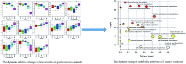
Cancer cachexia is a multifactorial metabolic syndrome characterized by a severe loss of body weight and lean body mass. Metabolic dysfunction is the primary hallmark of muscle atrophy. Herein, we studied dynamic metabolic profiles in serum and intact muscle. High-resolution magic angle spinning was employed for intact gastrocnemius muscle analysis and a dynamic metabolic model was established using C26 colon carcinoma-bearing mice from procachexia to the refractory cachexia period. When an integrated analysis of the 13 metabolites from the intact muscle gastrocnemius and 43 metabolites from the serum was performed, five distinguishable metabolic features were identified, including low blood glucose, elevated ketone bodies, decreased branched-chain amino acids, increased neutral amino acids, and high 3-methylhistidine and creatine. The metabolic hubs reveal potential biomarkers for the early detection of cachexia and indicate the underlying metabolic pathway reprogramming of muscle atrophy.


1. Introduction

Cachexia is a multifactorial metabolic syndrome that occurs in up to 80% of advanced cancer patients and contributes to approximately 30% of cancer mortality.1,2 It is associated with a reduction in quality of life, the response to therapeutic modalities and treatment tolerance.3 Lean body mass depletion is the most paradigmatic contribution to the severe body weight loss observed in cachexia, and it indicates a poor prognosis.4 Recent developments have shed light on the central role of maintaining skeletal muscle mass in the prevention and treatment of cachexia.5 Research has shown that the prevention of muscle wasting not only reverses the symptoms of cachexia but also dramatically prolongs survival, without influencing the level of fat loss or tumor growth.6 The loss of skeletal muscle cannot be reversed by appetite stimulants or nutritional support,7,8 and there is no pharmacological treatment that can sufficiently prevent ongoing skeletal muscle wasting. Therefore, the regulatory mechanisms underlying skeletal muscle metabolism are a topic of great interest.

The depletion of skeletal muscle in cancer cachexia is due to muscle fiber atrophy,9 which results from increased protein catabolism (hypercatabolism) or decreased protein synthesis (hypoanabolism).10 The presence of hypoanabolism suggests a shortage of the essential substrates for the net accumulation of muscle protein and the failure of the normal stimuli for muscle protein synthesis,11 whereas hypercatabolism potentially involves the excessive production of catabolic stimuli and activation of the ubiquitin-proteasome pathway. The mechanisms of hypoanabolism and hypercatabolism suggest possible metabolic hubs and altered pathways for skeletal muscle research.12 However, the underlying mechanisms that trigger and exacerbate cachexia-associated metabolic disorders remain elusive.

Although previous studies conducted in animal models and in humans have found that blood hyperlipidemia, hypoglycemia and decreasing levels of branched-chain amino acids (BCAAs) are associated with cancer cachexia,13–15 the specificity of these markers has been challenged, and the distinct metabolites in the skeletal muscle remain unknown. Moreover, metabolic dysfunction occurs before the symptoms of muscle atrophy, and the regulation of metabolism underlies the potential etiology and consequences of the subsequent weight loss. However, direct study of the skeletal muscles is unachievable with the currently available biochemical examinations and magnetic resonance imaging. However, integrated analysis of the skeletal muscle and serum could reveal biomarkers for the diagnosis of muscle atrophy and cancer cachexia. Metabolite changes in the skeletal muscle are associated with physiological dysfunction; therefore, altered metabolites might reveal the mechanisms underlying protein catabolism and synthesis.

In the present study, an approach consisting of the integrated analysis of serum and intact muscle metabonomics was employed to identify the early biomarkers of muscle atrophy and to reveal the metabolic regulatory mechanism underlying cancer cachexia. The gastrocnemius muscle was evaluated using high-resolution magic-angle spinning (HR-MAS) NMR spectroscopy. To identify metabolic profiles that were not affected by the cancer, a dynamic metabolic approach was employed by acquiring samples at multiple time points, from non-cachexia to the refractory cachexia period, from C26 colon carcinoma-bearing mice. The distinct metabolite hubs in the altered pathways could not only be used for the early detection of cancer cachexia but could also reveal the potential metabolic mechanism underlying cancer cachexia.

2. Materials and methods

2.1 Murine model of cancer cachexia

All of the procedures involving animals and their care in this study were approved by the animal care committee of our institution, in accordance with the institutional requirements and the Chinese government guidelines for animal experiments. Male BALB/c mice were purchased from Shanghai SLAC Laboratory Animal Co., Ltd. (Shanghai, China). The mice were maintained in pathogen-free conditions with a constant temperature (24 + 2 °C) and humidity (relative humidity of 55 ± 15%) and a 12[thin space (1/6-em)]:[thin space (1/6-em)]12 dark-light cycle. There was free access to water and identical standard food (when permitted) for all of the subjects.

The murine C26 colon carcinoma model for inducing cachexia was established as previously described.16,17 Briefly, BALB/c mice were randomly divided into groups and were subcutaneously injected with a suspension of 106 C26 cells in the left flank. The control mice were treated with an equivalent volume of PBS.

2.2 Experimental design

Two studies were performed. The first study assessed the relevance of the cancer cachexia model with the clinical features of body weight, muscle weight, and tumor size. Beginning on the day on which the mice were injected with the C26 adenocarcinoma cells, total body weight and food intake were measured daily. Tumor length and width were measured using digital calipers once the tumor was palpable, and in vivo tumor weight was calculated using the following equation: 0.52 × length × (width)2. Body weight was calculated as the total body weight minus the tumor weight. A second study was performed to reveal the dynamic metabolic profile of cancer cachexia. A total of eight groups were designed and every group had six to eight mice. The body and muscles were measured as the first experiment. Moreover, serum and the intact gastrocnemius muscle were acquired from the same mice following euthanasia via the inhalation of carbon dioxide, which was performed at multiple time points from procachexia to the refractory cachexia period. On every other day, from the ninth day to the twenty-first day, blood was collected in tubes. The heart, epididymal adipose tissue, gastrocnemius muscle and tibialis anterior muscle were dissected, weighed and quickly frozen in liquid nitrogen. The mice were then skinned, and the tumors were removed to measure the carcass body weight.

2.3 Western blot analysis

The gastrocnemius muscles from the model animals were homogenized and solubilized in lysis buffer using a commercial kit (Beyotime Institute of Biotechnology, Jiangsu, China). Briefly, 50 mg of tissue was extracted with lysis buffer (containing phenylmethanesulfonyl fluoride) to obtain low molecular weight soluble protein. The precipitate was then extracted with a hypertonic lysis buffer. After evaluating the protein content using a bicinchoninic acid protein assay kit (Pierce, Rockford, USA), the protein was solubilized in loading buffer and boiled at 95 °C for 5 minutes. Equal amounts of the protein samples (20 mg) were loaded on a 10% SDS-polyacrylamide gel to separate the muscle ring finger-1 (MuRF1) protein and on an 8% SDS-polyacrylamide gel to isolate the myosin heavy chain (MHC) protein. Subsequently, the proteins were electrophoretically separated and transferred onto PVDF membranes (Millipore Corporation, Bedford, USA) for western blot analysis. The blots were then incubated with the primary antibodies anti-MHC (ab51263, 1[thin space (1/6-em)]:[thin space (1/6-em)]1000 dilution, Abcam, Cambridge, USA) and anti-MuRF1 (ab77577, 1[thin space (1/6-em)]:[thin space (1/6-em)]1000 dilution, Abcam, Cambridge, USA), according to the manufacturer’s specifications. The proteins were detected using a peroxidase-conjugated secondary antibody (1[thin space (1/6-em)]:[thin space (1/6-em)]5000 dilution in Tris-buffered saline with Tween-20) with a chemiluminescence system and ImageQuant software.

2.4 1H-NMR spectroscopy of the serum

The plasma samples were prepared for NMR analysis by mixing 200 μL of plasma with 400 μL of PBS (containing 10% v/v D2O). All of the spectra were recorded using a Bruker Avance II 600 NMR spectrometer operated at a 600.13 MHz 1H resonance frequency. To attenuate the broad NMR signals from slowly tumbling molecules (such as proteins), a standard Carr–Purcell–Meiboom–Gill (CPMG) pulse sequence was used to record the 1D spin-echo spectra. A pre-saturation solvent was also employed to suppress the water peaks. Briefly, the CPMG pre-saturation pulse sequence worked using the equation −RD − 90° − (t − 180° − t)n − ACQ, where RD is the relaxation delay of 2 s; 90° and 180° represent the RF pulses that trip the magnetization vector; t is the spin-echo delay of 400 μs; n represents the number of loops (which was 80 in our experiment); and ACQ is the data acquisition period of 1.36 s.18 In our experiment, the data points were acquired using 128 transients, and the number of time domain points was 32k.

2.5 1H-MAS NMR spectra of the gastrocnemius muscle

The intact gastrocnemius muscle tissue (approximately 10 mg of tissue) was also evaluated using NMR spectroscopy. To eliminate the line broadening resulting from the macroscopic magnetic field homogeneities and the microscopic susceptibility differences in the intact tissue samples, a 4 mm HR-MAS 1H/13C probe (Bruker Biospin) was equipped with a Bruker Avance II 600 NMR spectrometer to obtain the HR-MAS NMR spectra. The samples were rinsed with D2O for about 10 minutes and immediately transferred into an MAS rotor using tweezers. The measurements were obtained at a constant 303k to reduce any metabolic degradation. To remove the spectral-broadening effects due to the interaction of solids, such as dipolar couplings and chemical shift anisotropy, the pulse sequence of 1D NOESY (noesypr1d) was used with the relaxation delay (RD) − 90° − t1 − 90° − tm − 90° acquired free induction decay (FID). The RD represents a relaxation delay of 1.5 s for the selective irradiation of water resonance. The 90° pulse length was 5.2 μs, and t1 was fixed at 4 μs. Following a mixing time (tm) of 150 ms, a second pulse was performed, and the saturation of the residual water signal was achieved by irradiating during the recycle delay at a δ equal to 4.70 ppm. The data points were recorded using a 5 kHz spinning speed with 256 transient acquisitions. The FIDs were multiplied by an exponential weighting factor corresponding to a line broadening at 0.3 Hz. After, the Fourier transformation, phase correction and baseline correction were carefully performed using the TopSpin software package (Bruker Biospin, Rheinstetten, Germany), version 3.0. The 1H chemical shifts referred to the methyl doublet signal of lactate (δ 1.33).

2.6 Data reduction of the NMR data

The corrected NMR spectra, corresponding to the chemical shift range of δ 0.2–10.0, were imported into AMIX 3.9.5 (Bruker Biospin, Rheinstetten, Germany), and all of the spectra were reduced into integral regions of equal lengths of 0.005 ppm. Regions of δ 4.7–5.1 that contained the resonance from residual water were set to zero. To reduce the concentration differences between the samples, the data were normalized to the total spectral area (100%).

2.7 Metabonomics analysis

The dataset was analyzed by pattern recognition methods using the software packages Simca-P, version 11.5 (UmetricsAB, Umeå, Sweden), and MetaboAnalyst, version 2.0 (http://www.metaboanalyst.ca). The dataset was arranged with the samples as observations and the peak areas of the chemical shifts as the response variables. Before multivariate statistics were performed, the response variables were centered and scaled to Pareto variance. The base weight was computed as 1/sqrt (standard deviation of the response variables). Moreover, to make the skewed distributions more symmetrical, log transformations were used for nonlinear conversions of the data.

To explain the maximum variation between the samples, a principal component analysis (PCA) bilinear decomposition method was used to view the clusters within the multivariate data. Moreover, a partial least squares-discriminant analysis (PLS-DA) was applied to explain the maximum separation between the defined class samples in the data. Three parameters, R2X, R2Y and Q2, were used for the evaluation of the models. R2X explains the cumulative variation in the response variables, and R2Y is the latent variable of the sum of the squares of all of the Xs and Ys. Q2 reflects the cumulative cross-validated percentage of the total variation that can be predicted by the current latent variables. High coefficient values of R2Y and Q2 represent good discrimination and predictive ability, respectively. The specific metabolites between classes were interpreted using variable importance in the projection (VIP) and correlation coefficients. The variables with a high VIP are considered to be statistically significant; therefore, VIP statistics were applied for metabolic pathway analysis.

2.8 Metabolite identification

Based on the statistical results of the metabonomics analysis, the discriminating peaks were prioritized for identification. The NMR signals were compared with reference spectra from the HMDB database and with Chenomx NMR Suite metabonomics software (Chenomx, Inc., Alberta, Canada), based on the coupling constant and the splitting model. The signal assignments were conducted using the 2D J-resolved, COSY, and TOCSY spectra. The overlaps or slight shifts in their positions were confirmed by re-recording the 1H NMR spectra of the serum after the addition of small quantities of the respective standard compounds.

2.9 Metabolic pathway analysis

To identify the most relevant metabolic pathways involved in cancer cachexia, metabolomic pathway analysis was employed to perform a pathway enrichment analysis and pathway topology analysis. The pathway enrichment analysis used GlobalTest and GlobalAncova to analyze the concentration values with high sensitivity in order to identify subtle changes involved in the same biological pathway. However, a relative betweenness centrality was used for the metabolite importance measurement and the established global network topology analysis for its ability to order the metabolic pathways according to their positions. Changes in a more important node of the network triggered a more severe impact on the pathway than changes that occurred in marginal or relatively isolated positions.

2.10 Statistical methods

The data are expressed as the means ± SDs. The calculated means were statistically analyzed using Origin software, version 8.1 (OriginLab Corp., Northampton, MA, USA). The differences between the two groups were analyzed using Student’s two-sided t test, and differences involving more than two groups were analyzed using one-way ANOVA, followed by Tukey’s post hoc test. The level of significance was set at p < 0.05.

3. Results

3.1 CT26 tumor induced cancer cachexia results in ongoing loss of body weight and skeletal muscle in mice

The growth of C26 colon carcinoma resulted in impaired body weight gain that was associated with a progressive loss of skeletal muscle weight. On day 13 following tumor implantation, the body weights of the mice differed from the control values by at least 5%, which was statistically significant (Fig. 1). To explore the changes in body composition associated with cancer cachexia, the mice were sacrificed, and the weights of the organs, skeletal muscle, fat and the wet and dry carcass were determined. The lean body weight loss is presented relative to the significant loss of skeletal muscle. In particular, the mass of the gastrocnemius and tibialis anterior muscles decreased by more than 20% on day 13 and reached approximately 45% on day 21. The ongoing loss of wet carcass weight was consistent with that for dry carcass weight, and the correlation coefficient was 96.88% (Table 1). The decreased dry carcass weight was mainly due to muscle wasting, and these results indicated that the body weight loss was due to muscle atrophy in the murine cachexia model.
image file: c5ra19004e-f1.tif
Fig. 1 Line charts showing changes in carcass weight (A), dry carcass weight (B), C26 colon carcinoma size (C), the intact gastrocnemius muscle (D), and the protein expression of MuRF1 (E) and MHC (F). The carcass weight decreased significantly from day 15 following the implantation of the tumor. The dry carcass weight decreased relative to the wet carcass. Moreover, the gastrocnemius muscle also showed a wasting trend that was related to the expression of MHC.
Table 1 The body characteristics of tumor-bearing mice from procachexia to the refractory cachexia perioda
  D9 D11 D13 D15 D17 D19 D21 Control
a The data are shown as the mean ± SD. D9, D11 and so on are the days after tumor implantation. Each group included 6 mice, with the exception of 8 mice in the control group. The control mice were sacrificed on day 7, which corresponded to the model animals. Values that were significantly different from the control group (indicated by *p < 0.05) were determined by Tukey’s post hoc test followed by one-way ANOVA.
Heart (mg) 126.33 ± 15.40 107.67 ± 4.03* 127.5 ± 26.01 112.50 ± 20.73* 116.33 ± 20.35* 102.83 ± 33.08* 110.67 ± 22.62* 124.00 ± 25.49
Lung (mg) 138.17 ± 12.53* 139.17 ± 13.76* 141 ± 13.64* 145.51 ± 12.39* 143.01 ± 17.13* 143.5 ± 12.23* 131.17 ± 11.09* 117.67 ± 9.97
Liver (mg) 1225 ± 81.99* 1226.5 ± 70.53* 1351 ± 60.97* 1332 ± 182.71* 1303.5 ± 416.04* 1256.67 ± 199.52* 1221.5 ± 231.6* 869.33 ± 55.44
Spleen (mg) 131.67 ± 19.48* 157.50 ± 29.95* 215.83 ± 21.67* 220.67 ± 27.43* 286.67 ± 43.26* 284.00 ± 71.88* 210.83 ± 114.83* 80.33 ± 13.95
Kidney (mg) 364.5 ± 28.33* 348.83 ± 20.41* 353.33 ± 34.85* 345.62 ± 64.24* 338.33 ± 24.96* 314.00 ± 27.73 313.83 ± 40.67 306.83 ± 26.72
Gastrocnemius muscle (mg) 233.83 ± 11.48* 228.00 ± 15.13* 248 ± 24.75* 227.00 ± 16.75* 195.67 ± 49.56* 213.17 ± 18.86* 183.17 ± 21.55* 322.67 ± 119.41
Anterior tibial muscle (mg) 77.51 ± 6.09* 86.33 ± 16.51* 72.5 ± 35.39* 80.67 ± 14.88* 99.67 ± 68.45* 62.50 ± 32.10* 60.17 ± 5.91* 112.01 ± 34.39
Epididymal adipose (mg) 466.5 ± 79.32* 412.17 ± 51.94* 432.17 ± 60.14* 328.21 ± 67.11* 199.52 ± 92.57* 137.67 ± 105.16*   560.67 ± 108.28
Tumor weight (mg) 476 ± 331.78 864.17 ± 473.13 2853.67 ± 1060.09 3631.17 ± 701.68 5130.07 ± 0.94 7811.24 ± 1.14 9522.41 ± 0.84  
Carcass (g) 14.83 ± 0.46 14.68 ± 0.31 14.38 ± 0.63 13.45 ± 0.86* 12.75 ± 1.25* 12.65 ± 0.5* 11.83 ± 1.59* 15.63 ± 0.62
Dry body weight (g) 5.40 ± 0.29 4.88 ± 0.16* 4.82 ± 0.27* 4.42 ± 0.55* 4.37 ± 0.36* 3.93 ± 0.29* 3.47 ± 0.42* 5.61 ± 0.21


MHC expression was significantly decreased during the cachexia period (17 d and 21 d). MHC is degraded by the ubiquitin-proteasome system, as suggested by the increase in the muscle-specific E3 ubiquitin ligase MuRF1 from day 13, which has previously been associated with decreased MHC content.19,20 The dynamic loss of muscle weight and increase in MuRF1 indicated the progression of the cancer cachexia over the duration of the sampling.

3.2 Disorders of serum metabolic profiling is a hallmark of cancer cachexia

The typical 1H NMR spectra of the serum demonstrated a unique metabolic profile along with the metabolite assignments (Fig. 2). The spectra contained very high-intensity signals from glucose, lactate, creatine, alanine and VLDL/LDL. Moreover, numerous signals from the dynamic cachexia mouse model showed marked changes in the levels of the endogenous metabolites compared with the control mice, including pyruvate, acetate, 2-oxoglutarate, phenylalanine, glycine, leucine, valine, isoleucine, glutamine, choline, succinate, acetate, carnitine, and citrate.
image file: c5ra19004e-f2.tif
Fig. 2 The typical 1H-NMR spectra of the serum from the dynamic cachexia mouse model. The metabolites are assigned and marked. The overlapping peaks were identified by adding a reference substance to the sample.

To illustrate the differences in the metabolic profiles, the NMR spectra dataset was subjected to PCA. The scores plot revealed distinct separation of the tumor-bearing model (G1–G7) from the control mice (G8). Moreover, the samples from the procachexia groups (G1–G2) were clustered together, and the samples from the later cachexia period (G5–G7) were clustered in a different region. Though the discrimination was visible, the cumulative R2X was 0.433 and Q2 was 0.278 with 4 principal components. Therefore, a supervised PLS analysis was also applied to the data after Pareto scaling, and it showed well-separated clusters (Fig. 3). Three principal components were given and the cumulative R2X, R2Y and Q2 were 0.663, 0.997, 0.925 respectively. The high values of R2Y and Q2(cum) represent good discrimination and predictive ability. The G1 and G2 groups were clustered closely to the control group, suggesting that the homeostasis during the initial cancer cachexia period was similar to that of the control group. The G5, G6 and G7 groups were clustered in other regions, indicating a typical cancer cachexia metabolic profile. The datasets were analyzed using MetPA and showed similar score-loading plots (Fig. S1 in the ESI). The significantly different metabolites, as well as the p values of the dynamic mouse model, were calculated (Table S1 and Fig. S2 in the ESI). The relative intensity levels are partially shown in Fig. 4, including increased glutamate, 3-hydroxybutyrate, citrate, isocitrate, malate, arginine, creatine, carnitine, taurine, TMAO, 3-methylhistidine, glycine, and phenylalanine levels and decreased glucose, 2-oxoglutarate, succinate, ornithine, leucine and valine levels.


image file: c5ra19004e-f3.tif
Fig. 3 The PLS-DA scores plot of the eight groups, based on the serum NMR spectra. G1–G7 represent the groups from day 9 to day 21 (every other day), and G8 is the control group. The control group (G8) was separated from the tumor-bearing mouse groups (G1–G7), and the non-cachexia mouse groups (G1–G2) were distributed over a time period different from the cachexia period (G5–G7). The development of severe cachexia occurred dynamically.

image file: c5ra19004e-f4.tif
Fig. 4 The normalized concentration of the examined serum metabolites from non-cachexia to severe cachexia. G1–G7 represent the groups from day 9 to day 21 (every other day).

3.3 MAS NMR reveals the distinct metabolic features for skeletal muscle atrophy during cachexia

Because muscle atrophy is the primary factor contributing to body wasting in cachexia, an analysis of the skeletal muscle metabolites could provide a direct clue for biomarker research. Unlike the serum metabolic spectra, there was a low concentration of glucose, and the most abundant metabolite was creatine, which was relatively low compared with the control group and which increased from procachexia to cachexia (Fig. 5). The overall spectra revealed endogenous metabolites without degradation or lysis. Subtle changes in specific metabolites in the skeletal muscle were observed, including in taurine, 3-methylhistidine, creatine, inosine, carnosine, and 3-hydroxybutyrate (Fig. 6). This finding showed that the MAS NMR spectra could highlight the original perturbations of the endogenous metabolites in the controls and the dynamic mouse model.
image file: c5ra19004e-f5.tif
Fig. 5 The typical HR-MAS NMR spectra of the intact gastrocnemius muscle tissue from the dynamic cachexia mouse model. Unlike the metabolic profiles of the serum, the intact tissue contained high levels of creatine and taurine and low glucose levels.

image file: c5ra19004e-f6.tif
Fig. 6 The normalized concentration of the examined muscle metabolites from non-cachexia to severe cachexia. M1 to M4 represent the groups from day 9 to day 21 (every three days), and M0 is the control group.

PCA revealed complete separation between the cachexia group and the control group in the scores plot (Fig. S3 in the ESI). Five principal components were given and the R2X was 0.822. Similar to the serum samples, the gastrocnemius muscles of the cachexia mice showed a dynamic metabolic profile, whereas the procachexia group clustered closely to the control group. To find the corresponding different metabolites, PLS-DA was employed and two principal components were given with R2X, R2Y and Q2(cum), being 0.605, 0.929, and 0.881 respectively. The S-plot (Fig. S4 in the ESI) displayed a compromised insulin action and altered intermediate metabolism of fats and amino acids. Based on multiple tests, the dynamic changes included increased phenylalanine, 3-methylhistidine, inosine, taurine, creatine, carnitine, glycine, 3-hydroxybutyrate, and lactate and decreased leucine, valine, isoleucine, and glucose (Fig. 6 and Table S2 in the ESI). In particular, changes in the muscle concentrations of select essential amino acids and their derivatives, particularly the BCAAs, the sulfur amino acids and phenylalanine were apparent, in addition to muscle atrophy, both of which often occurred before the onset of clinically diagnosed cachexia.

3.4 Integrated analysis of serum and intact muscle metabonomics revealed the five distinguishable metabolic features of cancer cachexia

MetPA presented 21 altered metabolic networks (Fig. 7), with a detailed score of impact and −log(p) (Table S3 in the ESI). Valine, leucine and isoleucine biosynthesis were the most impacted pathways, which were calculated using pathway topology analysis. Three matched metabolites of valine, leucine, and isoleucine were gradually reduced, and the difference was significant. Additionally, the ketone body synthesis and degradation pathway was enhanced, with a 0.60 impact factor. The ketone bodies were primarily metabolized from lipids and emerged as a compensatory mechanism for glucose metabolism. The cachexia syndrome resulted in the synthesis and release of ketone bodies into the blood, causing the acetoacetate levels to rise gradually and inducing hypolipidemia during the later cachexia period. The high ketone levels were toxic and prompted ketoacidosis, indicating ketone body metabolism and pyruvate metabolism in cancer cachexia.
image file: c5ra19004e-f7.tif
Fig. 7 Cancer cachexia triggers metabolic pathway reprogramming. The X axis represents pathway impact, and the Y axis represents the −log(p). Biosynthesis of BCAA was the most important impacted pathway and −log(p) > 1.3 denoted statistically significant differences between pathways.

The cachexia pathway that stood out the most was phenylalanine biosynthesis. As an aromatic amino acid, phenylalanine is primarily metabolized in the liver. The presence of phenylalanine indicates impaired hepatic function and negative regulation of food intake, corresponding to the biochemical parameters of transaminase from other reports.21–23

Among the marked endogenous metabolite perturbations, the pathway analysis further showed subtle changes in taurine and hypotaurine metabolism, glycine, serine and threonine metabolism, glycerolipid metabolism, glyoxylate and dicarboxylate metabolism, the citrate cycle (TCA cycle), alanine, aspartate and glutamate metabolism, glycolysis and gluconeogenesis, inositol phosphate metabolism, cysteine and methionine metabolism, arginine and proline metabolism, and glycerophospholipid metabolism. Interestingly, the metabolic intermediates of the amino acids, such as creatine, carnitine, taurine, TMAO and 3-methylhistidine, steadily increased during the ongoing progress of cancer cachexia. An additional multigroup analysis of the compounds revealed that the gradual increases in the intermediate metabolites occurred along with the accumulation of nitrogen and the aggravation of muscle wasting.

Multivariate data analysis of serum and intact muscle metabonomics from multiple stages in the murine cachexia model indicated that there were distinguishable profiles of serum and muscle metabolites. However, similar differences appeared in the composition and concentration of the metabolite hubs. After the integrated analysis of the metabolic profiles obtained from serum and intact muscle, the distinct metabolic characteristics included low blood glucose, elevated ketone bodies, decreased BCAAs, increased phenylalanine and elevated amino acid intermediates.

4. Discussion

Currently, dual-energy X-ray absorptiometry is considered to be the gold standard for assessing muscle wasting; however, this method provides little chemical information and has a limited ability to detect early wasting of skeletal muscle.24 In fact, metabolic disorder occurs prior to the wasting observed in cachexia, and metabolites can be used for the early detection of cancer cachexia.25,26 Previous metabolomic research into cancer cachexia was based on the NMR analysis of serum; however, the blood metabolites were not sufficient to provide detailed functional data on the biochemistry of muscle wasting. Instead, intact muscle can reveal tissue-specific metabolic processes, as well as allow for the integration of data collected from multiple experiments. The major issue with the analysis of intact muscle is the anisotropic electronic and magnetic interactions between the nuclei in intact biological tissues, which cannot be completely averaged, resulting in broad lines in a conventional NMR spectrum.27 MAS based NMR spectra provided narrow lines and well-resolved samples, and the intact muscle tissue could be analyzed nondestructively.28 Thus, in our study, the metabolic disorders of intact gastrocnemius muscle were analyzed with HR-MAS NMR technology.

In the present study, we performed integrative metabolic profiling of the serum and intact skeletal muscle tissue of mice with cachexia using 1H-NMR-based metabonomics methods to identify early biomarkers of muscle atrophy and cancer cachexia and to examine the metabolic regulation of protein catabolism and synthesis. Forty-three distinguishing metabolites were found to be altered significantly in the serum, and thirteen metabolites were altered in the gastrocnemius muscle during the dynamic progression of cancer cachexia. Although there have been metabolomic reports on the identification of serum and urine biomarkers in cancer cachexia,13,14 the present study revealed the specifically altered metabolites in intact skeletal muscle tissues in a dynamic manner.

Consistent with previous reports, decreased levels of glucose and increased levels of lactate were observed in both the serum and intact gastrocnemius muscle. Tumor-bearing mice require glucose at a faster rate than healthy animals. However, the glucose is metabolized and enters the glycolytic pathway, leading to greater pyruvate formation. The majority of the pyruvate is then disposed of via non-oxidative pathways and is diverted into lactate and alanine, rather than being oxidized via the citric acid cycle.29 Excessive lactate production exacerbates energy wasting and induces higher glucose uptake and utilization.30 Reports have shown that altered glucose metabolism is associated with whole-body protein turnover.23 Skeletal muscle is the primary target of glucose uptake, and the disruption of glucose metabolism and its intermediate metabolites bears some similar characteristics to tumor cell metabolism. Thus, its specificity for the evaluation of muscle wasting was limited.

Elevated ketone bodies were also observed in the blood and showed changing tendencies. The increase in ketobodies in the gastrocnemius muscle can also be viewed as a characteristic of muscle wasting. Although neither the ATP synthesis efficiency nor the mitochondrial uncoupling in the skeletal muscles of cachectic animals were shown to be altered,31 compensatory energy transduction was increased in the presence of dysregulated glucose metabolism. In this instance, the adipose tissue was broken down to catabolize circulating triacylglycerol. Lipolysis not only regulates the homeostasis of systemic energy production but also leads directly to the loss of adipose tissue. Recent studies in animal models of cachexia and in humans have confirmed that the lipolysis of triacylglycerol is the primary factor contributing to fat loss.32 Triacylglycerol is catalyzed to release fatty acids and to generate quantities of acetyl-CoA, which overwhelms the transfer ability of carnitine acyltransferase. Subsequently, ketone biosynthesis is enhanced, yielding 3-hydroxybutyrate and acetoacetate. Reports have shown that skeletal muscle maintains an increased usage of 3-hydroxybutyrate and lactate,33 indicating that in cachexia, ketone bodies can be utilized as metabolic fuel for energy metabolism.

Additionally, the most impacted pathway of muscle wasting was the biosynthesis of BCAAs, accompanied by a gradual reduction in valine, leucine, and isoleucine concentrations in both the serum and the gastrocnemius muscle. Consistent evidence has shown that BCAAs are not only an important energy substrate,34,35 but they also act as precursors to improve nitrogen retention and protein synthesis.36,37 Research has shown that leucine can stimulate muscle protein anabolism,11 inhibit catabolism,38 and modulate glucose homeostasis.34 An infusion of leucine can promote the synthesis of glutamine and alanine, which are then exported to the liver and are metabolized into glucose. Moreover, BCAAs are primarily catabolized in the skeletal muscle; therefore, they are proposed for the treatment of catabolic disease states.

Interestingly, the aromatic amino acid phenylalanine steadily increased in the serum. High levels of phenylalanine can interfere with the production of serotonin and other amino acids, as well as nitric oxide, which thus indicates metabolic reprogramming in cancer cachexia.22 In a low-calorie environment, to maintain homeostasis, large amounts of neutral amino acids are released and metabolized as an alternative energy-generating pathway. This process results in high circulating concentrations of glutamine and aspartate. High levels of neutral amino acids have been reported to promote an anabolic state by inhibiting proteolysis and directly stimulating protein synthesis.39 High protein turnover wastes the essential amino acids, which cannot be supplied during cachexia. In the presence of amino acid hypermetabolism and accelerated protein catabolism, amino acid intermediates, such as 3-methylhistidine and creatine, were also found to be elevated in our study.

3-Methylhistidine showed specificity for the indication of the wasting of muscle protein. When actin and myosin are synthesized, histidine residues are methylated to construct the cytocontractile apparatus in skeletal muscle fibers. However, during myofibrillar breakdown, 3-methylhistidines are released, and they cannot be recycled for intermediary metabolism or protein synthesis. Based on its biology, serum or urine 3-methylhistidine represents gross protein degradation and serves as a valuable biomarker for monitoring myofibrillar protein degradation.40,41 Moreover, elevation of 3-methylhistidine could be detected before the symptom of muscle wasting, suggesting the presence of increased muscle protein catabolism.

Creatine was another elevated amino acid intermediate found in the wasting muscle. Creatine is produced from arginine, glycine, and methionine in the kidneys and liver. After being transported into the blood, creatine is mainly taken up by the skeletal muscle through an active transport system.42 In fact, a large proportion of the total body creatine is distributed in the skeletal muscle and is stored as phosphocreatine. In the state of catabolic hypermetabolism and disrupted muscle metabolism, large pools of creatine circulate in the blood. Therefore, the accumulation of serum creatine and excessive activated creatine kinase are both markers for dysregulated muscle function.43

In conclusion, the present study reports an integrated metabonomics analysis of serum and intact gastrocnemius muscles from mice with cancer cachexia. Five distinguishable metabolic features of cachexia were identified: low blood glucose, elevated ketone bodies, decreased BCAAs, increased neutral amino acids and high amino acid intermediates. The consistent metabolic profiles of the serum and intact muscle imply a feasible and practicable diagnostic approach for predicting skeletal muscle loss or other features of at-risk clinical populations through the detection of serum metabolites. Moreover, the altered pathways could reveal the potential metabolic mechanism of cancer cachexia.

Nevertheless, limitations must be considered in the present research. Firstly, the complex metabolic syndrome of cancer cachexia cannot currently be distinguished by a single biomarker; however, a comprehensive evaluation of the five features described above is indicated as a potential biomarker for the early detection of cancer cachexia. Secondly, all of the results were acquired using the classic cancer cachexia model. Although further clinical studies are needed to confirm the results, this dynamic model allowed us to directly identify the metabolic profile of intact muscle in a dynamic manner.

Conflict of interest

The authors declare that they have no conflicts of interest.

Acknowledgements

The present study was supported by grants from the Natural Science Foundation of China (No. 81503155) and the College Subject of Shanghai Jiao Tong University Affiliated Sixth People’s Hospital (No. ynlc201420).

References

  1. K. Fearon, F. Strasser, S. D. Anker, I. Bosaeus, E. Bruera, R. L. Fainsinger, A. Jatoi, C. Loprinzi, N. MacDonald and G. Mantovani, Lancet Oncol., 2011, 12, 489–495 CrossRef.
  2. K. Kalantar-Zadeh, C. Rhee, J. J. Sim, P. Stenvinkel, S. D. Anker and C. P. Kovesdy, J. Cachexia Sarcopenia Muscle, 2013, 4, 89–94 CrossRef PubMed.
  3. K. C. Fearon, N. Engl. J. Med., 2011, 365, 565–567 CrossRef CAS PubMed.
  4. J. A. Langius, S. Bakker, D. H. Rietveld, H. M. Kruizenga, J. A. Langendijk, P. J. Weijs and C. R. Leemans, Br. J. Cancer, 2013, 109, 1093–1099 CrossRef CAS PubMed.
  5. S. Dodson, V. E. Baracos, A. Jatoi, W. J. Evans, D. Cella, J. T. Dalton and M. S. Steiner, Annu. Rev. Med., 2011, 62, 265–279 CrossRef CAS PubMed.
  6. X. Zhou, J. L. Wang, J. Lu, Y. Song, K. S. Kwak, Q. Jiao, R. Rosenfeld, Q. Chen, T. Boone, W. S. Simonet, D. L. Lacey, A. L. Goldberg and H. Q. Han, Cell, 2010, 142, 531–543 CrossRef CAS PubMed.
  7. I. Bosaeus, Support. Care Cancer, 2008, 16, 447–451 CrossRef PubMed.
  8. L. Santarpia, F. Contaldo and F. Pasanisi, J. Cachexia Sarcopenia Muscle, 2011, 2, 27–35 CrossRef PubMed.
  9. M. J. Tisdale, J. Supportive Oncol., 2005, 3, 209–217 CAS.
  10. V. E. Baracos, Cancer, 2001, 92, 1669–1677 CrossRef CAS.
  11. K. Fearon, J. Arends and V. Baracos, Nat. Rev. Clin. Oncol., 2013, 10, 90–99 CrossRef CAS PubMed.
  12. V. E. Baracos, Nutrition, 2000, 16, 1015–1018 CrossRef CAS.
  13. H. der-Torossian, S. Asher, J. Winnike, A. Wysong, X. Yin, M. Willis, T. O’Connell and M. Couch, Metabolomics, 2013, 9, 730–739 CrossRef CAS.
  14. T. M. O’Connell, F. Ardeshirpour, S. A. Asher, J. H. Winnike, X. Yin, J. George, D. C. Guttridge, W. He, A. Wysong and M. S. Willis, Metabolomics, 2008, 4, 216–225 CrossRef.
  15. Y. Quanjun, Y. Genjin, W. Lili, L. Bin, L. Jin, Y. Qi, L. Yan, H. Yonglong, G. Cheng and Z. Junping, Mol. BioSyst., 2013, 9, 3015–3025 RSC.
  16. Y. Quan-Jun, B. Jun, W. Li-Li, H. Yong-Long, L. Bin, Y. Qi, L. Yan, G. Cheng and Y. Gen-Jin, RSC Adv., 2015, 5, 11766–11775 RSC.
  17. Y. Tanaka, H. Eda, T. Tanaka, T. Udagawa, T. Ishikawa, I. Horii, H. Ishitsuka, T. Kataoka and T. Taguchi, Cancer Res., 1990, 50, 2290–2295 CAS.
  18. O. Beckonert, H. C. Keun, T. M. Ebbels, J. Bundy, E. Holmes, J. C. Lindon and J. K. Nicholson, Nat. Protoc., 2007, 2, 2692–2703 CrossRef CAS PubMed.
  19. N. Ebner, L. Steinbeck, W. Doehner, S. D. Anker and S. von Haehling, J. Cachexia Sarcopenia Muscle, 2014, 5, 27–34 CrossRef PubMed.
  20. G. Zhang, B. Jin and Y. P. Li, EMBO J., 2011, 30, 4323–4335 CrossRef CAS PubMed.
  21. M. Lima, S. Sato, R. T. Enos, J. W. Baynes and J. A. Carson, J. Appl. Physiol., 2013, 114, 824–828 CrossRef CAS PubMed.
  22. B. K. Ubhi, J. H. Riley, P. A. Shaw, D. A. Lomas, R. Tal-Singer, W. MacNee, J. L. Griffin and S. C. Connor, Eur. Respir. J., 2012, 40, 345–355 CrossRef CAS PubMed.
  23. H. Norrelund, H. Wiggers, M. Halbirk, J. Frystyk, A. Flyvbjerg, H. E. Botker, O. Schmitz, J. O. Jorgensen, J. S. Christiansen and N. Moller, J. Intern. Med., 2006, 260, 11–21 CrossRef CAS PubMed.
  24. V. E. Baracos, J. Cachexia Sarcopenia Muscle, 2011, 2, 71–73 CrossRef PubMed.
  25. R. P. Sah, S. J. Nagpal, D. Mukhopadhyay and S. T. Chari, Nat. Rev. Gastroenterol. Hepatol., 2013, 10, 423–433 CrossRef CAS PubMed.
  26. T. Agustsson, P. Wikrantz, M. Ryden, T. Brismar and B. Isaksson, Nutrition, 2012, 28, 851–855 CrossRef PubMed.
  27. J. Z. Hu, Methods Mol. Biol., 2011, 708, 335–364 CAS.
  28. O. Beckonert, M. Coen, H. C. Keun, Y. Wang, T. M. Ebbels, E. Holmes, J. C. Lindon and J. K. Nicholson, Nat. Protoc., 2010, 5, 1019–1032 CrossRef CAS PubMed.
  29. G. P. Bongaerts, H. K. van Halteren, C. A. Verhagen and D. J. Wagener, Med. Hypotheses, 2006, 67, 1213–1222 CrossRef CAS PubMed.
  30. J. A. Tayek, J. Am. Coll. Nutr., 1992, 11, 445–456 CrossRef CAS.
  31. C. M. Julienne, J. F. Dumas, C. Goupille, M. Pinault, C. Berri, A. Collin, S. Tesseraud, C. Couet and S. Servais, J. Cachexia Sarcopenia Muscle, 2012, 3, 265–275 CrossRef PubMed.
  32. M. Lafontan, C. Moro, C. Sengenes, J. Galitzky, F. Crampes and M. Berlan, Arterioscler., Thromb., Vasc. Biol., 2005, 25, 2032–2042 CrossRef CAS PubMed.
  33. H. D. Mulligan and M. J. Tisdale, Biochem. J., 1991, 277(Pt 2), 321–326 CrossRef CAS.
  34. S. H. Adams, Adv. Nutr., 2011, 2, 445–456 CrossRef CAS PubMed.
  35. V. E. Baracos and M. L. Mackenzie, J. Nutr., 2006, 136, 237S–242S CAS.
  36. P. L. de Campos-Ferraz, I. Andrade, W. das Neves, I. Hangai, C. R. Alves and A. H. Lancha Jr, J. Cachexia Sarcopenia Muscle, 2014, 5, 105–110 CrossRef PubMed.
  37. W. L. Morrison, J. N. Gibson and M. J. Rennie, Eur. J. Clin. Invest., 1988, 18, 648–654 CrossRef CAS.
  38. Z. Aversa, A. Bonetto, P. Costelli, V. G. Minero, F. Penna, F. M. Baccino, S. Lucia, F. Rossi Fanelli and M. Muscaritoli, Int. J. Oncol., 2011, 38, 713–720 CAS.
  39. G. Wu, Amino Acids, 2009, 37, 1–17 CrossRef CAS PubMed.
  40. M. Sheffield-Moore, E. L. Dillon, K. M. Randolph, S. L. Casperson, G. R. White, K. Jennings, J. Rathmacher, S. Schuette, M. Janghorbani, R. J. Urban, V. Hoang, M. Willis and W. J. Durham, J. Cachexia Sarcopenia Muscle, 2014, 5, 19–25 CrossRef CAS PubMed.
  41. N. Aranibar, J. D. Vassallo, J. Rathmacher, S. Stryker, Y. Zhang, J. Dai, E. B. Janovitz, D. Robertson, M. Reily, L. Lowe-Krentz and L. Lehman-McKeeman, Anal. Biochem., 2011, 410, 84–91 CrossRef CAS PubMed.
  42. S. A. Stimpson, S. M. Turner, L. G. Clifton, J. C. Poole, H. A. Mohammed, T. W. Shearer, G. M. Waitt, L. L. Hagerty, K. S. Remlinger, M. K. Hellerstein and W. J. Evans, J. Appl. Physiol., 2012, 112, 1940–1948 CrossRef CAS PubMed.
  43. S. S. Patel, M. Z. Molnar, J. A. Tayek, J. H. Ix, N. Noori, D. Benner, S. Heymsfield, J. D. Kopple, C. P. Kovesdy and K. Kalantar-Zadeh, J. Cachexia Sarcopenia Muscle, 2013, 4, 19–29 CrossRef PubMed.

Footnotes

Electronic supplementary information (ESI) available. See DOI: 10.1039/c5ra19004e
Contributed equally for the manuscript.

This journal is © The Royal Society of Chemistry 2015
Click here to see how this site uses Cookies. View our privacy policy here.