DOI:
10.1039/C5RA18701J
(Paper)
RSC Adv., 2015,
5, 97066-97075
Preparation and characterization of pullulanase debranched starches and their properties for drug controlled-release
Received
12th September 2015
, Accepted 27th October 2015
First published on 28th October 2015
Abstract
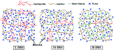
Debranched starches (DBSs) with different degrees of debranching (low, L-DBS; moderate, M-DBS; high, H-DBS) were prepared and investigated. After pullulanase modification, the starch granules became more porous and many small particles containing short glucan chains were generated. DBSs adopt a single-helical V-type crystalline structure with low crystallinity. L-DBS samples contained fewer (20.70%) and longer (degree of polymerization, DP: 21.92) linear short glucan chains than their counterparts (M-DBS: 40.92%, 20.05 DP; H-DBS: 55.52%, 18.52 DP). Pullulanase enzymatic hydrolyzate for DBS samples with higher degrees of debranching inclined towards retrogradation at 20 °C. DBSs with higher degrees of debranching could form a hydrogel with higher G′ and G′′ values, indicating these samples formed a stronger gel network. L-DBS could hold more water and its digestibility was higher. The in vitro test showed that DBS is a good candidate to control drug release for over 12 h. Furthermore, the drug release profiles from both DBS-based and HPMC-based tablets showed an anomalous transport mechanism. The drug release from these four matrices was controlled by a combination of drug diffusion and matrix erosion. The drug release properties from DBS-based tablets were considerably influenced by the degree of debranching. The in vitro drug release profile of M-DBS was similar to that of HPMC (f2 = 60.75), while L-DBS and H-DBS differed from HPMC (f2 < 50). In summary, DBS is a good hydrogel candidate, and it can be used as an excipient in oral tablets to control drug release.
1 Introduction
Starch is the second most abundant carbohydrate in nature after cellulose.1 It is composed of linear amylose and branched amylopectin. Starch is a cheap, nontoxic, biocompatible and biodegradable material. Starch modification technologies, including physical, chemical and enzymatic modifications, can endow starch derivatives with new functional properties. Now, starch and its derivatives have been widely used in food, paper and textiles, agriculture, plastic, consumer items, as well as the pharmaceutical industry.2–6
Debranching enzymes, like pullulanase and isoamylase, can be utilized to hydrolyze 1,6-α-D-glucosidic bonds at branching points.7 These enzymatically modified starches contain many linear, low-molecular-weight, recrystallizable glucan chains. These short glucan chains are available for chain realignment via hydrogen bonding and hydrophobic interactions, leading to chain aggregation and gel network formation.8
Most of the previous research has focused on the nutritional characteristics of debranched starches. Researchers confirmed that debranched starch (DBS) is an important source for the production of resistant starch (RS) and slowly digestible starch (SDS).8–11 Temperature-cycling storage or low-temperature incubation can be used to induce retrogradation and recrystallization, thereby improving RS and SDS yields.9,11–13
Retrograded starch,14 starch acetate,15 cross-linked starch,16–18 carboxymethyl starch19–21 and grafted starch22,23 have been introduced into the pharmaceutical industry and used as excipients in oral tablets to control drug release attributed to their hydrogel properties. These modifications can rectify the deficiencies of native starch used as extended-release excipients, like low compactibility, elastic compression and enzymatic degradation by α-amylase.24 Debranched starches are hydrophilic and can form hydrogels in aqueous media.25–28 Thus, based on previous research, it is feasible to use debranched starches as hydrophilic excipients to extend drug release.
The fine structure of debranched starches can influence the realignment and retrogradation of short glucan chains. Various degrees of debranching generate differences in structural and hydrogel properties. Thus, debranched starches with different degrees of debranching would perform differently in forming hydrogels with different properties and potential applications. In this work, we focus on investigating the influence of the degree of debranching on the hydrophilic properties and drug release properties. Simultaneously, this work aims to explore the applications of debranched starches as excipients with different drug controlled-release properties.
2 Experimental
2.1 Materials
Maize starch was provided by Zhucheng Xingmao Corn Development Co., Ltd. (amylose content: 27.59% ± 0.01%, batch no. 79696, Shandong, P. R. China). Pullulanase (Promozyme D2, EC 3.2.1.41, enzyme activity: 1350 NPUN g−1 (one Pullulanase Unit Novo), density: 1.20 g mL−1) was obtained from Novozymes (batch no. ATS20036, Tianjin, P. R. China). Pancreatin (batch no. SLBC2100V) was purchased from Sigma-Aldrich Chemical Co. (St. Louis, MO, USA). Propranolol hydrochloride was purchased from Wuhan Xinjialing Biotechnology Co., Ltd. (batch no. 121002, Hubei, P. R. China). All other chemicals used in this study were of analytical grade.
2.2 Preparation of pullulanase debranched starch (DBS)
Maize starch (10% w/w, based on the dry weight of starch) was suspended in 0.01 M acetic acid buffer (pH 5.5) and boiled in a 250 mL pressure tube (ACE Glass, Vineland, NJ, USA) with constant stirring for 15 min. The tube was then transferred to a hot-air oven for 1 h at 130 °C to gelatinize the maize starch. When the starch product was cooled to 55 °C, pullulanase was added, and the hydrolysis process was maintained for various periods at 55 °C. DBS samples with a low debranching degree (L-DBS) and moderate debranching degree (M-DBS) were hydrolyzed for 0.5 h and 4 h, respectively, using 10 μL mg−1 enzyme (based on dry basis starch). Samples with a high debranching degree (H-DBS) were prepared by hydrolyzing for 4 h with 30 μL mg−1. Thereafter, the hydrolyzate was precipitated by slowly adding the solution to 1 L of anhydrous ethanol with continuous stirring. The mixture was stored at room temperature overnight, and the precipitate was collected after centrifugation at 4500 rpm for 15 min (RJ-LD-IIB, Ruijiang Instruments Co., Ltd., Jiangsu, P. R. China), followed by washing three times with 200 mL anhydrous ethanol. The modified starch was finally vacuum-dried (DZG-6050, Senxin Experimental Instrument Co., Ltd., Shanghai, P. R. China) at 40 °C, and DBS was obtained after grinding and passing through a 100 mesh sieve.
2.3 Gel permeation chromatography
The molecular weight distribution (MWD) of starch samples was determined by gel permeation chromatography (GPC) using Cai’s29 method with modifications. Starch samples (30 mg, dry basis) were dissolved in 90% DMSO (3 mL) containing 50 mmol L−1 NaNO3 and constantly stirred in boiling water for 24 h. Samples were passed through a 0.45 μm organic filter and then injected into a Shimadzu HPLC/GPC instrument (CTO-20A, Shimadzu Corporation, Kyoto, Japan) equipped with an RID-10A refractive index detector, three Phenogel columns, specifically Styragel HR3 (molecular weight, MW: 500–30
000), Styragel HR4 (MW: 5000–600
000), Styragel HMW7 (MW: 500
000–1 × 10⁸) (Waters, Inc., Torrance, CA, USA), and a differential refractive index detector. The eluent system was composed of 90% (v/v) DMSO containing 0.5 mmol L−1 NaNO3, and the flow rate was 0.8 mL min−1. The column oven temperature was maintained at 80 °C. Dextran standards (Sigma-Aldrich, Co., St. Louis, MO, USA) of different MWs were used for method calibration. The data were processed using XPS Peak fit software, and after peak fitting the GPC data were used to characterize the MWD of the starch samples.
2.4 Scanning electron microscopy
Starch samples were coated with Au/Pd before examining the micromorphology under a high-resolution field-emission scanning electron microscope (SEM, S4800, Hitachi, Ibaraki, Japan) at an accelerating voltage of 1 kV. Images were obtained at 1200×, 2500×, 5000× and 10
000× magnifications.
2.5 Particle size analysis
The particle size distribution of DBS samples was determined using a Microtrac S3500 laser particle size analyzer (Microtrac Inc., North Largo, FL, USA) by dry method. The data were analyzed by Microtrac Flex software (version 10.3.3).
2.6 X-ray diffraction analysis
X-ray diffraction (XRD) analysis was conducted using an X-ray diffractometer (D8 Advance, Bruker AXS Co., Karlsruhe, Germany). Starch samples were scanned at 3° min−1 from 5° to 40° (2θ) at 35 kV and 20 mA with Cu-Kα radiation (λ = 1.5406 Å). The data were analyzed using MDI Jade 5.0 software (Materials Data Inc., Livermore, CA, USA).
2.7 Brabender viscosity analysis
A Brabender® viscograph (803200 series Brabender® OHG; Duisburg, Germany) was used to investigate the viscosity changes during pasting, pullulanase hydrolysis and retrogradation.
Native starches (45 g, dry basis) were dispersed in HAc–NaAc (0.01 mol L−1, pH 5.5). The concentration of starch slurries was 10% (w/w), and the total mass was 450 g. At the pasting stage, the temperature of the slurry rose from 50 °C to 95 °C at a rate of 1.5 °C min−1 and kept at 95 °C for 30 min. Then, the temperature was reduced from 95 °C to 55 °C at a rate of 1.5 °C min−1. Pullulanase was added into the starch paste when the temperature reached 55 °C. During the pullulanase hydrolysis phase, the temperature was held at 55 °C for 0.5 h, 4 h, 4 h for L-DBS, M-DBS, H-DBS whose pullulanase dosages were 450 μL, 450 μL, 1350 μL, respectively. Then, the system was rapidly heated from 55 °C to 95 °C at a rate of 5 °C min−1, and the mixture was held at 95 °C for 15 min to terminate the reaction. The enzymatic hydrolyzate was cooled to 20 °C at a rate of 1.5 °C min−1 and maintained at 20 °C for 3 h with the aim to investigate the retrogradation of debranched starch.
2.8 Rheological properties
The rheological properties of DBS samples were measured according to the method mentioned in our previous work.25,28 The tests were taken on a TA AR-G2 rotational rheometer (TA Instruments, New Castle, DE, USA) with a 60 mm flat-plate system. The flat gap was 1 mm. The starch slurry (10%, w/w) was prepared for strain sweep, frequency sweep and time sweep. The edge of the flat-plate was covered by silicon oil to prevent water evaporation during measurements.
The strain sweep was measured at a constant frequency of 1 rad s−1 over the strain range of 0.1–100% at 30 °C to determine the linear viscoelastic region of the samples. The frequency sweep was conducted at an appropriate strain within the linear viscoelastic region at 30 °C over the angular frequency range of 0.1–100 rad s−1. The time sweep was measured at 30 °C and the frequency of 1 rad s−1 for 1 h at a strain within the linear viscoelastic region. The storage modulus (G′) and loss modulus (G′′) were used to evaluate the rheological properties of DBS samples.
2.9 Water holding capacity (WHC)
The WHC of DBSs was measured by the modified method of Onofre.30 DBSs (40 mg, dry basis, W0) mixed with 1.5 mL distilled water were added to a 2 mL centrifuge tube (W1: weight of the tube). Then, the centrifuge tube was incubated in a hot-air oven at 37 °C for 1 h. The tube was immediately centrifuged at 15
000 rpm for 10 min (TG16G; Kaida Scientific Instruments Co., Ltd., Hunan, P. R. China). Then, the supernatant was carefully removed, and the tube was accurately weighted again (W2: weight of the gel and tube). The WHC value was calculated according to eqn (1). |
 | (1) |
2.10 Digestibility
The digestibility of DBS samples was measured according to Gurruchaga.31,32 The starch samples (150 mg, dry basis) were homogeneously dispersed in 100 mL of phosphate buffer solution (simulated intestinal fluid, SIF, pH 6.8). The reaction mixture was poured into a stoppered 250 mL Erlenmeyer flask and 20 glass beads were added. The suspension was first incubated at 37 °C for 15 min. Pancreatin was added to the suspension. Pancreatin (0.045 g) was prepared by suspending it in 4 mL of distilled water, and vigorously and constantly agitating in a 5 mL centrifuge tube using a vortex shaker for 10 min. The pancreatin dispersion was centrifuged at 4000 rpm for 15 min and a 10 μL aliquot of the supernatant was added to the starch suspension. Samples (2 mL) were withdrawn from the starch suspension at various times (0 h, 1 h, 2 h, 6 h, 12 h and 24 h) and centrifuged (3000 rpm, 5 min). The supernatant (1 mL) was then used to determine the reducing sugar content by 3,5-dinitrosalicylic acid (DNS) according to the method of Miller.33 An identical sample without pancreatin was used as a control.
2.11 Tablet preparation
DBS samples (80%, w/w) and the drug model (propranolol hydrochloride, 20%, w/w) were dry-blended in a sealed plastic box, and 250 mg of the mixture was directly compressed at 15 kN using a 10 mm, flat-faced, tooling on a hydraulic press (769 YP-15A, Tianjin Keqi High & New Technology Co., Tianjin, P. R. China).
2.12 In vitro release test
The in vitro drug release properties of DBS-based tablets were measured using a United States Pharmacopeia XXIII dissolution apparatus 2 (paddle apparatus) according to Onofre with minor modifications.30 The drug dissolution tests were performed in dissolution media (900 mL) at a paddle rotation speed of 50 rpm at 37 °C. The drug release analysis was continuously performed in different dissolution media without replacement. In the first stage, drug release was measured in simulated gastric fluid (SGF, pH 1.2 hydrochloric acid solution with 0.05 mol L−1 NaCl) for 2 h. Then, the pH of the medium was adjusted to 6.8 by adding an appropriate amount of anhydrous Na3PO4. Pretreated pancreatin (0.45 g) was then added to the SIF medium. The pH was increased to 7.4 by adding anhydrous Na3PO4 (simulated colon fluid) after 4 h and maintained for up to 24 h. Samples were withdrawn at 0 h, 1 h, 2 h, 6 h, 12 h and 24 h, and the ultraviolet absorption at 289 nm was evaluated.
3 Results and discussion
3.1 Debranching degree analysis
DBSs with various degrees of debranching were prepared under different pullulanase hydrolysis conditions. L-, M-, and H-DBSs were used to investigate the influence of the debranching degree on the structural and hydrogel properties of DBSs. GPC was used to evaluate the debranching degree of the DBS samples and the results are shown in Table 1.
Table 1 Molecular weight and fractions of L-MW and H-MW molecules in DBSs
Starch samples |
MW (Da) |
Peak 1a |
Peak 2b |
Peak area (%) |
MW (g mol−1) |
Peak area (%) |
MW (g mol−1) |
Peak 1 represents larger molecules (H-MW). Peak 2 represents smaller molecules (L-MW). |
L-DBS |
1555 299 |
79.30 |
1960 429 |
20.70 |
3552 |
M-DBS |
176 855 |
54.08 |
324 276 |
45.92 |
3248 |
H-DBS |
144 518 |
44.48 |
321 168 |
55.52 |
3001 |
1,6-α-D-Glucosidic bonds exist at the branching points of starches, and can be selectively hydrolyzed by pullulanase leading to the formation of linear short glucan chains.34 Peak 1 represents the larger molecule distributions containing amylopectin, partially debranched amylopectin and large amylose, while peak 2 (smaller molecule distributions) is composed of smaller amylose and glucan.12
The area fractions of peak 2 were 20.70%, 45.92%, and 55.52% for L-DBS, M-DBS and H-DBS samples. The MW of peak 2 indicated that the average chain length (CL) of the smaller amylose and glucan for L-DBS, M-DBS and H-DBS were 21.92 DP (MW: 3552 g mol−1), 20.05 DP (MW: 3248 g mol−1) and 18.52 DP (MW: 3001 g mol−1), respectively (Table 1).
The GPC results indicated that DBSs with a higher degree of debranching contained more and shorter glucan chains. These lower MW molecules can endow modified starches with novel properties and they are beneficial for holding water, forming hydrogels, as well as retrogradation and RS/SDS formation.8,12,29,35,36 Debranched starches with various degrees of debranching would influence the structural and functional properties.
3.2 Morphological properties
SEM images of DBS samples are shown in Fig. 1. The starch granule structure was thoroughly destroyed after debranching. Starch fragments and a porous structure formed and strengthened with increasing degrees of debranching. The realignment and aggregation of glucan chains contributed to the formation of starch fragments, while chemical desiccation, such as washing with anhydrous ethanol, resulted in the formation of a porous structure, generating products with a noticeably increased specific surface area.37 These new structural properties are beneficial for starches to capture water. Thus, DBS samples would perform better at holding water to form hydrogels.
 |
| Fig. 1 SEM images of starch samples with different magnifications. (A): native starch (NS); (B): L-DBS; (C): M-DBS; (D): H-DBS. | |
3.3 Particle size distribution
The particle size of starch is closely related to its physicochemical and functional properties, like pasting properties, rheological properties as well as digestibility.38–41 Particle size distribution was analyzed using a Microtrac S3500 laser particle size analyzer by dry method. The results are shown in Fig. 2. Fig. 2 shows that smaller particles (smaller than 10 μm in diameter) exist in DBS samples with higher degrees of debranching.
 |
| Fig. 2 Particle size distribution of NS and DBSs. | |
The D50 values (particle size diameter when passing particle percent reaches 50%) are 52.85 μm, 19.05 μm and 11.13 μm for L-, M- and H-DBS samples, respectively. These results compared with the results of GPC (Table 1) and SEM (Fig. 1) confirmed smaller particles containing many short glucan chains generated after pullulanase modification. In addition, the generation of particles larger than 100 μm in diameter was mainly caused by granule swelling and caking during the preparation of the DBS samples. These small particles containing short glucan chains are beneficial for the hydrophilic properties, which are a prerequisite for holding water to form hydrogels.42,43
3.4 Crystalline characterization
The XRD results are illustrated in Fig. 3. Native maize starch had a double-helical A-type crystalline structure, whereas DBSs adopted a single-helical V-type crystalline structure. The crystallinity of DBS samples was lower than that of native maize starch (26.16%) indicating that DBSs possessed a more amorphous structure. In contrast, the crystallinity of L-DBS was the lowest, while that of M-DBS was slightly higher than that of its counterparts.
 |
| Fig. 3 XRD diffraction patterns of NS and DBSs. | |
Although the crystalline structure of native starch is destroyed during gelatinization at high temperatures (130 °C), short glucan chains generated by pullulanase hydrolysis can induce retrogradation and recrystallization.13 The formation of an inclusion complex between the linear short glucan chains and anhydrous alcohol causes striking changes in the crystalline structure.44,45
In contrast, the single-helical V-type crystalline structure possesses larger hollow structures beneficial for accommodating small molecules.46,47 The increase in amorphous starch also contributes to the formation of gel networks.14 After association, the amorphous and non-ordered starch chains developed into a non-ordered gel network, which is resistant to the erosion of gastrointestinal enzymes.48–50 The combination of these new crystalline structures plays important roles in hydrogel applications and the delivery of targeting substances.
3.5 Hydrolysis and retrogradation properties of pullulanase enzymatic hydrolyzate
A 803200 Brabender® viscograph was used to investigate the viscosity changes during pasting, pullulanase hydrolysis and retrogradation aiming to investigate the effect of the fine structure of starch molecules on the aging of pullulanase enzymatic hydrolyzate. The results are shown in Fig. 4.
 |
| Fig. 4 Brabender® viscosity curve. (A) Pasting stage; (B) pullulanase hydrolysis stage; (C) enzyme inactivation and retrogradation stage. | |
Pullulanase was able to reduce the viscosity of starch paste immediately. The pullulanase hydrolysis rate was higher during the preparation of H-DBS compared to that of L- and M-DBS (Fig. 4B). The pullulanase enzymatic hydrolyzate performed differently during retrogradation (Fig. 4C). The viscosity rapidly rose when the temperature cooled down below 40 °C. The phenomenon was more obvious for the H-DBS sample containing more short glucan chains. After aging at 20 °C for 3 h, the Brabender® viscosity of H-DBS sample was higher than that of the other two samples. After the enzymatic modification by pullulanase, the chain length, molecular weight as well as the ratios of amylose to amylopectin and crystalline to amorphous structure are tremendously altered. These factors can influence the retrogradation of starch paste.51 Pullulanase enzymatic hydrolyzate containing more short glucan chains was apt to retrogradation, while L-DBS tended to form a more stable system in retrogradation. Thus, the viscosity increased with the degrees of debranching in the retrogradation stage. These results indicate that short glucan chains are beneficial for the realignment and retrogradation of DBS samples.
3.6 Rheological properties
A dynamic rheometer can be used to investigate the viscoelastic or rheological properties of starch samples.52–54 The rheological properties of DBS samples were investigated with the aim of studying the hydrogel properties, and the results are illustrated in Fig. 5.
 |
| Fig. 5 Rheological properties of DBSs. | |
The linear viscoelastic region of the DBS slurry was identified during strain sweep tests. The results indicated that the strain value of 0.5% was within the linear ranges for all the three DBS samples. Thus, the frequency sweep and time sweep were tested with this strain value during hydrogel studies.
DBS samples performed differently during the frequency sweep and time sweep. The moduli (G′ and G′′) of the DBS samples increased with the increasing frequency. The DBS sample with a higher degree of debranching had higher G′ and G′′ values during both the frequency sweep and time sweep. The starches comprising more amylose had higher G′ and G′′ values.55 The G′ and G′′ values of DBS samples containing shorter glucan chains are also higher.
Short glucan chains generated from the decomposition of amylopectin tend to reassociate and hold water to form gel network. The content and mean DP values of glucan chains influence the behavior of forming hydrogel. The hydrogel properties were further studies by water holding capacity and digestibility.
3.7 Water holding capacity
The ability to hold water is a prerequisite to form hydrogels for hydrophilic materials.56,57 The WHC values of DBSs are shown in Fig. 6.
 |
| Fig. 6 Water holding capacity (WHC) of DBSs. | |
After debranching modification, shorter glucan chains are generated. Short amylose or glucan chains tend to form an insoluble gel network holding less water, while the longer chains can capture more water to form softer hydrogels.56 Highly branched amylopectin is less prone to reassociation.57 Therefore, DBS samples with a lower debranching degree possess higher WHC values.
A schematic diagram about the influence of glucan chains on the structural properties of DBS-based hydrogels is illustrated in Scheme 1 based on the results of the rheological properties and WHC values.
 |
| Scheme 1 Schematic diagram of gel network formed by DBSs. | |
There are fewer (20.70%) short glucan chains in the L-DBS samples, and the glucan CL (21.92 DP) is longer than that of DBS samples with higher debranching degrees (M-DBS: 40.92%, 20.05 DP; H-DBS: 55.52%, 18.52 DP) (Table 1). In the L-DBS sample, the longer glucan chains tend to form larger blocks via hydrogen bondings and hydrophobic interactions. Shorter amylose and glucans possess better mobility and are the most likely to align and aggregate to form smaller blocks. These blocks hold less water. Thus, the gel network of DBS samples with higher degrees of debranching is more compact and stronger.
3.8 Digestibility
Intestinal α-amylase plays an important role in applying DBSs as extended-release excipients in oral tablets to control drug release. Starch samples are apt to be hydrolyzed by α-amylase in the small intestine. Therefore, it is necessary to investigate the digestibility of DBS samples. The curves in Fig. 7 demonstrate that DBSs can resist the hydrolysis of intestinal α-amylase compared to native starch.
 |
| Fig. 7 Digestibility of NS and DBSs. | |
Furthermore, the hydrolysis values of L-DBS samples are substantially greater than M-DBS and H-DBS. Thus, DBS with a higher degree of debranching can more effectively resist hydrolysis by gastrointestinal enzymes.
Gel networks formed by the realignment and aggregation of glucan chains contribute greatly to the resistance to digestive enzymes. This is because the spatial conformation of the gel networks prevents the erosion and degradation by water and enzymes, thereby leading to low enzymatic hydrolysis of DBS.31 As Fig. 6 and Scheme 1 show, L-DBS can hold more water, and the gel network of L-DBS was softer than that of M-DBS and H-DBS. A higher water holding capacity results in softer hydrogels and higher digestibility; thus, offering greater opportunities for the enzyme to gain access to the gel interior of L-DBS. Therefore, M-DBS and H-DBS were more resistant to digestive enzymes than L-DBS.
3.9 Drug release study
Based on the above results, DBS samples can hold water to form hydrogels and resist the degradation by gastrointestinal enzymes. Thus, we explored the feasibility of using DBSs as excipients to control drug release in oral tablets. Propranolol hydrochloride (PH) was used as the drug model. Hydroxypropyl methylcellulose (HPMC), a kind of cellulose candidate, is hydrophilic and performs excellently in forming hydrogels to control drug release. The purpose of this research was to prepare a modified starch which performed similarly to HPMC in extending drug release. Thus, in the in vitro tests, HPMC was used as a control. The in vitro drug release data are illustrated in Fig. 8 and Tables 2 and 3.
 |
| Fig. 8 In vitro drug release of DBS-based tablets. | |
Table 2 Similarity factors (f2) of drug release from DBS-based and HPMC based tablets
Comparison |
f2 |
Results |
L-DBS/HPMC |
42.83 |
Dissimilar |
M-DBS/HPMC |
60.75 |
Similar |
H-DBS/HPMC |
46.65 |
Dissimilar |
Table 3 Mathematical modeling and drug release kinetics of DBS-based and HPMC-based tablets
Tablets |
L-DBS |
M-DBS |
H-DBS |
HPMC |
N |
0.6147 |
0.5354 |
0.5379 |
0.6266 |
r2 |
0.9982 |
0.9930 |
0.9970 |
0.9997 |
radj2 |
0.9977 |
0.9916 |
0.9963 |
0.9996 |
F |
2205.36 |
711.44 |
1344.61 |
20 223.3 |
The similarity factor (f2) and Peppas equation were employed to evaluate the in vitro drug release properties (Tables 2 and 3). The similarity factor (f2) is defined by the US Food and Drug Administration (FDA) in order to evaluate the similarity between two different drug release profiles (eqn (2)):
|
 | (2) |
where
Rt and
Tt are the percentages of drug released at time
t. An
f2 value between 50 and 100 indicates similarity between two drug release profiles.
58
The drug release data (ranging from 5% to 60%) were fitted to the Peppas equation59 (eqn (3)) to explore the kinetics and mechanism of drug release:
|
 | (3) |
where
Mt/
M∞ is the fraction of drug released at time
t, and
k and
n are the kinetic constant and diffusional exponent, respectively. Values of
n < 0.45, 0.45 <
n < 0.89,
n > 0.89 indicate Fickian diffusion, anomalous (non-Fickian) transport, and case-II transport,
60 respectively.
The in vitro drug release data in Fig. 8 indicate that both DBS-based and HPMC-based tablets can extend drug release for more than 12 h, and that there is no initial burst effect during drug release. According to f2 values (Table 2), the in vitro drug release profile of M-DBS was similar to that of HPMC (f2 = 60.75), while L-DBS and H-DBS differed from HPMC (f2 < 50).
The Peppas equation was used to investigate the mechanism and kinetics of the drug release profiles from DBS-based and HPMC-based tablets, with the fitting constants listed in Table 3. All matrices provided n values between 0.5354 and 0.6266, revealing that the drug release mechanism from these four matrices was controlled by a combination of drug diffusion and matrix erosion (anomalous transport).61
The physical appearance of DBS-based and HPMC-based tablets during in vitro dissolution was also studied (Fig. 9). The gel layer that formed on the surface of the DBS-based tablets was different. During the in vitro dissolution tests, tablets with a higher degree of debranching tended to form cracks on the surface. Cracks that formed on the surface of M-DBS and H-DBS matrices could decrease the path length for drug diffusion and increase the available area for Fickian release, leading to an increase in drug release from DBS-based tablets at later stages.62 This property is essential for maintaining a constant drug release and increasing the bioavailability of propranolol hydrochloride. The gel network of the L-DBS matrices was soft, and easily degraded by the dissolution medium. During the in vitro test, the L-DBS-based tablet was severely degraded, while M-DBS-based and H-DBS-based tablets were slightly eroded. This result was related to the WHC and digestibility of DBS samples.
 |
| Fig. 9 Physical appearance of DBS-based and HPMC-based tablets during in vitro dissolution tests. | |
The evaluation of pH values of dissolution medium on the drug release properties is an essential part in the investigation of this extended-release systems. Thus, the drug release profiles in pH 1.2 (simulated gastric fluid, SGF) and pH 6.8 (simulated intestinal fluid, SIF) dissolution media is shown in Fig. 10. The results infers that there are no burst effects at the initial stages of the drug release profile, which can lead to a fluctuation of drug plasma concentration and do harm to the treatment of many illnesses, like hypertension. Furthermore, the low pH medium can increase the drug release slightly. However, the drug release profiles from pH 1.2 medium and pH 6.8 medium are similar judging from the f2 values (H-DBS: 72.85; M-DBS: 81.61; L-DBS: 77.27).
 |
| Fig. 10 Influence of the pH values of the release medium on the drug release process from DBS-based tablets. | |
The above results indicate that DBS is a good candidate to control drug release for over 12 h. The drug release properties from DBS-based tablets were considerably influenced by the degree of debranching. Furthermore, by controlling the degree of debranching and the tabletting technology (e.g. compression pressure, drug loading, magnesium stearate, etc.), controlled-release tablets with different drug release properties could be obtained.
4 Conclusions
The above results indicate that the structural and hydrogel properties of DBSs are influenced by the degree of debranching. These differences were attributed to the MWD and CL of DBS. More linear glucan chains and small fragments were released at higher degrees of debranching. The glucan CL of the H-DBS sample was the shortest (18.52 DP, MW: 3001 g mol−1), followed by M-DBS (20.05 DP, MW: 3248 g mol−1) and L-DBS (21.92 DP, MW: 3552 g mol−1). Pullulanase enzymatic hydrolyzate containing shorter glucan chains inclined towards retrogradation. The glucan CL of L-DBS was longer leading to the formation of larger blocks which were capable to hold more water. The L-DBS was more easily degraded by the dissolution medium. The in vitro drug release data showed that the drug release profiles of M-DBS-based and HPMC-based tablets were similar. DBS matrices with a lower degree of debranching were likely degraded and eroded by the dissolution medium. In contrast, more cracks formed on tablets with a higher degree of debranching. The drug release from both DBS-based and HPMC-based tablets was controlled by the combination of drug diffusion and matrix erosion (anomalous transport). All four matrices were able to control drug release over 12 h. Thus, DBS was a good tablet matrix to extend drug release. Further studies on in vivo drug release and the tabletting technology are necessary to investigate the drug release properties of DBS-based tablets to prepare DBS-based tablets with different drug release properties.
Acknowledgements
This research was financially supported by the National Natural Science Foundation of China (No. 31571794 and No. 31371787), the Key Program of the National Natural Science Foundation of China (No. 31230057) and the Twelfth Five-Year National Key Technology Research and Development Program of the Ministry of Science and Technology of China (No. 2012BAD37B01).
References
- B. Zhang, Q. Huang, F.-x. Luo and X. Fu, Food Hydrocolloids, 2012, 28, 174–181 CrossRef CAS.
- M. W. Meshram, V. V. Patil, S. T. Mhaske and B. N. Thorat, Carbohydr. Polym., 2009, 75, 71–78 CrossRef CAS.
- X. D. Zhang and W. L. Li, J. Appl. Polym. Sci., 2003, 88, 1563–1566 CrossRef CAS.
- A. N. Jyothi, Compos. Interfaces, 2010, 17, 165–174 CrossRef CAS.
- M. Casas, C. Ferrero, M. V. de Paz and M. R. Jiménez-Castellanos, Eur. Polym. J., 2009, 45, 1765–1776 CrossRef CAS.
- Y. Hong, G. Liu and Z. Gu, Drug Delivery, 2014, 1–9 CAS.
- R. F. Tester, J. Karkalas and X. Qi, J. Cereal Sci., 2004, 39, 151–165 CrossRef CAS.
- H. S. Guraya, C. James and E. T. Champagne, Starch - Stärke, 2001, 53, 131–139 CrossRef CAS.
- C.-W. Chiu and M. Henley, US Pat. No. 5409542, 1995.
- S. I. Shin, H. J. Choi, K. M. Chung, B. R. Hamaker, K. H. Park and T. W. Moon, Cereal Chem., 2004, 81, 404–408 CrossRef CAS.
- R. A. Gonzalez-Soto, R. Mora-Escobedo, H. Hernandez-Sanchez, M. Sanchez-Rivera and L. A. Bello-Perez, Food Res. Int., 2007, 40, 304–310 CrossRef CAS.
- L. Cai, Y.-C. Shi, L. Rong and B. S. Hsiao, Carbohydr. Polym., 2010, 81, 385–393 CrossRef CAS.
- Y. H. Leong, A. A. Karim and M. H. Norziah, Starch - Stärke, 2007, 59, 21–32 CrossRef CAS.
- H. S. Yoon, J. H. Lee and S. T. Lim, Carbohydr. Polym., 2009, 76, 449–453 CrossRef CAS.
- O. Korhonen, H. Kanerva, M. Vidgren, A. Urtti and J. Ketolainen, J. Controlled Release, 2004, 95, 515–520 CrossRef CAS PubMed.
- V. Lenaerts, I. Moussa, Y. Dumoulin, F. Mebsout, F. Chouinard, P. Szabo, M. A. Mateescu, L. Cartilier and R. Marchessault, J. Controlled Release, 1998, 53, 225–234 CrossRef CAS PubMed.
- P. Ispas-Szabo, F. Ravenelle, I. Hassan, M. Preda and M. A. Mateescu, Carbohydr. Res., 2000, 323, 163–175 CrossRef CAS PubMed.
- F. Onofre, Y.-J. Wang and A. Mauromoustakos, Carbohydr. Polym., 2009, 76, 541–547 CrossRef CAS.
- Y. J. Wang, E. Assaad, P. Ispas-Szabo, M. A. Mateescu and X. X. Zhu, Int. J. Pharm., 2011, 419, 215–221 CrossRef CAS PubMed.
- M. Lemieux, P. Gosselin and M. A. Mateescu, Int. J. Pharm., 2009, 382, 172–182 CrossRef CAS PubMed.
- J. Tatongjai and N. Lumdubwong, Carbohydr. Polym., 2010, 81, 377–384 CrossRef CAS.
- I. Silva, M. Gurruchaga and I. Goni, Starch - Stärke, 2011, 63, 808–819 CrossRef CAS.
- A. V. Singh and L. K. Nath, Carbohydr. Polym., 2012, 87, 2677–2682 CrossRef CAS.
- H. J. Prado, M. C. Matulewicz, P. R. Bonelli and A. L. Cukierman, Carbohydr. Res., 2009, 344, 1325–1331 CrossRef CAS PubMed.
- G. Liu, Y. Hong and Z. Gu, Carbohydr. Polym., 2013, 98, 995–1001 CrossRef CAS PubMed.
- J. J. Kasica and C. Wai-Chiu, Enzymatically debranched starches as tablet excipients, US Pat. No. 5468286, 1995.
- G. Liu, Y. Hong and Z. Gu, Food Hydrocolloids, 2014, 45, 351–360 CrossRef.
- Y. Hong, G. Liu and Z. Gu, Food Res. Int., 2014, 67, 212–218 CrossRef.
- L. Cai and Y.-C. Shi, Carbohydr. Polym., 2010, 79, 1117–1123 CrossRef CAS.
- F. Onofre and Y. J. Wang, Int. J. Pharm., 2010, 385, 104–112 CrossRef CAS PubMed.
- J. Alias, I. Goñi and M. Gurruchaga, Polym. Degrad. Stab., 2007, 92, 658–666 CrossRef CAS.
- I. Silva, M. Gurruchaga and I. Goñi, Carbohydr. Polym., 2009, 76, 593–601 CrossRef CAS.
- G. L. Miller, Anal. Chem., 1959, 31, 426–428 CrossRef CAS.
- D. J. Manners, Carbohydr. Polym., 1989, 11, 87–112 CrossRef CAS.
- L. Cai and Y.-C. Shi, J. Agric. Food Chem., 2013, 61, 10787–10797 CrossRef CAS PubMed.
- M. Miao, B. Jiang and T. Zhang, Carbohydr. Polym., 2009, 76, 214–221 CrossRef CAS.
- G. Te Wierik, J. Bergsma, A. Arends-Scholte, T. Boersma, A. Eissens and C. Lerk, Int. J. Pharm., 1996, 134, 27–36 CrossRef CAS.
- H. Dengate and P. Meredith, Starch - Stärke, 1984, 36, 305–309 CrossRef CAS.
- D. Peterson and R. Fulcher, Food Res. Int., 2001, 34, 357–363 CrossRef.
- S.-H. Park, J. D. Wilson and B. W. Seabourn, J. Cereal Sci., 2009, 49, 98–105 CrossRef CAS.
- G. J. Al-Rabadi, R. G. Gilbert and M. J. Gidley, J. Cereal Sci., 2009, 50, 198–204 CrossRef CAS.
- J. Jia, B. Wang, A. Wu, G. Cheng, Z. Li and S. Dong, Anal. Chem., 2002, 74, 2217–2223 CrossRef CAS PubMed.
- W. F. Lee and S. H. Yen, J. Appl. Polym. Sci., 2000, 78, 1604–1611 CrossRef CAS.
- H. Thérien-Aubin and X. Zhu, Carbohydr. Polym., 2009, 75, 369–379 CrossRef.
- P. Bulpin, E. Welsh and E. Morris, Starch - Stärke, 1982, 34, 335–339 CrossRef CAS.
- S. Immel and F. W. Lichtenthaler, Starch - Stärke, 2000, 52, 27 CrossRef.
- A. Imberty, A. Buléon, V. Tran and S. Péerez, Starch - Stärke, 1991, 43, 375–384 CrossRef CAS.
- H. W. Hsein-Chih and A. Sarko, Carbohydr. Res., 1978, 61, 7–25 CrossRef.
- R. E. Cameron, C. M. Durrani and A. M. Donald, Starch - Stärke, 1994, 46, 285–287 CrossRef CAS.
- M. Gidley, D. Cooke, A. Darke, R. Hoffmann, A. Russell and P. Greenwell, Carbohydr. Polym., 1995, 28, 23–31 CrossRef CAS.
- R. Tester, Special Publications of the Royal Society of Chemistry, 1997, vol. 205, pp. 163–171 Search PubMed.
- S. Hsu, S. Lu and C. Huang, J. Food Sci., 2000, 65, 215–220 CrossRef CAS.
- M. L. Tsai, C. F. Li and C. Y. Lii, Cereal Chem., 1997, 74, 750–757 CrossRef CAS.
- C. Y. Lii, M. L. Tsai and K. H. Tseng, Cereal Chem., 1996, 73, 415–420 CAS.
- L. Kaur, N. Singh and N. S. Sodhi, Food Chem., 2002, 79, 183–192 CrossRef CAS.
- T. Aberle, W. Burchard, R. Hanselmann, E. Michel, R. W. Klingler and G. Galinsky, Macromol. Symp., 1997, 47–63 CrossRef CAS.
- O. B. Wurzburg, in Modified starches-properties and uses, CRC Press Inc., Boca Raton, 3 edn, 1986, vol. 2, ch. 1, pp. 1–3 Search PubMed.
- U. FDA, Food and Drug Administration, Center for Drug Evaluation and Research (CDER), 1997 Search PubMed.
- N. Peppas, Pharm. Acta Helv., 1985, 60, 110 CAS.
- P. L. Ritger and N. A. Peppas, J. Controlled Release, 1987, 5, 37–42 CrossRef CAS.
- P. Colombo, R. Bettini, P. L. Catellani, P. Santi and N. A. Peppas, Eur. J. Pharm. Sci., 1999, 9, 33–40 CrossRef CAS PubMed.
- S. Pohja, E. Suihko, M. Vidgren, P. Paronen and J. Ketolainen, J. Controlled Release, 2004, 94, 293–302 CrossRef CAS PubMed.
|
This journal is © The Royal Society of Chemistry 2015 |
Click here to see how this site uses Cookies. View our privacy policy here.