Ashish Kumar,
Amit Kumar,
Mrigendra Dubey,
Arnab Biswas and
Daya Shankar Pandey*
Department of Chemistry, Faculty of Science, Banaras Hindu University, Varanasi-221 005, UP, India. E-mail: dspbhu@bhu.ac.in; dsprewa@yahoo.com; Fax: +91 542 2368174; Tel: +91 542 6702480
First published on 30th September 2015
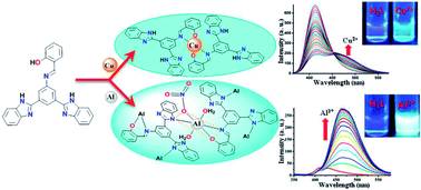
The synthesis is described of a new bis-benzimidazole appended Schiff base ligand [2-(3,5-bis(1H-benz-imidazol-2-yl)-phenyliminomethyl)phenol] (H3L) followed by its thorough characterization by elemental analyses, spectroscopic studies (FT-IR, 1H, 13C NMR, ESI-MS, UV/vis, fluorescence) and X-ray single crystal analyses. The excellent selectivity of H3L towards Cu2+ and Al3+ via distinct responses in mixed aqueous conditions was established by spectroscopic studies. The selective detection of Cu2+ due to the creation of complex 1 in which Cu2+ occupied only the salen-type N2O2 coordination site and the strong binding with Al3+ through both benzimidazole and salen moieties to yield complex 2 was confirmed by various studies. The disparate detection of these cations by H3L was substantiated by comparative studies, performed under analogous conditions, of the precursor compound 3,5-bis(1H-benzimidazol-2-yl)-aniline (BBA). The distinct interaction of H3L with Cu2+ and Al3+ and the sensing mechanisms were investigated in detail by spectroscopic studies and were supported by theoretical studies (Density Functional Theory). It has been clearly shown that the use of appropriate substituents may facilitate the design and development of probes suitable for the real time detection of more than one analyte.
Lately, directional efforts have been made towards the development of molecular sensors based on colorimetric/fluorescence methods15 employing photoinduced electron transfer (PET),16 internal charge transfer (ICT),17 chelation enhanced fluorescence (CHEF)18 and deprotonation mechanisms (ESIPT)19 for the detection of Cu2+ and Al3+. Being non-destructive, rapid, easy to carry out, and highly sensitive, these methods are advantageous over methods like atomic absorption,20 inductively coupled plasma mass and emission spectroscopy,21 and voltammetry.22,23 However, the previously developed sensors, had limitations vis-à-vis solubility, real time efficiency, bulky structures as well as the expensive and complicated synthesis of the probe materials. Therefore, it has been a challenging task to develop a multi-analyte detection technique which follows an easy and straightforward sensing approach whilst eliminating the limitations of previously developed systems. In this direction, a novel Schiff base [2-(3,5-bis(1H-benzimidazole-2-yl)-phenyliminomethyl)phenol, (H3L)] possessing a regular N,O-binding site on a phenyl ring (salen) and two independent benzimidazole –NH units has been designed and synthesized starting from a newly designed precursor 3,5-bis(1H-benzimidazol-2-yl)aniline (BBA).
The strategic dissimilarity of the functional groups (N,O and –NH) controls the reactivity of the probe (H3L) to achieve the distinct binding modes for Cu2+ and Al3+ which are reflected by diverse photophysical changes. The present work deals with the exploration of a multi-analyte detection system by rational detailing of the sensing interactions in the competitive milieu of two dissimilar sites. In this contribution we describe the synthesis and characterization of H3L and its potential application in the selective detection of Cu2+ and Al3+ via distinct routes attained through the strategic choice of different functional groups. To our knowledge, this is the first time that this approach has been applied to develop a probe that accomplishes the selective detection of Cu2+ and Al3+ with admirable cost efficiency.
Casymm), 1474, (s, νstr hetero-ring), 740 (vs, νbreathing hetero-ring). 1H NMR (300 MHz, dmso-d6, δH, ppm): δ 12.91 (s, 2H, H![[8 with combining low line]](https://www.rsc.org/images/entities/char_0038_0332.gif)
and ![[8 with combining low line]](https://www.rsc.org/images/entities/char_0038_0332.gif)
), 8.12 (s, 1H, H
), 7.63 (2H, H
and H
), 7.52–7.47 (broad, 4H, H![[4 with combining low line]](https://www.rsc.org/images/entities/char_0034_0332.gif)
, ![[4 with combining low line]](https://www.rsc.org/images/entities/char_0034_0332.gif)
, H![[7 with combining low line]](https://www.rsc.org/images/entities/char_0037_0332.gif)
and ![[7 with combining low line]](https://www.rsc.org/images/entities/char_0037_0332.gif)
), 7.23–7.18 (m, 4H, H![[5 with combining low line]](https://www.rsc.org/images/entities/char_0035_0332.gif)
, ![[5 with combining low line]](https://www.rsc.org/images/entities/char_0035_0332.gif)
, H![[6 with combining low line]](https://www.rsc.org/images/entities/char_0036_0332.gif)
and ![[6 with combining low line]](https://www.rsc.org/images/entities/char_0036_0332.gif)
), 5.56 (s, 2H, H
). 13C NMR (75.45 MHz, dmso-d6, δC, ppm): δ 151.6, 149.4, 135.0, 131.4, 125.2, 122.4, 121.5, 118.6, 113.1, 111.2, 105.6. ESI-MS (relative intensity), calcd, found (m/z): 326.1406, 326.1401 [(M + H)+, 45%]. UV/vis [c, 1.0 × 10⁻⁵ M; MeOH/H2O, 9
:
1, v/v MeOH/H2O; λmax, nm (ε, M−1 cm−1)]: 345 (7.75 × 10³), 300 (4.10 × 10⁴).
:
MeOH (50/50, v/v) solution. Yield: 1.7 g, 79%. Anal. calcd (%) for C27H19N5O: C, 66.85; H, 5.61; N, 4.10; found: C, 66.79; H, 5.59; N, 4.02%. IR (KBr pellets, cm−1): 1617–1607 (s, νstr C
Casymm), 1574 (s, νstr C
N), 1464 (s, νstr hetero-ring), 733 (vs, νbreathing hetero-ring). 1H NMR (300 MHz, dmso-d6, δH, ppm): δ 13.20 (s, 2H, H![[8 with combining low line]](https://www.rsc.org/images/entities/char_0038_0332.gif)
and ![[8 with combining low line]](https://www.rsc.org/images/entities/char_0038_0332.gif)
), 12.90 (s, 1H, H![[1 with combining low line]](https://www.rsc.org/images/entities/char_0031_0332.gif)
), 9.22 (s, 1H, H
), 8.99 (s, 1H, H
), 8.30 (s, 2H, H
and H
), 7.80 (d, J = 7.5 Hz, 1H, H1
), 7.72 (d, J = 7.2 Hz, 2H, H![[4 with combining low line]](https://www.rsc.org/images/entities/char_0034_0332.gif)
and ![[4 with combining low line]](https://www.rsc.org/images/entities/char_0034_0332.gif)
), 7.58 (d, J = 7.2 Hz, 2H, H![[7 with combining low line]](https://www.rsc.org/images/entities/char_0037_0332.gif)
and ![[7 with combining low line]](https://www.rsc.org/images/entities/char_0037_0332.gif)
), 7.47 (t, J = 7.8 Hz, 1H, H![[1 with combining low line]](https://www.rsc.org/images/entities/char_0031_0332.gif)
), 7.29–7.20 (m, 4H, H![[5 with combining low line]](https://www.rsc.org/images/entities/char_0035_0332.gif)
, ![[5 with combining low line]](https://www.rsc.org/images/entities/char_0035_0332.gif)
, H![[6 with combining low line]](https://www.rsc.org/images/entities/char_0036_0332.gif)
and ![[6 with combining low line]](https://www.rsc.org/images/entities/char_0036_0332.gif)
), 7.04 (m, 2H, H![[1 with combining low line]](https://www.rsc.org/images/entities/char_0031_0332.gif)
and H![[1 with combining low line]](https://www.rsc.org/images/entities/char_0031_0332.gif)
). 13C NMR (75.45 MHz, dmso-d6, δC, ppm): δ 164.7, 164.6, 160.9, 160.5, 151.8, 150.5, 149.8, 149.7, 136.7, 134.1, 132.8, 132.3, 131.6, 129.8, 123.1, 122.9, 122.4, 120.5, 119.8, 119.7, 119.5, 117.4, 117.0, 113.5, 113.1. ESI-MS (relative intensity), calcd, found (m/z): 430.1668, 430.1666 [(M + H)+, 25%]. UV/vis [c, 1.0 × 10⁻⁵ M; 9
:
1 (v/v) MeOH/H2O; pH ∼ 7.2; λmax, nm (ε, M−1 cm−1)]: 355 (1.00 × 10⁴), 305 (4.24 × 10⁴).
:
2) in polyphosphoric acid (PPA) at ∼220 °C under vigorous stirring afforded the precursor 3,5-bis(1H-benzimidazol-2-yl)-aniline (BBA) with ∼67% yield (Fig. S1, ESI†). BBA reacted with salicylaldehyde in methanol at a ratio of 1
:
1 under refluxing conditions to afford H3L with a reasonably good yield (79%) (Fig. S2, ESI†). A simple strategy adopted for the preparation of BBA and H3L is illustrated below in Scheme 1. The characterization of both the precursor BBA and ligand H3L was achieved by carrying out elemental analyses and spectroscopic studies (IR, NMR, UV/vis, fluorescence, ESI-MS) (Fig. S3, ESI†), and the structure of H3L was authenticated by X-ray single crystal analysis.
The 1H NMR spectrum of BBA was expected to exhibit singlets due to benzimidazole (–NH) and aromatic amine (–NH2) along with resonances due to aromatic and benzimidazole ring protons. As expected, it displayed singlets at δ 12.91 (2H; H![[8 with combining low line]](https://www.rsc.org/images/entities/char_0038_0332.gif)
and ![[8 with combining low line]](https://www.rsc.org/images/entities/char_0038_0332.gif)
) and 5.56 ppm (2H; H
) assignable to –NH and –NH2 protons. Aromatic protons other than the benzimidazole ring resonated as singlets at δ 8.12 (1H; H
) and 7.63 ppm (2H; H
and H
) while the benzimidazole ring protons appeared at δ 7.52–7.47 (broad, 4H; H![[4 with combining low line]](https://www.rsc.org/images/entities/char_0034_0332.gif)
, H![[4 with combining low line]](https://www.rsc.org/images/entities/char_0034_0332.gif)
, H![[7 with combining low line]](https://www.rsc.org/images/entities/char_0037_0332.gif)
and H![[7 with combining low line]](https://www.rsc.org/images/entities/char_0037_0332.gif)
) and 7.23–7.18 ppm (m, 4H; H![[5 with combining low line]](https://www.rsc.org/images/entities/char_0035_0332.gif)
, H![[5 with combining low line]](https://www.rsc.org/images/entities/char_0035_0332.gif)
, H![[6 with combining low line]](https://www.rsc.org/images/entities/char_0036_0332.gif)
and H![[6 with combining low line]](https://www.rsc.org/images/entities/char_0036_0332.gif)
) (Fig. S1a, ESI†). Likewise, the 1H NMR spectrum of H3L was expected to show signals due to –NH and –OH protons associated with benzimidazole and salen moieties in the downfield side at a 2
:
1 ratio. Expectedly, the –NH and –OH protons resonated in the downfield side at δ 13.20 (2H, H![[8 with combining low line]](https://www.rsc.org/images/entities/char_0038_0332.gif)
![[a with combining low line]](https://www.rsc.org/images/entities/char_0061_0332.gif)
![[, with combining low line]](https://www.rsc.org/images/entities/char_002c_0332.gif)
) and 12.90 ppm (1H, H![[1 with combining low line]](https://www.rsc.org/images/entities/char_0031_0332.gif)
) at a ratio of ∼2
:
1 [with respect to the signal at δ 9.22 ppm (s, 1H, H
) due to aldimine (–CH
N–)]. Assignment of the –NH proton resonance for H3L in the extreme downfield side was supported by an analogous signal (vide supra) of the precursor BBA. Further, doublets at δ 7.72 and 7.58 ppm may be attributed to benzimidazole protons H![[4 with combining low line]](https://www.rsc.org/images/entities/char_0034_0332.gif)
, ![[4 with combining low line]](https://www.rsc.org/images/entities/char_0034_0332.gif)
(adjacent to –NH–) and H![[7 with combining low line]](https://www.rsc.org/images/entities/char_0037_0332.gif)
, ![[7 with combining low line]](https://www.rsc.org/images/entities/char_0037_0332.gif)
(adjacent to ring
N–) while a broad multiplet at δ 7.294–7.209 ppm, may be attributed to H![[5 with combining low line]](https://www.rsc.org/images/entities/char_0035_0332.gif)
, ![[5 with combining low line]](https://www.rsc.org/images/entities/char_0035_0332.gif)
, H![[6 with combining low line]](https://www.rsc.org/images/entities/char_0036_0332.gif)
and ![[6 with combining low line]](https://www.rsc.org/images/entities/char_0036_0332.gif)
protons (Fig. S2a, inset, ESI†). The phenyl ring protons excluding benzimidazole and salen, resonated at δ 8.99 (1H, H
) and 8.30 (2H, H
and H
) ppm as distinct singlets. On the other hand, resonances pertaining to salen ring protons appeared at δ 7.80 (d, 1H, H![[1 with combining low line]](https://www.rsc.org/images/entities/char_0031_0332.gif)
), 7.47 (t, 1H, H![[1 with combining low line]](https://www.rsc.org/images/entities/char_0031_0332.gif)
) and 7.04 (m, 2H, H![[1 with combining low line]](https://www.rsc.org/images/entities/char_0031_0332.gif)
and H![[1 with combining low line]](https://www.rsc.org/images/entities/char_0031_0332.gif)
) ppm (Fig. S2a, ESI†). Thus, 1H NMR spectral data affirmed the formation and formulation of BBA and H3L. 13C NMR spectral data further supported their formations (Fig. S1b and S2b, ESI†).
The composition of BBA and H3L was further substantiated by ESI-mass spectrometric studies. ESI-MS of these compounds exhibited molecular ion peaks at m/z 326.1401 [(M + H)+, 45%] and 430.1666 [(M + H)+, 25%], respectively which strongly supported their proposed formulations (Fig. S3, ESI†).
| H3L | |
|---|---|
| Identification code | SHELXL |
| Empirical formula | C29H21N5O3 |
| fw | 487.508 |
| Temperature (K) | 293(2) |
| Wavelength (Å) | 1.54184 |
| Crystal system | Tetragonal |
| Space group | I41/acd |
| Unit cell dimensions | |
| a (Å) | 28.2864(11) |
| b (Å) | 28.2864(11) |
| c (Å) | 13.3571(7) |
| α (deg) | 90 |
| β (deg) | 90 |
| γ (deg) | 90 |
| Volume (Å3) | 10 687.3(10) |
| Z | 16 |
| Density (cald) (mg m−3) | 1.212 |
| Absorbance coefficient (mm−1) | 0.658 |
| F(000) | 4064.0 |
| Crystal size (mm3) | 0.30 × 0.15 × 0.04 |
| θ max for data collection (deg) | 66.270° |
| Data completeness | 99.60% |
| Absorbance correction | Multi-scan |
| Refinement method | Full-matrix least-squares on F2 |
| Data/restraints/parameter | 2335/31/185 |
| GOF on F2 | 1.044 |
| Final R indices [I > 2σ(I)] | R1 = 0.0789, wR2 = 0.2152 |
| R indices (all data) | R1 = 0.0931, wR2 = 0.2301 |
![]() | ||
| Fig. 1 (a) ORTEP view of H3L (50% ellipsoid probability and hydrogen atoms omitted). (b) DFT optimized structure of H3L supporting the crystal structure. | ||
The molecular arrangements of subsequent layers in the crystallographic ‘ab’ plane extended along the ‘c’ axis to define an anti-parallel arrangement (Fig. S5a, ESI†). Various bond distances and angles fall within the acceptable range and are comparable with other closely related systems (Table S1, ESI†).26 Furthermore, the crystal lattice exhibited one methanol as a free solvent which is involved in H-bonding interactions (two) with benzimidazole nitrogen. The respective distances lie within the reported range (∼2.699 and 2.744 Å; Fig. S5b, ESI†).27
:
1, v/v; pH ∼ 7.2) exhibit a weak and a prominent band at 355 and 305 nm, respectively. The low energy (LE) band at 355 (ε, 1.00 × 10⁴ M−1 cm−1) and the high energy (HE) band at 305 nm (ε, 4.24 × 10⁴ M−1 cm−1) have been tentatively assigned to n–π* and π–π* transitions. To explore its interaction with metal ions, a solution of H3L was treated with various cations viz., Li+, Na+, K+, Mg2+, Ca2+, Fe3+, Mn2+, Co2+, Ni2+, Cu2+, Ag+, Zn2+, Cd2+, Hg2+, Al3+ and Pb2+ (nitrate salts; 10.0 equiv.). It has been observed that the spectral features of H3L exhibited substantial changes only with Cu2+ and Al3+, while the other tested cations resulted in negligible variation (Fig. 2a). The addition of Cu2+ (10.0 equiv.) to a solution of H3L led to the emergence of a new band at ∼394 nm (ε, 6.9 × 10³ M−1 cm−1) with loss of the band at ∼355 nm. In addition, an insignificant hypso- (Δλ, ∼2 nm) and hyperchromic shift (Δε, 3.10 × 10² M−1 cm−1) was also noted for the HE band. Conversely, the addition of Al3+ (10.0 equiv.) to a solution of H3L resulted in the emergence of a new band at 370 nm (ε, 7.30 × 10³ M−1 cm−1) with a decrease in the optical density for the LE band. The HE band displayed a substantial hypo- and hypsochromic shift (Δλ, 7 nm; Δε, 2.80 × 10³ M−1 cm−1). Also, a small but noticeable hump emerged at ∼276 nm (ε, 2.98 × 10⁴ M−1 cm−1) which was a visible difference from the spectral features of H3L + Cu2+ (Fig. 2b).
The observed differences in the spectral features of H3L upon the addition of Cu2+ and Al3+ simply demonstrated their significant interaction with the salen and/or benzimidazole moieties of the probe. The considerable change in the LE band (λ, 355 nm) accompanied by the appearance of a new band at 394 nm and the negligible effect on the HE band in the presence of Cu2+ most probably occurs due to the formation of complex 1 via azomethine nitrogen and phenolate oxygen in N2O2 fashion [CuII–salen complex].28 In contrast, the disparity shown by both the LE and HE bands in the presence of Al3+ may be attributed to its interaction through both the aldimine and benzimidazole moieties.29 Based on these differences it has been suggested that Cu2+ and Al3+ may adopt distinct binding modes with H3L.
Although these suggestions are consistent with the spectroscopic annotations, to confirm this viewpoint precursor BBA (c, 1.0 × 10⁻⁵ M) was treated with Cu2+ and Al3+ under analogous conditions. Since, BBA is short of an N,O- chelating site and has only two benzimidazole moieties along with a free amine group, a comparative absorption study using BBA as the probe may be quite informative and throw light on the competing interaction of Cu2+/Al3+ with a benzimidazole moiety. This study demonstrated that there is hardly any change in the features of the absorption spectrum of BBA upon addition of Cu2+ (∼20.0 equiv.), whilst there is a considerable change upon the addition of Al3+ (∼20.0 equiv.). This suggests that Al3+ indeed interacts with the benzimidazole moiety, however, a comparatively large amount of Al3+ was required to exhibit the said changes resulting in a lower sensitivity for BBA (Fig. S6, ESI†). From this study it has been substantiated that Cu2+ binds only with salen while Al3+ binds through both the benzimidazole and salen moieties of H3L and exhibits a dissimilar interaction mode. Thus, we conclude that the probe has dual selectivity towards Cu2+ and Al3+ in mixed aqueous media.
To gain a deep insight into the mechanism of interaction, UV/vis titration studies were undertaken. The addition of Cu2+ (0.1 equiv., 1.0 × 10⁻³ M; 3.0 μL) to a solution of H3L (c, 1.0 × 10⁻⁵ M; MeOH/H2O, 9
:
1, v/v; pH ∼ 7.2) displayed hypochromism for the band at 355 nm (ε, 9.3 × 10³ M−1 cm−1) with the emergence of a new band at 394 nm (ε, 2.5 × 10³ M−1 cm−1). Further, saturation was achieved by addition of ∼1.0 equiv. of Cu2+, which resulted in a significant decrease in the optical density for the band at 355 nm along with a final increase in the optical density of the new band at 394 nm (ε, 6.9 × 10³ M−1 cm−1). The spectral variations accompanied by the clear isosbestic point (λ, 368 nm) indicated the ratiometric conversion of H3L to a third species (other than H3L and Cu2+). This species may be CuII–salen complex (1) which may be responsible for sensing (Fig. 3a).30
Similarly, in the titration involving H3L with Al3+ (c, 1.0 × 10⁻⁵ M; MeOH/H2O, 9
:
1, v/v; pH ∼ 7.2) saturation occurred upon the addition of ∼8.0 equiv. of the metal ion. A prominent band was observed at ∼370 nm which signified a hypso- and hypochromic shift (Δλ, 7 nm; ε, 7.3 × 10³ M−1 cm−1) for the HE band (Fig. 4a). The moderately high amount of Al3+ required relative to Cu2+ may be due to the larger number of Al3+ ions involved in the interaction with H3L. Here, the final species at saturation (limit of quantification ∼1.0–0.5 to ∼1.0–8.0 equiv. of H3L/Al3+) can be considered to adopt an intricate structure (2) rather than a simple AlIII–salen complex (3). The formation of 3 or any species parallel to it may not be sterically stable due to the bulkiness of the benzimidazole groups. Furthermore, a hump at ∼276 nm exhibited by H3L + Al3+ suggested that Al3+ is involved in some other interactions relative to that occurring in H3L + Cu2+. These additional aspects can essentially be related to the interaction between Al3+ and the benzimidazole moieties provided that Al3+ most likely binds with the benzimidazole groups of different H3L molecules.
This interpretation of the interactions occuring between H3L and analytes (Cu2+ and Al3+) confirmed that the mode of interaction of H3L with Cu2+ and Al3+ in aqueous media is discrete due to the presence of two different interaction sites which is a necessary criterion for dual selective detection.
To explore the suitability of H3L as a selective chemosensor for Cu2+ and Al3+, solutions containing H3L + Cu2+ (∼1.0 equiv.) and H3L + Al3+ (∼8.0 equiv.) were treated with interfering cations. The changes which occurred were negligible (Fig. 3b and 4b). Moreover, the mutual interference of Al3+ and Cu2+ was also verified by the insignificant changes (Fig. S7, ESI†) which were observed following the addition of Al3+ (∼10.0 equiv.) to H3L + Cu2+ (1.0 equiv.) and Cu2+ (∼10.0 equiv.) to a solution of H3L + Al3+ (8.0 equiv.). However, the addition of a large excess of Al3+ (+50.0 equiv. approx.) to a solution of H3L + Cu2+ (1.0 equiv.) + Al3+ (10.0 equiv.) resulted in some changes near the HE band (λ, ∼300 nm). This may be due to interaction between Al3+ and the free benzimidazole moiety which indirectly supports the binding of Cu2+ with only the salen core. On the other hand, no change was observed in the spectra of H3L + Al3+ (8.0 equiv.) in the presence of other analytes which confirmed that binding occurs between Al3+ and salen as well as the benzimidazole moieties.
:
1, v/v; pH ∼ 7.2), whereas in the case of BBA (c, 1.0 × 10⁻⁶ M; MeOH/H2O, 9
:
1, v/v) a strong band was observed at the same wavelength (λem, ∼415 nm) with a large Stokes shift of ∼115 nm (Fig. S8, ESI†). The salen core probably enhances the process of photoinduced electron transfer (PET) due to increased conjugation which significantly diminishes the fluorescence intensity for H3L relative to BBA. The effect was also studied of various metal ions (Li+, Na+, K+, Mg2+, Ca2+, Fe3+, Mn2+, Co2+, Ni2+, Cu2+, Ag+, Zn2+, Cd2+, Hg2+, Al3+ and Pb2+; nitrate salts; 10.0 equiv.) on the fluorescence intensity of H3L. It was observed that only Cu2+ and Al3+ produce a significant change in the spectra while other metal ions were ineffective (Fig. 5a). The addition of Cu2+ (10.0 equiv.) to a solution of H3L resulted in a decrease in fluorescence intensity for the band at ∼415 nm with the emergence of a new weak band at ∼485 nm. This is quite interesting as Cu2+ is known to be a fluorescence quencher which commonly induces a “fluorescence-off” signal.31
To gain a better understanding of the Cu2+ induced spectral changes, fluorescence titration studies were performed using 1.0 × 10⁻⁵ M solution of H3L in the presence of Cu2+ (clear changes were not observed with 1.0 × 10⁻⁶ M solution of H3L). A gradual decrease in the intensity of the band at ∼415 nm upon the addition of Cu2+ with the emergence of a new band at ∼485 nm was demonstrated. Saturation was attained by addition of up to ∼1.0 equiv. of Cu2+ (Fig. 5b). The observed changes can be ascribed to the continuous consumption of H3L leading to the creation of complex 1 which is detectable to the naked eye under UV radiation (λ, 365 nm) (Fig. 5b, inset). The formation of 1 was also confirmed by ESI-MS of the isolated species (H3L + Cu2+, ∼1.0 equiv.). A molecular ion peak for 1 was observed at m/z 920.2478 [calcd 920.2397, (M + H)+] (Fig. S15a, ESI†).
From the fluorescence studies that were carried out on H3L (c, 1.0 × 10⁻⁶ M; MeOH/H2O, 9
:
1, v/v; pH ∼ 7.2) it was shown that H3L displays an excellent selectivity for Al3+ (10.0 equiv.) relative to the tested metal ions (10.0 equiv.) including Cu2+ and a new strong band (λem, 466 nm; Fig. 5a) was observed. Here, distinct fluorescence signals due to the two types of interactions between H3L and Al3+ were not observed. Thus, the pertinent role of benzimidazole and aldimine moieties towards Al3+ was investigated by titration studies. In the titration experiments carried out using H3L (c, 1.0 × 10⁻⁶ M) a small but gradual decrease in the fluorescence intensity for the band at 415 nm was observed with the emergence of a new band and isosemissive point at 466 and ∼425 nm, respectively. The intensity of the new band was greatly enhanced (∼20 folds) and attained saturation upon the addition of ∼8.0 equiv. of Al3+ (Fig. 6a). Noticeably, the enhancement in intensity for the band at 466 nm was considerably high relative to the decrease in the intensity of the band at 415 nm. Rather, it appeared that the band at 415 nm attained a steady intensity after a significant decrease until the complementary band (466 nm) acquired saturation. This remarkable feature was ascribed to binary spectral behaviour (ratiometric change followed by a segment of constant intensity). This was a strong indication that interaction had occurred between both benzimidazole and salen moieties with Al3+ leading to an enormous increase in the fluorescence intensity. The binary spectral behaviour was further confirmed by titration studies at a slightly higher concentration (c, 1.0 × 10⁻⁵ M) as spectral changes were clearly visible at this concentration particularly in the isosemissive region and corroborated well with the unusual spectral behaviour (Fig. S9, ESI†). This type of spectral conduct undoubtedly advocated the occurrence of two types of interaction between H3L and Al3+, involving salen (N,O) and benzimidazole (–NH) moieties.
To validate the above conclusions, the precursor BBA (c, 1.0 × 10⁻⁶ M) was treated with Al3+ as it was expected to show changes exclusively due to the benzimidazole moieties. A new band at ∼465 nm was present in the fluorescence spectrum of BBA (λex, 300; λem, 415 nm) in the presence of a large excess of Al3+ (∼50 equiv.). This band was similar to the one obtained for H3L + Al3+ (λem, 466 nm), but with a negligible increase in the fluorescence intensity (Fig. S10, ESI†). This not only signifies the importance of the aldimine moiety in the enhancement of fluorescence by H3L in the presence of Al3+ (fluorescence-on response) but also, confirmed the involvement of the benzimidazole moiety in the interaction with the metal centre. Further, the ultimate spectral features of BBA + Al3+ (∼50 equiv.) resembled those of H3L + Al3+ (∼8.0 equiv.) to some extent. Thus at this instance, one can imagine that H3L may undergo hydrolytic cleavage in the presence of Al3+ to generate BBA (amine counterpart of H3L) in the solution itself. But, this presumption was discarded on the basis of 1H NMR titration and ESI-MS studies (vide infra) as these provided apt evidence regarding the existence of the aldimine linkage after the partial/complete addition of Al3+. Additionally, since a large excess of Al3+ (∼50 equiv.) is required to bring about the spectral changes of BBA, it can be specified that BBA + Al3+ (∼50 equiv.) results in a different species (relative to H3L + Al3+, 8.0 equiv.; 2). In conclusion, the comparative fluorescence studies on H3L and BBA proved that there are distinct binding modes for H3L with Cu2+ and Al3+. The studies also offered a reasonable explanation regarding the role of ‘binary fluorescence spectral behaviour’ in the fluorescence ‘turn-on’ detection by H3L particularly for Al3+.
Further, to explore the applicability of the probe as a selective chemosensor, an interference study was performed (H3L; c, 1.0 × 10⁻⁶ M). Successive addition of the interfering cations (10.0 equiv.) to a solution of H3L + Cu2+ (∼1.0 equiv.) and H3L + Al3+ (∼8.0 equiv.) exhibited insignificant changes (Fig. 6b). Interference was examined more precisely by measuring the background effect of the individual cations. The fluorescence intensity of H3L (λex, 300 nm; λem, 466 nm) was measured by addition of individual cations (10.0 equiv.) followed by the addition of Al3+ (10.0 equiv.; bar diagram, Fig. 7). These results not only supported the excellent selectivity of H3L towards Al3+ over the tested metal ions/Cu2+, but also, demonstrated the insignificant background effect of individual cations on H3L.
Job’s plot analyses revealed a 2
:
1 stoichiometry for H3L/Cu2+ and a 1
:
1 stoichiometry for H3L/Al3+ (Fig. S11, ESI†). The former one is consistent with the above discussions however, the latter (1
:
1 stoichiometric ratio) may be in contradiction with the formation of a distinct monomeric species from H3L–Al3+ interactions. Instead, the 1
:
1 ratio indicates that in complex (2) Al3+ binds with the salen core and only one benzimidazole moiety from H3L. Further, if the coordinated Al3+ adopts an octahedral geometry, the remaining three coordination sites are most probably occupied by NO3− and H2O molecules. Consequently, a polymeric species is likely formed which may involve Al3+ and/or some uniform distinct species in equilibrium with the polymeric species. The 1
:
1 stoichiometry may also be supported by the 1H NMR titration between H3L and Al3+ wherein only ∼1.0 equiv. of Al3+ was sufficient to complete the loss of the –NH and –OH protons (vide infra). The association constant for H3L in the presence of Al3+ was estimated following the Benesi–Hildebrand method (KAl = 1.8 × 10⁵ mol−1: Fig. S12, ESI†).32 It indicated a pretty good sensitivity of H3L for Al3+ and is in agreement with earlier reports.33 Furthermore, the limits of detection (LOD) of Cu2+ and Al3+ using H3L were also estimated and were found to be 4.2 × 10⁻⁸ M and 8.1 × 10⁻⁷ M, respectively. These merely defined the lowest amount of Cu2+ and Al3+ that can be precisely measured using H3L (c, 1.0 × 10⁻⁵ M), and are in the order of 10−7 M and 10−6 M, respectively which is good enough for practical applications.
In the light of the above discussions we conclude that H3L exhibits dissimilar fluorescence changes in the presence of Cu2+ and Al3+ due to the formation of 1 and 2, respectively through discrete interactions involving the benzimidazole and salen moieties. These interactions significantly alter the electron transfer phenomenon in the system and induce a substantial change in the fluorescence intensity, especially upon the addition of Al3+. The annotations from the fluorescence spectral studies are consistent with the results obtained from UV/vis studies and confirm the usability of H3L as a dual selective chemosensor for Cu2+ and Al3+ (Fig. 8).
![]() | ||
Fig. 8 The changes observed in (a) color and (b) fluorescence of c, 1.0 × 10⁻⁵ M [MeOH/H2O, 9 : 1, v/v] solutions of H3L with 10.0 equiv. of the tested cations. | ||
![[8 with combining low line]](https://www.rsc.org/images/entities/char_0038_0332.gif)
and H![[8 with combining low line]](https://www.rsc.org/images/entities/char_0038_0332.gif)
, δ 13.20 ppm) and –OH (1H; H![[1 with combining low line]](https://www.rsc.org/images/entities/char_0031_0332.gif)
, δ 12.90 ppm) protons in 2
:
1 ratio, thus any interaction involving these groups can be easily followed. The addition of Al3+ (0.25 equiv.) to a solution of H3L resulted in a radical loss of signal associated with –NH and a small decrease in the intensity for the –OH proton (Fig. 9). Evidently, the disappearance of the –NH proton before –OH represents an unusual behaviour as phenolate is expected to be highly reactive toward M+. To highlight the direct involvement of benzimidazole an expanded view between δ 7.00–8.00 ppm is depicted in Fig. S14, ESI.† It shows that doublets assigned to H![[4 with combining low line]](https://www.rsc.org/images/entities/char_0034_0332.gif)
,
b and H![[7 with combining low line]](https://www.rsc.org/images/entities/char_0037_0332.gif)
, ![[7 with combining low line]](https://www.rsc.org/images/entities/char_0037_0332.gif)
appearing at δ 7.72 and 7.58 ppm undergo significant up- and downfield shifts of about Δδ ∼0.07 ppm, in the presence of Al3+ (0.25 equiv.) which merged into one signal. Besides, a very small but significant downfield shift (Δδ ∼ 0.010 ppm) has been noted for the multiplet at δ 7.294–7.209 ppm (H![[5 with combining low line]](https://www.rsc.org/images/entities/char_0035_0332.gif)
, ![[5 with combining low line]](https://www.rsc.org/images/entities/char_0035_0332.gif)
, H![[6 with combining low line]](https://www.rsc.org/images/entities/char_0036_0332.gif)
and ![[6 with combining low line]](https://www.rsc.org/images/entities/char_0036_0332.gif)
). Significant shifts in the resonances due to benzimidazole ring protons clearly demonstrates the instant interaction of Al3+ with the –NH moieties. Further additions of Al3+ (0.5–1.0 equiv.) apparently demonstrated obvious chemical shifts and signal broadening in the aromatic region (Fig. S14a, ESI†). On the other hand, the pertinent involvement of the salen core was simultaneously signified by a gradual decrease in the intensity of the –OH signal with an upfield shift (Δδ = 0.13 ppm) for the aldimine signal (1H; H
, δ 9.22 ppm). The inferior reactivity of the phenolate to benzimidazole may also be attributed to intramolecular H-bonding which most likely prevents the salen unit from interacting with the analyte (Al3+) prior to the benzimidazole moiety. The titration study thus significantly indicated that the –OH proton persists for a longer time in solution than the –NH proton which, in turn, supports the immediate binding of Al3+ to benzimidazole over the salen unit.
The contribution of –NH was substantiated by the 1H NMR spectra for BBA in the presence of Al3+ (∼10 equiv.), which displayed a significant downfield shift for benzimidazole aromatic signals with the complete loss of the –NH signal (Fig. S14b, ESI†). Moreover, significant support of the benzimidazole–Al3+ interaction was provided by monitoring the hydrogen–deuterium exchange in H3L (dmso-d6) in the presence of blank D2O (trace amount) which displayed a much smaller loss of the signal for –NH than for –OH (Fig. S15, ESI†). Under biased deuterium exchange conditions it significantly describes the requisite interaction of Al3+ with –NH of benzimidazole because in the H3L vs. Al3+ titration, the signal for –NH disappeared prior to that for –OH which should not occur if deuterium exchange dominates.
Thus, the 1H NMR titration study of H3L/Al3+ enabled us to elucidate which particular binding site is involved prior to the other site in a competitive environment. Besides this, although transformations were achieved using different analyte ratios, the study provided irrefutable evidence regarding the interaction of both benzimidazole and salen moieties with Al3+ and demonstrated that the former moiety is favoured over the latter. In addition, the study clearly demonstrates the different efficiency of the two distinct groups on the same probe towards the analyte which may be useful in the development of efficient multi-analyte detection tools in the future. Conversely, a well resolved 1H NMR spectrum could not be obtained for Cu2+ titration studies due to the paramagnetic Cu2+(d9) system.35
:
1 binding mode (which complies with Job’s analysis and 1H NMR studies). This adopted an octahedral geometry in the presence of appropriately coordinated NO3− and H2O molecules and supported the above mentioned spectral observations without any significant infringement (Fig. S17, ESI†). The occurrence of the coordinated NO3− was also verified by the FT-IR spectrum of complex 2 which displayed bands at ∼1440 and ∼1313 cm−1 due to asymmetric vibrations of the nitrate ion in the mono coordinated form.36 The efficacy of the structural optimizations for 1 and 2 demonstrated that these are the most likely formed species in the presence of Cu2+ and Al3+, respectively. Also, attempts to optimize model structures other than 2 (viz. complex 3) failed. Thus, the end species formed after sensing interactions had been substantiated by DFT studies, in turn, confirmed the viability of H3L as an efficient chemosensor for Cu2+ and Al3+.
Tentative mechanistic routes for the detection of Cu2+ and Al3+ were worked out on the basis of the results from the above spectral and theoretical studies. An aldimine chelated complex 1 having pendent benzimidazole arms displaying significant photophysical changes was created in the presence of Cu2+. However, with Al3+, both benzimidazole and salen moieties of H3L are involved in interactions leading to the formation of complex 2. Spectral (photophysical and 1H NMR) studies including binary fluorescence spectral behaviour and comparative studies on the precursor (BBA) clearly confirmed the involvement of both the functionalities (benzimidazole and salen) with Al3+. The creation of the end products 1 and 2 was substantiated by ESI-MS and their structures were verified by DFT. Furthermore, the formation of complexes 1 and 2 was also verified by obtaining PXRD patterns for H3L, 1 and 2 in the range 2Ө, 5–60°. Fairly different diffraction patterns were observed for 1 and 2 than for H3L which clearly indicate their formation (Fig. S18, ESI†). Therefore, the involvement of both functionalities has been reasonably attested and it has been established that H3L can be used as an efficient probe for the detection of Cu2+ and Al3+ in mixed aqueous media at room temperature (Fig. 10).
:
2 ratio (verified by Job’s plot analysis), while benzimidazole remains uncoordinated. Conversely, Al3+ interacts with both benzimidazole and salen moieties in a 1
:
1 ratio. The addition of Al3+ to a solution of H3L resulted in an enormous increase in the fluorescence intensity (20 fold) and the formation of complex 2, which was substantiated by spectral studies and supported by DFT. H3L has been found to be very selective and sensitive towards Cu2+ and Al3+ and showed insignificant effects due to other cations. This type of dual selective behavior accomplishing the detection of two metal ions of extreme biological interest has been accredited to the strategic design of a probe material with more than one interaction site of unequal potential. The consequence of this is to permit the exploration of new avenues in the field of multi-analyte detection via the design of molecules with more than one substituent.
Footnote |
| † Electronic supplementary information (ESI) available: 1H, 13C NMR spectra, ESI-MS, UV/vis and fluorescence spectra, crystal data file in CIF format and log files of DFT optimized structures. CCDC 1039505. For ESI and crystallographic data in CIF or other electronic format see DOI: 10.1039/c5ra18566a |
| This journal is © The Royal Society of Chemistry 2015 |