DOI:
10.1039/C5RA18489D
(Paper)
RSC Adv., 2015,
5, 93234-93242
Synthesis of chitosan-coated polyoxometalate nanoparticles against cancer and its metastasis
Received
9th September 2015
, Accepted 26th October 2015
First published on 26th October 2015
Abstract
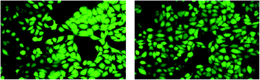
Three different Keggin-type polyoxometalates (POMs) [PW12O40]3−, [TiW11CoO40]7−, and [Ti2PW10O40]7−, were synthesized and then encapsulated in chitosan to prepare nanoparticles, CS–PW12, CS–TiW11Co, and CS–Ti2PW10. The synthesized nanoparticles were physicochemically characterized in terms of particle size, zeta potential, entrapment efficiency and in vitro release of the entrapped POM. The most efficient formulation was CS–TiW11Co, with a particle size of 105 ± 6 nm and an entrapment efficiency of 87 ± 12 (%). The CS–TiW11Co nanoparticles showed the highest activity when tested against tissue nonspecific alkaline phosphatase (TNAP) with IC50 = 102.0 ± 9.68 nM. The anticancer potential of the free POMs and their nanoparticles were also studied and CS–TiW11Co showed the highest inhibition (IC50 = 1.06 ± 0.09) on HeLa cells. To observe signs of apoptosis in HeLA cells, DAPI staining was performed after treatment with CS–TiW11Co nanoparticles. Furthermore, the reactive oxygen species (ROS) production was examined by H2DCF-DA dye under a fluorescence microscope. Our study revealed that CS–TiW11Co nanoparticles are very effective in cancer treatment and its associated metastasis especially in osteoblastic lesions with minimal adverse effects on normal cells (Vero cells).
Introduction
Polyoxometalates (POMs) are discrete, anionic metal-oxides with potential applications in catalysis, material science, and medicine.1–5 POMs are highly effective for the treatment of cancer, diabetes and infections associated with bacteria, viruses, and leishmania parasites.6 POMs have excellent potential for the treatment of almost all types of cancers including pancreatic cancer,7 leukemia,8 hepatocellular carcinoma,9 colon carcinoma,10 ovarian cancer,11 gastric cancer.12 The exact mechanism of POMs causing cancer cell death is still unknown13 but previous studies have shown that POMs have high potential to generate reactive oxygen species (ROS) which result into intracellular oxidative-stress causing hypoxia and cell death.14,15 Another mechanism to determine apoptosis is seen by the generation of DNA fragments of cancer cells when treated with different POMs.7,10,16,17
Furthermore, cancers originating from kidney, prostate, breast and lungs have a tendency to metastasize into bones characterized by osteolytic and osteoblast formation.18 Bisphosphonates are highly recommended to reduce pain associated with osteolytic bone metastasis,19 whereas to the best of our knowledge no drug has been recommended to reduce osteoblastic lesion. The tissue non-specific alkaline phosphatase (TNAP) enzyme is involved in the hydrolysis of pyrophosphate (PPi) to form hydroxyapatite (HA) which triggers the process of mineralization (osteoblast activity).20 POMs have shown an excellent potential to treat the abnormal functions of alkaline phosphatases commonly seen in hydroxyapatite deposition disease (HADD) that is associated with over-expression of alkaline phosphatase enzyme.21,22 In the light of the above mentioned role of TNAP it can be assumed that by blocking TNAP activity also osteoblast formation can be prevented during cancer metastasis.
There are two major reasons why POMs are not applied more frequently in medicine. Firstly, several POMs are not sufficiently stable at physiological pH, and secondly, due to the lack of selectivity they exhibit toxicity.14 In order to render POMs more stable and less toxic to the surrounding non-cancerous cells, a surface modification is required, which may be accomplished by encapsulating POMs in a biodegradable polymer. Encapsulation of target molecules in biodegradable polymers ensures high protection of the drug in a physiological environment and allows for delivery of the drug at the target site.23–27 The particle size is highly important for the determination of the fate of the nanoparticles. A 5–200 nm particle size range is highly efficient for targeting a specific organ because such particles can be effectively excreted from the kidneys and the reticuloendothelial system (RES) to reach the target site.28,29
Chitosan is a polysaccharide composed of glucosamine and N-acetyl glucosamine linked by (1 → 4) β-glucosidic bonds obtained from chitin, which is widely present in the shells of crabs and the exoskeleton of shrimps.30 Chitosan is very popular in the field of nanotechnology because of its inert nature, high availability, biocompatibility, low immunogenicity, and mucoadhesive nature.31
The objective of the present study was to evaluate the anticancer potential of three novel POMs enclosed in chitosan. These nanoparticles were prepared by ionotropic gelation technique and characterized by different physicochemical tools (FTIR, particle size, zeta potential, entrapment efficiency and dissolution studies). The entrapment efficiency and in vitro release of the POMs from the nanoparticles was also determined. The POMs were further tested against TNAP, whereas the anticancer activity was tested on HeLa cells. In addition, apoptosis and ROS production were observed with a fluorescence microscope.
Results and discussion
Preparation of chitosan/POM nanocomplexes
The three Keggin-type POMs include phosphotungstates [PW12O40]3−, cobalt (Co) substituted titanium (Ti) containing polyanion [TiW11CoO40]7−, and Ti containing tungstates [Ti2PW10O40]7− were synthesized by following previously reported methods.35–37 The selected POMs were highly stable at physiological pH (7.4). The nanoparticles were prepared by using the ionotropic gelation technique, in which the chitosan and the POM served as cation and anion, respectively.
Characterization of the POMs and chitosan–POM nanoparticles
The POMs used here were characterized by FTIR and NMR (31P) spectroscopy (see Fig. 2 and 3). It is evident from SEM analysis that the prepared chitosan–POM nanoparticles were slightly irregular in shape with particle size not more than <200 nm (see Fig. 4). The size distribution of the nanoparticles (Fig. 5) was determined by dynamic light scattering (DLS). Electrophoretic light scattering was used to evaluate the stability and the surface charge of the nanoparticles. The particle diameter and the zeta potential of CS–PW12 and CS–Ti2PW10 were determined as 164 ± 9 nm and 190 ± 7 nm, as well as +44.8 ± 3.8 mV and +47.4 ± 6.1 mV, respectively. The best formulation amongst the studied compounds was CS–TiW11Co, with a particle size diameter and zeta potential of 105 ± 6 nm and +52.0 ± 5.2 mV, respectively (Table 1).
 |
| Fig. 1 Polyhedral representation of polyanions [PW12O40]3− (left), [Ti2PW10O40]7− (middle), and [TiW11CoO40]7− (right). Yellow phosphorous (P), light green titanium (Ti) and pink colour represents cobalt (Co) element. | |
 |
| Fig. 2 31P NMR spectra of [PW12O40]3− (left) and [Ti2PW10O40]7− (right) in H2O/D2O. | |
 |
| Fig. 3 FTIR spectra of (1): (A) chitosan (CS), (B) {PW12}, (C) CS–PW12 nanoparticles; (2): (A) chitosan (CS), (B) {Ti2PW10}, (C) CS–Ti2PW10 nanoparticles; (3): (A) chitosan (CS), (B) {TiW11Co}, (C) CS–TiW11Co nanoparticles. | |
 |
| Fig. 4 Surface morphology of nanoparticles CS–PW12 (A), CS–Ti2PW10 (B), and CS–TiW11Co (C). | |
 |
| Fig. 5 Size distribution of chitosan–POM nanoparticles, CS–TiW11Co (A); CS–PW12 (B) and CS–Ti2PW10 (C). | |
 |
| Fig. 6 Dissolution profile of CS–POM nanoparticles at physiological pH 7.4. | |
Table 1 Characterization of chitosan–POM nanoparticles
Code |
Formula |
CS/POM ratio |
Particle size ± SD (nm) |
Zeta potential ± SD (mV) |
Entrapment efficiency (%) |
CS–TiW11Co |
Chitosan–[TiW11CoO40]7− |
4 : 2 |
105 ± 6 |
+52.0 ± 5.2 |
87 ± 12 |
CS–PW12 |
Chitosan–[PW12O40]3− |
4 : 2 |
164 ± 9 |
+44.8 ± 3.8 |
83 ± 10 |
CS–Ti2PW10 |
Chitosan–[Ti2PW10O40]7− |
4 : 2 |
190 ± 7 |
+47.4 ± 6.1 |
91 ± 7 |
The overlaid FTIR spectra of CS, the three polyanions, and CS–POM nanoparticles are shown in Fig. 3. The characteristic band of {PW12} at 1633 cm−1 represents crystal waters, whereas for CS the spectral bands at 1625 cm−1 and 1081 cm−1 are due to N–H bending (amines) and C–O–C stretching, respectively.32,33 The nanocomplex formed showed bands at 1639 cm−1 and 1087 cm−1 which indicate the presence of both chitosan and {PW12}. Similarly, the CS–TiW11Co nanocomplex showed bands at 1614 cm−1 and 1071 cm−1, which are slightly different from the peaks in the chitosan and {TiW11Co} spectra (Fig. 3). The confirmation of the nanocomplex CS–Ti2PW10 was obtained when the 1638 cm−1 and 1085 cm−1 peaks appeared (Fig. 3).
Entrapment and release of drug from nanoparticles
The amount of drug entrapped in the chitosan-coated POM nanoparticles was determined spectrophotometrically. It was evident from the data that the entrapment efficiency (EE) of the selected nanoparticles was excellent and had a linear relationship with particle size (Table 1). The maximum EE was seen for CS–Ti2PW10 (91 ± 7%), while CS–TiW11Co and CS–PW12 also had good percentages of drug entrapping, 87 ± 12 (%) and 83 ± 10 (%), respectively.
In order to understand the mechanism of POM release from the nanoparticles, in vitro dissolution studies were carried out for 96 h in PBS (pH 7.4) supplemented with lysozyme. The lysozyme was used to degrade the chitosan backbone by cleavage of the glycosidic bonds. When the release kinetics was studied and it became evident that the selected chitosan–POM nanoparticles followed concentration-dependent first order release kinetics with excellent regression coefficients R2 (see Table 2). Similarly, the mechanism of POM release from chitosan was determined by using the Higuchi and Peppas models. The release of the POMs from chitosan was governed by a diffusion mechanism. The Peppas model confirmed that the release mechanism starts with erosion of the polymer matrix followed by diffusion (n > 0.45) (Table 2).
Table 2 Release mechanism of POMs from CS in dissolution experiments
Code |
Zero order (R2) |
First order (R2) |
Higuchi model (R2) |
Peppas model (R2) |
N |
CS–PW12 |
0.748 |
0.999 |
0.998 |
0.997 |
0.607 |
CS–TiW11Co |
0.710 |
0.996 |
0.995 |
0.991 |
0.568 |
CS–Ti2PW10 |
0.789 |
0.998 |
0.996 |
0.995 |
0.582 |
Alkaline phosphatase inhibition studies
An inhibitor of tissue non-specific alkaline phosphatase (TNAP) enzyme may become a future drug for the treatment of malignant tumors, which mainly target bones. The activities of unbound POMs and chitosan-coated POMs against TNAP were studied and showed excellent IC50 values in the nanomolar range. The results obtained from the colorimetric assay confirmed that the chitosan–POM nanoparticles were highly effective in TNAP inhibition (Fig. 7). The most potent formulation was CS–TiW11Co nanoparticles with IC50 = 102.0 ± 9.68 nM.
 |
| Fig. 7 The graphs represent the IC50 values for unbound POMs and chitosan–POM nanoparticles when tested against tissue non-specific alkaline phosphatase (TNAP). | |
Antiproliferation assay
Sulforhodamine B dye assay. The anticancer activities of the studied POMs were tested by a sensitive colorimetric assay, which uses sulforhodamine B dye for the determination of protein content in cancer cells. The unbound POMs and chitosan–POM nanoparticles were tested on HeLa cells and the IC50 value for each compound was determined. The results confirmed that the unbound POMs {PW12}, {TiW11Co}, and {Ti2PW10} showed less inhibition towards HeLa cells with IC50 values of 18.5 ± 4.72, 9.21 ± 2.64, and 16.0 ± 2.85 μM, compared to the chitosan-coated POMs CS–PW12, CS–TiW11Co, and CS–Ti2PW10 with IC50 values of 8.37 ± 1.84, 1.06 ± 0.09, and 5.36 ± 1.57 μM, respectively (Table 3). Amongst the tested compounds CS–TiW11CoO40 showed the highest inhibition of HeLa cells, which was then taken for further studies.
Table 3 Alkaline phosphatase and anticancer activities of POMs and CS–POM nanoparticles
Code |
Activity at TNAP (nM) (IC50 ± SEM) |
Anticancer activity on HeLa cells (μM) (IC50 ± SEM) |
Anticancer activity on Vero cells 10 (μM) (% inhibition ± SEM) |
{PW12} |
1622 ± 22 |
18.5 ± 4.72 |
18 ± 2 |
CS–PW12 |
820.5 ± 31.4 |
8.37 ± 1.84 |
15 ± 3 |
{TiW11Co} |
736.2 ± 12.7 |
9.21 ± 2.64 |
11 ± 4 |
CS–TiW11Co |
102.0 ± 9.68 |
1.06 ± 0.09 |
9 ± 3 |
{Ti2PW10} |
1050 ± 15 |
16.0 ± 2.85 |
14 ± 2 |
CS–Ti2PW10 |
301.7 ± 18.6 |
5.36 ± 1.57 |
13 ± 2 |
Levamisole |
1375 ± 29 |
|
|
Cisplatin |
|
17.6 ± 5.94 |
17 ± 5 |
Morphological evaluation of apoptosis by DAPI staining. The chitosan–POM nanoparticle CS–TiW11Co, which showed the lowest IC50 (1.06 ± 0.096 μM), was selected to observe apoptosis with fluorescence microscopy. After staining with DAPI apoptotic bodies with condensed chromatin were observed when HeLa cells were treated with the selected compound (Fig. 8). This observation confirmed that CS–TiW11Co nanoparticles kill HeLa cells by apoptosis.
 |
| Fig. 8 Morphological evaluation of untreated (left) and treated (right) Hela cells with CS–TiW11Co nanoparticles. The fluorescence microscope (Nikon Eclipse-Ni Japan) was used to get images at an excitation and emission wavelengths of 358 and 461 nm, respectively. | |
Determination of reactive oxygen species (ROS). The initiation of the production of ROS within HeLa cells by CS–TiW11Co was studied by using dichlorofluorescin diacetate (H2DCF-DA) dye and observed by fluorescence microscopy. Fig. 9 shows that CS–TiW11Co nanoparticle-treated HeLa cells exhibit disintegrated cell membranes and condensed cellular protein (DNA) that may be due to oxidation of lipids and proteins.34
 |
| Fig. 9 HeLa cells show the production of reactive oxygen species (ROS) when treated with CS–TiW11Co nanoparticles (right) and the ruptured cell membrane can be easily compared with untreated (left) HeLa cells. The image were captured at an excitation and emission wavelengths of 485 and 530 nm, respectively by using fluorescence microscope (Nikon Eclipse-Ni Japan). | |
Conclusions
In the present study cobalt- and titanium-containing heteropolytungstates of the Keggin-type and their chitosan nanoparticles were synthesized by the ionotropic gelation technique. The unbound POMs and chitosan–POM nanoparticles were characterized and then investigated for anticancer and tissue non-specific alkaline phosphatase (TNAP) inhibition activities. All tested compounds were active against TNAP and HeLa cells, and CS–TiW11Co showed the highest inhibition for TNAP (IC50 = 102.0 ± 9.68 nM), and also anticancer activity (IC50 = 1.06 ± 0.09 μM), and was the least toxic towards Vero normal cells (9 ± 3%). Furthermore, the apoptosis and ROS production by CS–TiW11Co was observed by fluorescence microscopy. Our work has demonstrated that CS–TiW11Co nanoparticles can serve as a potential drug for the treatment of cancer and bone metastasis.
Materials and methods
Chitosan (YC-100) (M ∼ 10
000 g mol−1), tissue non-specific alkaline phosphatase enzyme (TNAP), levamisole, Tris-HCl, Tris base, MgCl2, ZnCl2, DMEM high glucose, fetal bovine serum (FBS), penicillin, streptomycin and lysozyme of chicken egg white were purchased from Sigma-Aldrich, Steinheim, Germany. The human cervical adenocarcinoma (HeLa) cell line and African green monkey kidney (Vero) cell line were purchased from American Type Culture Collection (ATCC), USA.
Experimental
Synthesis of POMs
The selected POM salts Na3[PW12O40]·6H2O {PW12}, K4H3[Ti2PW10O40]·6H2O {Ti2PW10} and K6H2[TiW11CoO40]·12H2O {TiW11Co} (Fig. 1) were synthesized according to the published procedures and characterized by FT-IR (Fig. 3).35–37 Furthermore, polyanions [PW12O40]3− and [Ti2PW10O40]7− were characterized by 31P-NMR (Fig. 2).
Synthesis of chitosan–PW12 (CS–PW12)
CS–PW12 nanoparticles were synthesized by stirring 0.50 g (0.71 mM) of CTS in 70 mL distilled water for 5 min. The resultant solution was then filtered (solution A). Similarly, 1.83 g (0.6 mM) of {PW12} was dissolved in 30 mL distilled water (solution B). Solution B was then added dropwise to solution A, forming a gel-like precipitate which was separated by filtration and washed several times with water and then air-dried. The obtained product was characterized by FTIR (see Fig. 3).
Synthesis of chitosan–[Ti2PW10O40] (CS–Ti2PW10)
CS–Ti2PW10 nanoparticles were synthesized under the same conditions as CTS–PW12. However, solution B was prepared by dissolving 1.00 g (0.34 mM) of Ti2PW10 in distilled water. The obtained product was characterized by FTIR (see Fig. 3).
Synthesis of chitosan–[TiW11CoO40] (CS–TiW11Co)
CS–TiW11Co nanoparticles were synthesized under the same conditions as CTS–PW12. However, solution B was prepared by dissolving 1.11 g (0.34 mM) of {TiW11Co} in distilled water. The obtained product was characterized by FTIR (see Fig. 3).
Characterization of nanoparticles
The nanoparticles were characterized by investigation of particle size, zeta potential and surface morphology. Dynamic light scattering and electrophoretic light scattering techniques were used to determine the particle size distribution and zeta potential of the nanoparticles. Scanning electron microscopy (SEM) was used to observe the surface morphology of the nanoparticles. Fourier transform infrared (FTIR) spectroscopy was used to identify the vibrational bands.
Determination of entrapment efficiency
A previously described method with a slight modification was used to determine the entrapment efficiency of the CS–POM nanoparticles.14 Briefly, a predetermined weight of nanoparticles was centrifuged at 12
000 rpm for 40 min and the supernatant was investigated by UV-visible spectroscopy to determine the amount of non-entrapped POM in the formulation.
Entrapment efficiency (%) = (total POM in formulation − free POM/total POM in formulation) × 100 |
In vitro dissolution studies
To determine the release mechanism of the POMs from the nanoparticles, in vitro dissolution studies were carried out by following a previously reported method.14 The nanoparticles were centrifuged at 12
000 rpm for 40 min and the pellet obtained was redispersed in 900 mL phosphate buffered saline (PBS) at pH 7.4. A small amount of lysozyme (1.2 μg mL−1) was also added to promote chitosan degradation in PBS. The experiment was conducted on a magnetic stirrer with 75 rpm at 37 °C. Samples were taken at pre-determined time intervals and the amount of POM released in the medium was calculated by using a standardized curve. The release kinetics (zero order and first order) and release mechanism (Higuchi and Peppas models) were determined by following mathematical equations:
Zero order rate equation
Qo and
Qt represent the amount of drug release at (
to) and at a specified time (
t) whereas
ko is the zero order rate constant.
First order rate equation
|
ln M = −k1t + ln Mo
| (2) |
Mo is the initial amount of POM and
Mt is the remaining amount of POM at time
t and
k1 is the first order rate constant.
Higuchi equation
M is the amount of POM released at time
t and
kH is the Higuchi rate equation
Peppas model
|
ln(Mt/M∞) = ln kp + n ln t
| (4) |
(
Mt/
M∞) is the fraction of drug released at time
t and
n is the slope which determines the type of diffusion from the polymer matrix.
Enzyme inhibition studies against TNAP
Enzyme inhibition studies were carried out by following a previously reported spectrophotometric method.38 Briefly, TNAP was diluted in an assay buffer (pH 9.5) comprised of Tris-HCl (50 mM), MgCl2 (5 mM), ZnCl2 (0.1 mM), and glycerol (50%). The enzyme substrate p-nitrophenyl phosphate (p-NPP) was dissolved in the same buffer without glycerol. The experiment was started by mixing enzyme (10 μL) with test compound (10 μL) and kept at 37 °C for 10 min (pre-incubation). The enzyme substrate (p-NPP) was added and again incubated for 30 min. A yellow coloured product (p-nitrophenolate) was formed and the absorbance was measured at 405 nm using an ELISA plate reader (Bio-TekELx 800™, Instruments, Inc., USA). Each experiment was performed in triplicate and the results obtained were presented as IC50 values calculated by using Prism 5.0 (GraphPad Software, San Diego, CA, USA).
Cell lines and cell cultures
HeLa cells were cultured and maintained in DMEM high glucose medium supplemented with L-glutamine (2.0 mM), penicillin (100 U mL−1) and streptomycin (100 μg mL−1) and FBS (10%). The culture flasks were kept in a specialized incubator providing CO2 (5%) at a constant temperature of 37 °C. To conduct the antiproliferation assay, the HeLa cells were seeded in 96-well plates with a density of 20
000 cells per well.
Antiproliferation assay
A common, highly sensitive colorimetric assay utilizing sulforhodamine B dye, which binds with cellular proteins, was used to determine the degree of cytotoxicity.39 HeLa cells were harvested in 96-well plates and treated with different concentrations of the test compounds for 24 h. The plates were removed from the incubator and treated with ice-cold TCA (50%) and kept at 4 °C. The plates were gently rinsed with PBS and then treated with sulforhodamine B dye (0.4%) and kept again for 30 min at room temperature. In order to remove unbound dye the plates were treated with 1% acetic acid solution. The cell-bound dye was solubilized by adding Tris base (10 mM) and left for some time at room temperature. The absorbance was measured at 490 nm subtracting the background measurement at 630 nm.
Evaluation of apoptosis by fluorescence microscopy
Morphological evaluation of apoptosis with DAPI staining. HeLa cells (1.4 × 10⁴) were cultured in a 2-well sterile chamber slide, treated with the test compounds and incubated for 24 h. Formalin (4%) was used for cell fixation. After washing with PBS the cells were stained with DAPI (10 μg mL−1) for 10 min in the dark and examined under a fluorescence microscope (Nikon Eclipse-Ni Japan) with excitation and emission wavelengths of 358 and 461 nm, respectively.40
Determination of intracellular reactive oxygen species (ROS) production. The morphological evaluation of ROS production in treated HeLa cells was observed in order to determine characteristic changes in the cell membrane and condensed nuclei, due to the oxidation of lipids and proteins. HeLa cells (1.2 × 10⁶) were treated with the test compounds for 24 h and then fixed with formalin (4%) and stained with DCFH-DA. After 10 min of incubation at room temperature, the slide was studied under a fluorescence microscope with excitation and emission wavelengths of 485 and 530 nm, respectively.41
Acknowledgements
U. K. thanks Jacobs University and the German Science Foundation (DFG) for research support over the years. A. H. thanks Deutscher Akademischer Austauschdienst (DAAD) for a PhD fellowship. S. A. J. acknowledges Department of Science and Technology (DST), Government of India (GoI) for sanctioning leave to carry out research in Jacobs University, Bremen, Germany. J. I. is thankful to the Organization for the Prohibition of Chemical Weapons (OPCW), The Hague, The Netherlands and the Higher Education Commission of Pakistan for the financial support through Project No. 20-3733/NRPU/R&D/14/520.
References
- U. Kortz, A. Mueller, J. van Slageren, J. Schnack, N. S. Dalal and M. Dressel, Coord. Chem. Rev., 2009, 253, 2315–2327 CrossRef CAS.
- M. T. Pope and A. Müller, Angew. Chem., Int. Ed., 1991, 30, 34–48 CrossRef.
- N. V. Izarova, M. T. Pope and U. Kortz, Angew. Chem., Int. Ed., 2012, 51, 9492–9510 CrossRef CAS PubMed.
- M. T. Pope and U. Kortz, Encyclopedia of Inorganic and Bioinorganic Chemistry, 2012 Search PubMed.
- T. Yamase and M. Pope, Polyoxometalate chemistry for nano-composite design, Springer Science & Business Media, 2006 Search PubMed.
- J. T. Rhule, C. L. Hill, D. A. Judd and R. F. Schinazi, Chem. Rev., 1998, 98, 327–358 CrossRef CAS PubMed.
- A. Ogata, H. Yanagie, E. Ishikawa, Y. Morishita, S. Mitsui, A. Yamashita, K. Hasumi, S. Takamoto, T. Yamase and M. Eriguchi, Br. J. Cancer, 2008, 98, 399–409 CrossRef CAS PubMed.
- H. Thomadaki, A. Karaliota, C. Litos and A. Scorilas, J. Med. Chem., 2007, 50, 1316–1321 CrossRef CAS PubMed.
- Z. Dong, R. Tan, J. Cao, Y. Yang, C. Kong, J. Du, S. Zhu, Y. Zhang, J. Lu and B. Huang, Eur. J. Med. Chem., 2011, 46, 2477–2484 CrossRef CAS PubMed.
- L. Wang, K. Yu, B. B. Zhou, Z. H. Su, S. Gao, L. L. Chu and J. R. Liu, Dalton Trans., 2014, 43, 6070–6078 RSC.
- F. Zhai, X. Wang, D. Li, H. Zhang, R. Li and L. Song, Biomed. Pharmacother., 2009, 63, 51–55 CrossRef CAS PubMed.
- L. Wang, B. B. Zhou, K. Yu, Z. H. Su, S. Gao, L. L. Chu, J. R. Liu and G. Y. Yang, Inorg. Chem., 2013, 52, 5119–5127 CrossRef CAS PubMed.
- H. U. Gerth, A. Rompel, B. Krebs, J. Boos and C. Lanvers-Kaminsky, Anti-Cancer Drugs, 2005, 16, 101–106 CrossRef CAS PubMed.
- D. Menon, R. T. Thomas, S. Narayanan, S. Maya, R. Jayakumar, F. Hussain, V.-K. Lakshmanan and S. Nair, Carbohydr. Polym., 2011, 84, 887–893 CrossRef CAS.
- I. León, V. Porro, S. Astrada, M. Egusquiza, C. Cabello, M. Bollati-Fogolin and S. Etcheverry, Chem.-Biol. Interact., 2014, 222, 87–96 CrossRef PubMed.
- H. Yanagie, A. Ogata, S. Mitsui, T. Hisa, T. Yamase and M. Eriguchi, Biomed. Pharmacother., 2006, 60, 349–352 CrossRef CAS PubMed.
- T. Yamase, J. Mater. Chem., 2005, 15, 4773–4782 RSC.
- G. R. Mundy, Nat. Rev. Cancer, 2002, 2, 584–593 CrossRef CAS PubMed.
- G. D. Roodman, N. Engl. J. Med., 2004, 350, 1655–1664 CrossRef CAS PubMed.
- J. L. Millán, Mammalian alkaline phosphatases: from biology to applications in medicine and biotechnology, John Wiley & Sons, 2006 Search PubMed.
- R. Raza, A. Matin, S. Sarwar, M. Barsukova-Stuckart, M. Ibrahim, U. Kortz and J. Iqbal, Dalton Trans., 2012, 41, 14329–14336 RSC.
- S.-Y. Lee, A. Fiene, W. Li, T. Hanck, K. A. Brylev, V. E. Fedorov, J. Lecka, A. Haider, H.-J. Pietzsch and H. Zimmermann, Biochem. Pharmacol., 2015, 93, 171–181 CrossRef CAS PubMed.
- L. Zhang, J. M. Chan, F. X. Gu, J.-W. Rhee, A. Z. Wang, A. F. Radovic-Moreno, F. Alexis, R. Langer and O. C. Farokhzad, ACS Nano, 2008, 2, 1696–1702 CrossRef CAS PubMed.
- K. A. Whitehead, R. Langer and D. G. Anderson, Nat. Rev. Drug Discovery, 2009, 8, 129–138 CrossRef CAS PubMed.
- N. Bertrand and J.-C. Leroux, J. Controlled Release, 2012, 161, 152–163 CrossRef CAS PubMed.
- V. M. Pandya, U. Kortz and S. A. Joshi, Dalton Trans., 2015, 44, 58–61 RSC.
- H. S. Shah, R. Al-Oweini, A. Haider, U. Kortz and J. Iqbal, Toxicol. Rep., 2014, 1, 341–352 CrossRef CAS.
- E. Markovsky, H. Baabur-Cohen, A. Eldar-Boock, L. Omer, G. Tiram, S. Ferber, P. Ofek, D. Polyak, A. Scomparin and R. Satchi-Fainaro, J. Controlled Release, 2012, 161, 446–460 CrossRef CAS PubMed.
- S. M. Moghimi, A. C. Hunter and J. C. Murray, Pharmacol. Rev., 2001, 53, 283–318 CAS.
- M. R. Kumar, R. A. Muzzarelli, C. Muzzarelli, H. Sashiwa and A. Domb, Chem. Rev., 2004, 104, 6017–6084 CrossRef PubMed.
- M. Rinaudo, Prog. Polym. Sci., 2006, 31, 603–632 CrossRef CAS.
- G. Hungerford, F. Hussain, G. R. Patzke and M. Green, Phys. Chem. Chem. Phys., 2010, 12, 7266–7275 RSC.
- A. Anitha, V. D. Rani, R. Krishna, V. Sreeja, N. Selvamurugan, S. Nair, H. Tamura and R. Jayakumar, Carbohydr. Polym., 2009, 78, 672–677 CrossRef CAS.
- R. P. Rastogi, S. P. Singh, D.-P. Häder and R. P. Sinha, Biochem. Biophys. Res. Commun., 2010, 397, 603–607 CrossRef CAS PubMed.
- N. Chen and R. Yang, J. Catal., 1995, 157, 76–86 CrossRef CAS.
- W. Kraus, H. Stephan, A. Röllich and G. Reck, Acta Crystallogr., Sect. E: Struct. Rep. Online, 2005, 61, 35–37 Search PubMed.
- P. Domaille and W. Knoth, Inorg. Chem., 1983, 22, 818–822 CrossRef CAS.
- J. Iqbal, Anal. Biochem., 2011, 414, 226–231 CrossRef CAS PubMed.
- P. Skehan, R. Storeng, D. Scudiero, A. Monks, J. McMahon, D. Vistica, J. T. Warren, H. Bokesch, S. Kenney and M. R. Boyd, J. Natl. Cancer Inst., 1990, 82, 1107–1112 CrossRef CAS PubMed.
- R. V. Priyadarsini, R. S. Murugan, S. Maitreyi, K. Ramalingam, D. Karunagaran and S. Nagini, Eur. J. Pharmacol., 2010, 649, 84–91 CrossRef PubMed.
- S. Dhandayuthapani, P. Marimuthu, V. Hörmann, J. Kumi-Diaka and A. Rathinavelu, J. Med. Food, 2013, 16, 139–146 CrossRef CAS PubMed.
|
This journal is © The Royal Society of Chemistry 2015 |
Click here to see how this site uses Cookies. View our privacy policy here.