Eunae Kanga,
Jinwoo Leea,
Byoung Yeon Wonb,
Seongbeen Kima,
Sujeong Shinb,
Moon Il Kim*c and
Hyun Gyu Park*b
aDepartment of Chemical Engineering, POSTECH, San 31, Hyoja-dong, Nam-gu, Pohang, Gyungbuk 790-784, Republic of Korea
bDepartment of Chemical and Biomolecular Engineering (BK21+ Program), KAIST, 291 Daehak-ro, Yuseong-gu, Daejeon 305-701, Republic of Korea. E-mail: hgpark@kaist.ac.kr; Fax: +82-42-350-3910; Tel: +82-42-350-3932
cDepartment of BioNano Technology, Gachon University, 1342 Seongnamdae-ro, Sujeong-gu, Seongnam, Gyeonggi 461-701, Republic of Korea. E-mail: moonil@gachon.ac.kr; Fax: +82-31-750-8774; Tel: +82-31-750-8563
First published on 10th September 2015
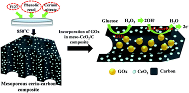
A conductive nanocomposite containing an immobilized oxidative enzyme in the pores of mesostructured ceria (CeO2)–carbon was developed as an efficient electrochemical biosensing platform. The construction of the nanocomposite began with the incorporation of CeO2 in a carbon matrix by the co-assembly of cerium nitrate, resol, and triblock copolymer via a facile evaporation-induced self-assembly method, which resulted in the formation of mesoporous ceria–carbon (denoted as Meso-CeO2/C). Glucose oxidase (GOx) was subsequently immobilized in the vacant pores of the Meso-CeO2/C by using glutaraldehyde crosslinking to prevent enzyme leaching from the matrix. H2O2 generated by the catalytic action of GOx in proportion to the amount of target glucose was rapidly converted into hydroxyl radicals by the catalytic activity of CeO2, which induced subsequent anodic oxidation of Ce3+ into Ce(OH)22+ or Ce(OH)4 with the anodic current. The constructed Meso-CeO2/C exhibited higher resolution in electrochemical detection of H2O2 than pure mesoporous carbon without ceria owing to the catalytic activity of ceria. The anodic current responses by the nanocomposite containing GOx in Meso-CeO2/C resulted in a linear increase in the concentration of target glucose (0.25–5 mM), which is suitable to measure the serum glucose, with excellent storage stability of over two months at room temperature. The biosensor also exhibited a high degree of precision and reproducibility when employing real human blood samples. Based on these results, we anticipate that this novel biosensing format can be readily extended to other oxidative enzymes for the detection of various clinically important target molecules.
Metal oxide nanostructures have recently become important materials in the field of biosensors owing to their high ionic conductivity, capacitive action, catalytic properties, and high isoelectric point. A number of oxide forms of inorganic metals such as zinc, iron, cerium, tin, zirconium, titanium, and magnesium have been found to exhibit interesting nanomorphological, biocompatible, and catalytic properties.15 Among these, ceria (CeO2) has been considered as a promising material for the fabrication of mediator-less electrochemical biosensors because of its ability to act as a redox couple.16 Furthermore, CeO2 is suitable for tight adsorption of most of the enzymes having an isoelectric point lower than ∼9, which is the isoelectric point of CeO2, by inducing electrostatic interaction without any further treatment. Moreover, the excellent electrical conductivity makes CeO2 an attractive matrix for biosensor applications. Based on these features, CeO2 has already been used to construct electrochemical biosensors to detect H2O2 or glucose by coupling it with GOx with excellent selectivity and sensitivity.16–18
In this work, in order to construct an electrochemical biosensor with the catalytic activity of ceria and the merits of electrically conductive carbon, a mesoporous ceria–carbon (denoted herein as Meso-CeO2/C) composite was synthesized through a simple “one-pot” assembly method. Typically, to fabricate a metal oxide/mesoporous carbon composite, a metal oxide precursor is backfilled into pre-synthesized mesoporous carbon.19 However, the “one-pot” approach eliminates many steps, thereby simplifying synthetic procedures.20,21 The constructed Meso-CeO2/C exhibited higher resolution for the electrochemical detection of H2O2 than pure mesoporous carbon without ceria owing to the catalytic activity of ceria, and glucose could be successfully quantified without any additional mediators by the successful incorporation of GOx in Meso-CeO2/C, proving its potential in the fabrication of reagentless glucose biosensors.
The amount of protein leaching from the nanostructure into the supernatant was measured using the bicinchoninic acid (BCA) assay (PIERCE, Rockford, IL) method. The final enzyme loading in the nanocomposite was calculated from the difference between the initial and leached enzyme amounts.
![]() | ||
Fig. 1 Synthetic scheme of Meso-CeO2/C and electrochemical biosensing mechanism of the conductive nanocomposite entrapping GOx in pores of Meso-CeO2/C. |
Three different amounts of CeO2, 20, 40, and 60 wt%, were loaded when Meso-CeO2/C was synthesized. TEM investigation revealed that Meso-CeO2/C showed clear distinctions with different ratios of carbon to ceria, as shown in Fig. 2. Highly ordered hexagonal arrays of mesopores are shown in Meso-CeO2 (20%)/C (Fig. 2a). The pore size and structure of Meso-CeO2 (20%)/C are similar to those of OMC synthesized without a ceria precursor because a small amount of hydrophilic ceria does not critically affect the co-assembly process.26 As the amount of ceria increased up to 60 wt%, the highly ordered mesoporous structure was replaced with a randomly oriented mesoporous structure with larger-sized pores. A large amount of cerium precursor easily aggregated during thermal treatment and generated the large ceria particles, which is typical of metal oxides. Therefore, large angular ceria particles could destroy the ordering of mesoporous materials. The SAXS patterns of the ceria–carbon material were investigated in order to obtain more detailed structural information. The SAXS pattern of Meso-CeO2 (20%)/C has two intense peaks at q = 0.44 and 0.76 nm−1 in the as-prepared condition. After heat treatment, the intensities of the two peaks decreased but did not disappear, indicating that the structural regularity of Meso-CeO2 (20%)/C was retained even after harsh thermal treatment (Fig. 2c). The SAXS profile of the 60 wt% sample (Fig. 2d) did not display prominent peaks, revealing deficiencies in the structural regulation in both the as-made and post-heat-treated samples, which is in accordance with the TEM images. The regularity of the ceria–carbon composite morphology gradually decreased as the amount of ceria increased. The TEM image and SAXS pattern of Meso-CeO2 (40%)/C shows that the structure was less ordered than the 20 wt% sample, as seen in Fig. S1.†
![]() | ||
Fig. 2 TEM images of (a) Meso-CeO2 (20%)/C and (b) Meso-CeO2 (60%)/C. SAXS traces of (c) Meso-CeO2 (20%)/C and (d) Meso-CeO2 (60%)/C. |
The nitrogen sorption isotherms of Meso-CeO2/C exhibited typical type IV isothermals with clear capillary condensation at P/P0 = 0.7–0.8, indicating uniform mesoporosity. A narrow pore-size distribution was obtained, approximately 4 nm and 13 nm for 20 wt% and 60 wt%, respectively, based on the Barrett–Joyner–Halenda (BJH) model. It is essential that sufficient pore volume be retained in order to accommodate GOx in the mesoporous structures; thus the 60 wt% sample, with its larger pore size, is more advantageous. The 20 wt% and 60 wt% Meso-CeO2/C materials have Brunauer–Emmett–Teller (BET) surface areas of 384.9 m2 g−1 and 224.6 m2 g−1, respectively, and total pore volumes of 0.25 cm3 g−1 and 0.38 cm3 g−1, respectively (Fig. 3a). Nitrogen physisorption was also carried out on the nanocomposite containing entrapped GOx in Meso-CeO2 (60%)/C (Fig. S2†). The pore volume of the nanocomposite was determined to be 0.23 cm3 g−1, which is less than 60% of the corresponding Meso-CeO2 (60%)/C, confirming that GOx molecules are immobilized inside the pore spaces of the Meso-CeO2 (60%)/C. The presence of GOx on Meso-CeO2 (60%)/C was also confirmed by the negative shift of zeta potential after the immobilization of GOx molecules, whereas the zeta potential of Meso-CeO2 (20%)/C did not show any significant change before and after the immobilization procedure (Fig. S3†). This observation clearly indicates that GOx molecules were efficiently immobilized within Meso-CeO2 (60%)/C having sufficiently large pore size (13 nm) but not within Meso-CeO2 (20%)/C presumably due to its pore size (4 nm) smaller than the size of GOx.
The XRD diffraction patterns display the presence of a crystalline phase of CeO2 (JCPDS: 34-0394). The characteristic diffraction peaks at 2θ = 28.5, 33.1, 47.5, and 56.3° correspond to the (111), (200), (220), and (311) planes, respectively, as shown in Fig. 3b. A typical carbon peak is also observed at approximately 2θ = 26°. The characteristic peaks of ceria are covered by broad carbon peaks for Meso-CeO2 (20%)/C owing to the highly loaded carbon. The 60 wt% samples exhibit obvious ceria crystalline phases with low-intensity carbon peaks. High resolution TEM images of Meso-CeO2 (20%)/C and Meso-CeO2 (60%)/C show that the wall of mesostructured composites is composed of cubic CeO2 nanocrystals (Fig. S4†). The crystallite size in Meso-CeO2 (60%)/C is evidently larger than that in Meso-CeO2 (60%)/C, which is consistent with the XRD result.
The potential of Meso-CeO2/C as an electrochemical biosensor toward the oxidation of H2O2 was investigated. The determination of H2O2 is of practical importance in chemical, biological, and many other fields.27–31 It also holds significant incentives for further applications in various oxidase-coupled biosensors.32,33 To demonstrate the electrocatalytic oxidation behavior of H2O2 by Meso-CeO2/C, CV experiments were conducted in the presence and absence of 10 mM H2O2 (Fig. 4a). When only chitosan-coated GCE was employed in the CV experiments as a control, an increase in current according to the addition of 10 mM H2O2 was observed starting from approximately 0.6 V (vs. Ag/AgCl), which is similar to the results for bare GCE without any coating.34,35 On the other hand, with a coating of Meso-CeO2/C mixed with chitosan on GCE, faster H2O2 oxidation was observed with increasing current starting from approximately 0.3 V, indicating the affirmative catalytic effect of Meso-CeO2/C on the oxidation of H2O2.17,18 The current peak intensity using Meso-CeO2/C in the presence of 10 mM H2O2 was approximately 10 times higher than that without H2O2, proving its potent capability to quantify H2O2 concentration with high resolution. MSU-F-C without CeO2 was also employed in the same CV experiments to see the effect of ceria on the oxidation of H2O2 (Fig. S5†).7 As a result, H2O2 oxidation from MSU-F-C started a little late (∼0.4 V) as compared to Meso-CeO2/C, and the current peak intensity with 10 mM H2O2 was only two times higher than that without H2O2, proving that the addition of ceria in mesoporous carbon enhanced the resolution for H2O2 determination because of the catalytic activity of ceria. Based on the CV results, the amperometric response of the electrode containing Meso-CeO2 (60%)/C and Meso-CeO2 (20%)/C obtained at 0.7 V (vs. Ag/AgCl) on sequential additions of 1 mM H2O2 resulted in a rapid increase (response time of 3 s) in anodic current (Fig. 4b). Although the initial background current for Meso-CeO2 (60%)/C was observed to be higher than that of Meso-CeO2 (20%)/C presumably due to higher conductivity originated from the highly loaded CeO2,17 the sensitivities of H2O2 detection for Meso-CeO2 (60%)/C and Meso-CeO2 (20%)/C were measured to be 198 nA mM−1 and 182 nA mM−1, respectively, indicating that detecting performance for H2O2 was not significantly varied from the loading amount of CeO2. Finally, we demonstrated our strategy by detecting glucose, a vital sugar in human blood at a normal concentration of 3–7 mM,36 employing the Meso-CeO2/C nanostructure containing immobilized GOx. As demonstrated above, Meso-CeO2 (60%)/C had a pore size (∼13 nm) large enough to accommodate GOx molecules (∼7 nm), whereas Meso-CeO2 (20%)/C had a relatively small pore size (∼4 nm) that could not accommodate the corresponding enzyme molecules. Thus, the catalytic action of GOx is expected to occur only with Meso-CeO2 (60%)/C. As shown in Fig. 5 and S6a,† the anodic current responses obtained rapidly from the cyclic voltammogram at 0.7 V (vs. Ag/AgCl) by employing the nanocomposite entrapping GOx in Meso-CeO2 (60%)/C resulted in a linear increase in the concentration of target glucose. The dynamic range of this system was from 0.25 to 5 mM, which is suitable to measure the serum glucose in both normal and patients with diabetes after appropriate dilution. The detection limit was approximately 0.1 mM based on a signal-to-noise ratio of 3. The sensitivity was measured to be 50.3 nA mM−1 for the nanocomposite using Meso-CeO2 (60%)/C. In the case of Meso-CeO2 (20%)/C, no oxidation current was measured owing to the absence of GOx, as expected (Fig. S6b†). The measured sensitivity and dynamic range of the current biosensor are among the best results reported for glucose biosensing based on nanostructured carbons.37,38 We further evaluated the selectivity of the biosensor toward the common interfering species for serum glucose detection including acetaminophen (AP), uric acid (UA), ascorbic acid (AA), and dopamine (DOP).39 Their concentrations were set as the real levels present in human serum. As a result, these substances have negligible effects on the current generated from the cyclic voltammogram at 0.7 V, demonstrating high specificity of the current biosensor (Fig. S7†). Long-term stability of the biosensor was also investigated by measuring its sensitivity from 1 to 5 mM glucose using the nanocomposite in Meso-CeO2 (60%)/C. The current response of the biosensor was revealed to maintain over 90% of the initial current response for 2 months at room temperature, indicating that the current biosensor is very robust for long-term use (Fig. 6).
![]() | ||
Fig. 6 Long-term stability of the biosensor stored at room temperature by measuring the sensitivity for 1–5 mM glucose using the nanocomposite in Meso-CeO2 (60%)/C. |
Encouraged by the above results, we evaluated the diagnostic capability of the nanocomposite system by using two independent human blood samples that contained representative levels of glucose corresponding to the normal and high stage of hyperglycemia (normal; ≤5.6 mM, borderline; 5.6–7 mM, and high; >7 mM).40 Blood samples that show the representative levels were selected based on the actual concentrations determined by using enzymatic methods (ADVIA 2400, Siemens Healthcare Diagnostics, USA). As a result, the serum glucose levels were quantified with excellent precisions yielding CVs in a range of 3.1–6.8% and recovery rates of 96–104% (Table 1), verifying the excellent reproducibility and reliability of the method. These observations show that the nanocomposite-based assay system should serve as a promising analytical tool to diagnose high level of glucose in clinical settings.
The key analytical performances of the nanocomposite in mesoporous ceria–carbon to detect H2O2 and glucose were summarized and compared with the recently published results based on the catalytic effect of CeO2 (Table S1†). It can be seen that the present biosensor is the sole electrochemical method that can detect clinical serum with high storage stability for glucose detection.
Footnote |
† Electronic supplementary information (ESI) available. See DOI: 10.1039/c5ra17808h |
This journal is © The Royal Society of Chemistry 2015 |