DOI:
10.1039/C5RA17484H
(Paper)
RSC Adv., 2015,
5, 92634-92647
Development of an efficient magnetically separable nanocatalyst: theoretical approach on the role of the ligand backbone on epoxidation capability†
Received
28th August 2015
, Accepted 22nd October 2015
First published on 22nd October 2015
Abstract
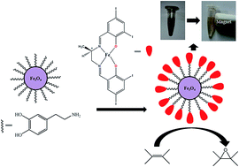
Three chiral Schiff base ligands H2L1, H2L2, H2L3 have been synthesized by treating (R)-1,2-diaminopropane separately with 3,5-dichlorosalicylaldehyde, 3,5-dibromosalicylaldehyde and 3,5-diiodosalicylaldehyde, respectively. Three new asymmetric FeIII complexes, namely, FeL1Cl (1), FeL2Cl (2), FeL3Cl (3) have been prepared from their corresponding ligands. The crystal structure of 2 reveals that the complexes are mononuclear in nature. Circular dichroism (CD) studies suggest that the ligands and their corresponding complexes contain an asymmetric center. The catalytic activity of these complexes toward the epoxidation of alkenes has been investigated in the presence of iodosylbenzene (PhIO), in two solvents CH3CN and CH2Cl2. The epoxide yield suggests that the order of their catalytic efficiency is 3 > 2 > 1. This trend as well as the role of substitution on the ligand backbone on alkene epoxidation has also been confirmed by density functional theory (DFT) calculations. For further adaptation, we attached our most efficient homogeneous catalyst, 3, with surface modified magnetic nanoparticles (Fe3O4@dopa) and thereby obtained the new magnetically separable nanocatalyst Fe3O4@dopa@FeL3Cl. This catalyst has been characterized and its olefin epoxidation ability investigated in similar conditions to those used for homogeneous catalysts. The enantiomeric excess of the epoxide yield reveals the retention of chirality of the active site of Fe3O4@dopa@FeL3Cl. The catalyst can be easily recovered by magnetic separation and recycled several times without significant loss of its catalytic activity.
Introduction
Oxygen activation and transfer by cytochrome P-450 has attracted the attention of organic chemists in particular, since it catalyses the mono-oxygenation of various compounds, both biotic and exobiotic, with high stereo- and regioselectivity under mild conditions. Groves and Nemo proposed that the oxygen transfer reaction proceeds through an oxohaem catalytic intermediate.1,2 From these basic results, many optically active complexes have been synthesized and found to be efficient catalysts for the epoxidation of olefins.3–5 The enantioselective epoxidation of olefins is one of the most important and challenging areas in organic synthesis because the resulting epoxide can be stereospecifically opened by nucleophiles to produce various optically active 1,2-difunctional compounds.6,7 Besides, the chiral epoxides play an eminent role as drug intermediates and chiral building blocks in the synthesis of optically active complex molecules.8–11
In the literature, transition metal Schiff base complexes have been extensively used as homogeneous catalysts for epoxidation.12–15 They show high efficiency since the active site is easily accessible, but their separation from the reaction mixture is really a difficult task. Heterogeneous catalysts appear to avoid this problems.16–18 In heterogeneous catalytic systems, the active catalytic sites and the reactants are in the different phases, so isolation and separation can be readily accomplished. Heterogenization is commonly achieved by entrapment or grafting of the active molecules on surfaces or inside the pores of a solid support, such as silica, alumina, organic nanotube etc.19,20 However, the active sites in heterogeneous catalysts are not as accessible as in a homogeneous system, and thus the activity of such catalysts become lowered. So, a catalytic system showing high activity and selectivity (like a homogeneous system) and ease separation and recovery (like a heterogeneous system) would be highly desirable. This goal can be achieved using magnetic nanoparticles (MNPs), which are able to bridge the gap between homogeneous and heterogeneous catalysis, preserving the desirable attributes of both systems.
The development and growth of nanotechnology has caused a continuous shift in every aspect of modern science. So far, it has changed the perspective of the scientific community towards catalysis and brought remarkable transformations in the synthetic chemical processes. MNPs nowadays have attracted immense scientific and technological interest due to their unique physical and chemical properties which make them an ultimate choice in the field of catalysis.21–23 The preparation and the use of MNPs in organic synthesis has become a subject of intense investigation as they offer advantages in clean and sustainable chemistry.24–27 The use of MNPs as catalysts in chemical synthesis has been extensively studied in recent years as their magnetic properties allow ready separation from the reaction mixture via the aid of an external magnet without cumbersome filtration and centrifugation techniques. The magnetic field-assisted separation can reduce energy consumption, catalyst loss and save the time in achieving catalyst recovery. Besides, these nanosized particles offer a large number of potential active sites for the reactants as a result of a large surface area to volume ratio which eventually results in higher yields.
Recent reports have shown that the mechanism of epoxidation of alkenes catalyzed by metal complexes, and the yield of products depends on several factors: the nature of the alkenes, oxidant, counter-ion of the catalyst, structure of the ligand moiety of catalyst and the added Lewis base.28 Steric and electronic effects of catalyst and olefinic substrates on epoxidation property have been investigated by several research groups also.29 However, the role of the halogen substituent present at the ligand back bone in transition metal Schiff base complexes on the epoxide yield is yet to be explored. Thus we are prompted to look at this issue. For this purpose we have designed and synthesized FeIII complexes of H2L1, H2L2 and H2L3 (Scheme 1), characterized them and investigated their catalytic efficiencies as epoxidation catalysts, in presence of terminal oxidant PhIO and in two solvents, CH3CN and CH2Cl2 under mild conditions. The order of their experimental catalytic efficiency has also been ratified by theoretical calculations. Finally, we attached the most efficient catalyst to surface modified nanoparticles (MNPs) and investigated its catalytic behavior. In this way we have developed an efficient magnetically separable asymmetric nanocatalyst for alkenes epoxidatation that preserve its activity after several catalytic cycles.
 |
| Scheme 1 Schematic diagram for the formation of the three ligands (H2L1, H2L2, H2L3). | |
Results and discussion
Preparation and characterization of complexes (1–3)
Three purposely selected Schiff bases H2L1, H2L2 and H2L3 have been prepared by treating 3,5-dichlorosalicylaldehyde, 3,5-dibromosalicylaldehyde and 3,5-diiodosalicylaldehyde separately with (R)-1,2-diaminopropane in ethanolic medium, respectively. Further treatment of H2L1, H2L2 and H2L3 with iron(III) chloride gives complexes 1, 2 and 3, respectively. We failed to get X-ray diffractable single crystals of 1 and 3; only a single crystal of 2 is obtained from DMF solvent. The complexes are characterized by regular physiochemical methods. All of them exhibit characteristic IR bands at the range of ∼1609–1638 cm−1 and ∼1498–1526 cm−1 assigned to C
N and skeletal vibrations (Fig. S1, ESI†), respectively. Electronic absorption spectra of the complexes in acetonitrile medium show multiple intense bands in the UV and visible regions (Fig. S2, ESI†). In these complexes, the absorption maxima observed in the near-UV regions (below 300 nm) are caused by π → π* transitions involving the phenolate units. Intense, high energy bands are also observed in the region between 300 and 350 nm, which are assigned to charge transfer transitions from the out-of-plane pπ orbital (HOMO) of the phenolate oxygen to the half-filled dx2−y2/dz2 orbital of high spin iron(III). The lowest energy bands (around 500 nm) are proposed to arise from charge-transfer transitions from the in-plane pπ orbital of the phenolate to the half-filled dπ* orbital of iron(III).30 Molar conductance of three complexes in acetonitrile medium are 2.3, 4.5, 5.8 Ω−1 cm2 M−1, which implies that all are nonelectrolyte in solution. Thermogram of the complexes (Fig. S3, ESI†) reveal that 1, 2 are stable up to 80 °C where 3 is stable upto 255 °C. Initial weight loss for both 1 and 2 are ∼3.1% (theoretical wt. loss are 3.4% and 2.6% for 1 and 2 respectively) may be due to loss of one absorbed water molecule. On further heating all three species generate Fe2O3 as the thermally stable end product (for complex 1, expt. wt loss = 58.33%, theo. wt loss = 56.51%; for complex 2 expt. wt loss = 69.74%, theo. wt loss = 67.5% and for complex 3 expt. wt loss = 75.3%, theo. wt loss = 73.82%). Circular dichroism (CD) measurements for all the ligands and their corresponding complexes have been performed. The CD spectra of ligand (H2L3) and 3 (FeL3Cl) are presented in Fig. 1 (as representative of all ligands and their corresponding complexes). Interestingly, the nature of CD spectra for FeL3Cl is the reverse of H2L3, suggesting the inversion of configuration after complexation.
 |
| Fig. 1 Circular dichroism (CD) spectra of the chiral ligand (H2L3) and the chiral complex 3 (FeL3Cl). | |
Description of crystal structure of complex 2
The X-ray structure of complex 2 has been depicted in Fig. 2. Selected bond lengths and bond angles are listed in Table S1 (ESI†). According to the X-ray structure the Schiff base ligand coordinates to one iron atom in a tetradentate mode. The metal center is five-coordinated and the geometrical index (τ5) is equal to 0.18, slightly shifted from zero, implying the geometry is slightly distorted square-pyramid. The two phenolic O atoms and the two imines N atoms of Schiff-base constitute the basal plane and one chlorine atom is occupying the axial position. Two Fe–O (phenolic) have almost similar bond distance, 1.888(6) and 1.890(6) Å, respectively. Two Fe–N (imine) distances of 2.093(8) and 2.088(8) Å, are also comparable whereas Fe–Cl bond distance is 2.222(3) Å. The methyl group on the 1,2-diaminopropane is disordered over two positions, C10a and C10b with refined occupancies of 0.65/0.35 respectively. This is intended to avoid steric clashes with the lattice dimethylformamide (DMF) molecule, which is also positionally disordered and refined with restraints on its geometry. The crystal is low diffracting (theta max = 21.80°) for the disorder detected. The atoms at lower occupancies (methyl C10b and DMF molecule) have been isotropically refined.
 |
| Fig. 2 Crystal structure of complex 2 with atom numbering scheme having 50% ellipsoid probability. | |
Optimization of the catalytic epoxidation conditions
In all cases, we have followed the same procedure to study the epoxidation reaction catalyzed by the three homogeneous catalysts (1–3). In a typical reaction, (E)-stilbene (30 mmol) (as representative), 3 (0.1 mmol) and PhIO (30 mmol) are mixed in 25 mL dichloromethane/acetonitrile and stirred for 4 h at room temperature (see Scheme 2). The progress of the reaction is monitored by TLC. After usual work up and chromatographic purification, the isolated yields of epoxide are found to be 74% in acetonitrile and 62% when dichloromethane used as a solvent. From this initial experiment we proceed to further optimization of the catalysis conditions, viz. amount of catalyst and terminal oxidant, along with the time required to obtain the maximum epoxide yield. To optimize the amount of catalyst, its concentration is varied between 0.05 and 0.2 mmol per 30 mmol of (E)-stilbene. Epoxide yield increases when the amount of catalyst is increased from 0.05 mmol to 0.1 mmol but the yield remains the same with further increment of catalyst amount up to 0.2 mmol. Then, the reaction has been studied with varying amounts of PhIO between 25 and 35 mmol per 30 mmol of (E)-stilbene. The yield of the epoxide also is studied by varying the time period between 2 and 6 h. We observe the epoxide yield attains the peak after 4 h of reaction and remain the same even after 6 h. An optimum of 87.84 mg (0.1 mmnol) of catalyst, PhIO (30 mmol) and 4 h reaction time are ideal for achieving the best yield. Table S2 (ESI†) represents the (E)-stilbene epoxide yields under different conditions using 3 as catalyst. The optimum reaction conditions thus determined have been followed in 1 and 2 catalyzed epoxidation reactions. The essential role played by the catalyst is evident from the extremely low (<2%) yield of epoxide found in a blank reaction carried out in absence of the catalyst.
 |
| Scheme 2 Model epoxidation reaction where (E)-stilbene is oxidized by iodosylbenzene, PhIO, in the presence of catalyst 3 (FeL3Cl). | |
Catalytic epoxidation with PhIO
Here we investigate the epoxidation of three alkenes [(E)-stilbene, (Z)-stilbene, or styrene] catalyzed by complexes 1–3 in CH3CN or CH2Cl2, with PhIO as oxidant (Table 1). From Table 1 three main features may be drawn. First, in all cases CH3CN solvent renders higher conversions and yields than CH2Cl2. Second, using the same catalyst for the epoxidation of the three olefins, all of them produce similar epoxide yields, with a very slight preference for (E)-stilbene. Finally, complex 3 emerges as the best catalyst for the epoxidation reaction under study, rendering yields close to 70% in CH3CN. However, complex 2 and complex 1 show average yields of 65% and 54% in CH3CN, respectively. The epoxidation process has also been monitored by UV-Vis spectroscopy. The isolated Fe(III) complex has two peaks at 306 and 487 nm. But, after addition of PhIO both disappear and a shoulder around 377 nm emerges (Fig. S4, ESI†). This observation clearly suggests the generation of higher valent FeV
O species as previously reported (see Scheme 3).15c,d The generation of FeV
O species is very common that were confirmed previously both experimentally an theoretically.31–36
Table 1 Epoxidation of alkenes catalyzed by the complexes 1–3 in CH3CN and CH2Cl2 with PhIO
Catalysta |
Substrate |
Solvent |
Conversion (%) |
TONb |
Yieldc (%) |
eed,f (%) |
Catalyst (0.1 mmol), alkenes (30 mmol), PhIO (30 mmol), and CH3CN or CH2Cl2 (25 mL), are stirred at room temperature for 4 h in air. TON = moles of substrate converted per mole of catalyst. Isolated epoxide yield. Determined by 1H NMR (300 Hz) in the presence of Eu(hfc). Trace = concentration < 5%. Configuration not determined. |
1 |
(E)-Stilbene |
CH3CN |
58 |
43.5 |
55 |
Tracee |
CH2Cl2 |
55 |
41.4 |
50 |
Tracee |
(Z)-Stilbene |
CH3CN |
61 |
45.9 |
55 (cis : trans = 70 : 30) |
18 (trans) |
CH2Cl2 |
52 |
39 |
48 (cis : trans = 80 : 20) |
15 (trans) |
Styrene |
CH3CN |
60 |
45 |
53 |
20 |
CH2Cl2 |
47 |
35.4 |
44 |
Tracee |
2 |
(E)-Stilbene |
CH3CN |
64 |
48 |
68 |
16 |
CH2Cl2 |
61 |
45.9 |
57 |
Tracee |
(Z)-Stilbene |
CH3CN |
61 |
45.7 |
67 (cis : trans = 77 : 23) |
25 (trans) |
CH2Cl2 |
57 |
42.9 |
54 (cis : trans = 60 : 40) |
21 (trans) |
Styrene |
CH3CN |
57 |
50.4 |
61 |
Tracee |
CH2Cl2 |
59 |
44.4 |
53 |
Tracee |
3 |
(E)-Stilbene |
CH3CN |
79 |
59.4 |
74 |
21 |
CH2Cl2 |
66 |
49.5 |
62 |
14 |
(Z)-Stilbene |
CH3CN |
78 |
58.5 |
72 (cis : trans = 90 : 10) |
27 (trans) |
CH2Cl2 |
74 |
55.5 |
67 (cis : trans = 75 : 25) |
22 (trans) |
Styrene |
CH3CN |
72 |
54 |
69 |
11 |
CH2Cl2 |
67 |
50.4 |
65 |
Tracee |
 |
| Scheme 3 Schematic presentation of the generation of Fe(V) oxo species during epoxidation of alkenes. | |
Theoretical investigation
Theoretical investigation on the effect of halogen substituent on the oxidation of iron complex catalysts.
Geometry and charge distribution of the non-oxidized Fe(III) complexes. All the initial complexes are electrically neutral and only differ in the halogen at the ortho and para positions of the two benzene rings of the Schiff base bonded to the Fe(III) center (Fig. 3). The non-oxidized initial complexes could present low (2S + 1 = 2) or high (2S + 1 = 6) spin depending of the distribution of the 5 most external iron electrons over the non-degenerate d orbitals of the metal in the complexes. We have computationally checked that high spin initial complexes are about 15 kcal mol−1 more stable in Gibbs energy than the corresponding low spin ones (Table S3, ESI†).
 |
| Fig. 3 Main DFT geometrical parameters of the initial halogen Fe(III) complexes. (UB3LYP/6-311+G(d,p) with LANL2DZ pseudopotential for Fe, Cl, Br, and I.) All distances in angstroms. | |
DFT geometrical parameters essentially match those provided by X-ray except for a slight lengthening of bond distances. Metal ion is bonded through stronger bonds to both O atoms (bond distances of 1.91 Å) than to both N atoms (average bond distances of 2.12 Å), forming average O–Fe–O and N–Fe–N bonds of 100.3° and 77.4°, respectively. The metal is also bonded to a chloride anion at a distance of 2.37 Å and placed slightly over a distorted plane of a square pyramid. Such similar geometries for the three initial complexes indicate that geometry is not a factor determining a different oxidation capability.
As oxidation is mainly and electronic issue, we also checked the NBO charge of some relevant atoms at each initial complex. The charges of the halogens in the aromatic rings become increasingly positive when moving from chlorine to bromine and iodine systems (−0.02 to +0.08 to +0.20, respectively for para halogens), as expected due to their diminishing electronegativity. The electron density that halogens donate makes the negative charge on atoms bonded to Fe(III) slightly higher as the atomic number of the halogen increases. Thus, as an average, O atoms charges go from −0.686 to −0.688 to −0.693, whereas those of N atoms vary from −0.521 to −0.522 to −0.525 for chlorine, bromine, and iodine systems, respectively. However, the slightly more negative charges of atoms bonded to Fe(III) do not make the metal more negatively charged since it lets the charge flow to chloride ligand (Fe charges goes from 1.219 to 1.220 and to 1.222 whereas that of chloride goes from −0.502 to −0.505 and to −0.511).
Geometry and charge distribution of the oxidized Fe(V) complexes. Assuming that the metal complexes become oxidized by iodosylbenzene, PhIO, during the epoxidation of alkenes, it is interesting to characterize the oxidized metal complexes through DFT calculations. The three oxidized complexes, complex 1-oxo, complex 2-oxo, and complex 3-oxo, have been optimized including the released chloride anion in such a way that the whole system is electrically neutral (Fig. 4). It is interesting to note that the presence of the oxygen atom at the apical position of the complex yields less distorted square pyramid geometries for the Fe(V) than that observed for Fe(III). N–Fe bonds (1.930 Å in average) are now stronger and closer in length to O–Fe bonds (1.917 Å in average), whereas O–Fe–O and N–Fe–N angels are both of about 85°. Concerning NBO charges, the same trend described for the initial complexes is found for the oxidized ones: heavier halogens more easily donate electron charge that goes by through O and N atoms bonded to Fe(V) and from the metal to the apical oxygen (oxygen charge evolves from −0.350 to −0.372 and to −0.385 when going from chlorine to bromine and iodine oxidized complexes).
 |
| Fig. 4 Main DFT geometrical parameters of the final oxidized Fe(V) complexes. (UB3LYP/6-311+G(d,p) with LANL2DZ pseudopotential for Fe, Cl, Br, and I.) All distances in angstroms. | |
Reaction energy for the intermediate oxidation of the catalyst by PhIO
As already said, initial Fe(III) complexes are assumed to be oxidized by PhIO ((a) in Fig. 5) to render the oxidized complexes just described and PhI ((b) at Fig. 5), being this oxidation an intermediate step in the epoxidation of alkenes. The oxidation of the catalyst is schematically represented by eqn (1). |
Complex + PhIO ⇔ complex-oxo + PhI
| (1) |
 |
| Fig. 5 Main DFT geometrical parameter of the oxidized ((a), PhIO) and reduced ((b), PhI) iodobenzine molecule. (UB3LYP/6-311+G(d,p) with LANL2DZ pseudopotential for I.) Distance in angstroms. | |
Theoretical results show that the high-spin initial complexes are about 15 kcal mol−1 more stable in Gibbs energy than low-spin ones, whereas oxidized complexes present similar energies for both spin states (Table S3, ESI†). With these data at hand and to fulfill the required spin-conservation along the reaction displayed in eqn (1), two situations are possible. On the one hand, an ancillary reaction involving a spin transition from a quartet to a sextet could happen associated to the oxidation of the catalyst to allow its spin crossover, or, on the other hand, the oxidation of the catalyst could take place in the doublet state. Few examples of spin flip during catalysis are already reported.37–39 The second assumption involves an energy supply from the environment to excite Fe(III) complexes from the most stable sextet to the doublet one.
Table 2 collects the electronic and Gibbs energy of the catalyst oxidation step (calculated as the summation of the energy of the corresponding products menus summation of the energy of the corresponding reactants in eqn (1)) for both hypotheses. For the first one, considering the electronic energy, only the products of oxidized complex 3 are more stable than the corresponding initial reactants. In terms of Gibbs energy in acetonitrile solution, all the oxidized products are less stable than the initial ones. However, both electronic and Gibbs energies agree to show that the yield of oxidized complexes is expected to increase from chlorine to bromine and iodine complexes. The second hypotheses, renders the same trend, now predicting exothermic processes in all the cases. As a consequence, what is most important for this study is that theoretical calculations based on any of the possible assumptions agree to point the iodine catalyst as the most effective one.
Table 2 Reaction electronic and Gibbs energy in acetonitrile solution calculated at B3LYP/6-31+G(d,p) with LANL2DZ psudopotential for Fe, Cl, Br, and I. Left values at each column for the reaction between the most stable spin-states of reactants and products; right values for the reaction in the doublet state (see text)
Complex |
ΔEe (kcal mol−1) |
ΔGsol (kcal mol−1) |
1 |
6.6 |
9.1 |
2 |
3.7 |
6.2 |
3 |
−0.6 |
2.1 |
Preparation and characterization of Fe3O4@dopa@FeL3Cl
A schematic diagram of Fe3O4@dopa@FeL3Cl formation is presented in Scheme 4. Firstly, the preparation of magnetic nanoparticles Fe3O4 and then their surface modification has been done following the procedure earlier reported by R. S. Varma et al. in 2009.40 For the preparation of Fe3O4@dopa@FeL3Cl, 1 g of FeL3Cl is added in the dispersed acetonitrile solution of amine-functionalized nano-Fe3O4 (500 mg). The mixture is stirred for 12 h at room temperature. The product is allowed to settle, washed several times with acetonitrile, and dried under vacuum at 60 °C for 2 h. Newly prepared Fe3O4@dopa@FeL3Cl has been characterized by the following process.
 |
| Scheme 4 Schematic representation of the preparation of Fe3O4@dopa@FeL3Cl particles. | |
Fourier-transform infrared spectra (FTIR) of Fe3O4, Fe3O4@dopa and Fe3O4@dopa@FeL3Cl are represented in Fig. 6. The peaks at around 583 cm−1 and 636 cm−1 are the characteristic absorptions of the Fe–O bond, which confirmed the presence of iron oxide, as previously reported.41 For Fe3O4@dopa a peak around 1485 cm−1 arises, which may be due to the vibration of the benzene ring present at dopamine moiety. Several new peaks are generated for Fe3O4@dopa@FeL3Cl, along with the characteristic peak of Fe3O4. The peak at 1526 cm−1 may be assigned as the skeleton vibration of the complex, the sharp peak at 1624 cm−1 may be due to the C
N vibration of incorporated complex moiety. The FTIR spectra allow to conclude that the desired surface medication of MNPs has been successfully done.
 |
| Fig. 6 Fourier-transform infrared spectra (FTIR) spectra of Fe3O4 NPs, Fe3O4@dopa and Fe3O4@dopa@FeL3Cl. | |
The degree of crystallinity of magnetic Fe3O4, Fe3O4@dopa and Fe3O4@dopa@FeL3Cl are obtained from PXRD measurements (Fig. 7). The PXRD data of the synthesized magnetic nanoparticles show diffraction peaks at 2θ = 29.69°, 35.19°, 42.61°, 56.5°, 59.5° and 62.36° which can be assigned to the (220), (311), (400), (422), (511) and (440) planes of Fe3O4, respectively, indicating that the Fe3O4 particles in the nanoparticles are pure Fe3O4 with a cubic spinel structure. These match well with the standard Fe3O4 sample. The same peaks are observed in both of the Fe3O4@dopa and Fe3O4@dopa@FeL3Cl in PXRD patterns, indicating that the resultant nanoparticles contain pure Fe3O4 with a spinel structure and that the grafting process does not induce any phase change of Fe3O4 as previously mentioned by other group previously.42
 |
| Fig. 7 Power X-ray diffraction (PXRD) spectra of Fe3O4 NPs, Fe3O4@dopa and Fe3O4@dopa@FeL3Cl. | |
Solid state UV spectrum of Fe3O4 is very similar to a previously reported one.43 The existence of Fe3O4 in the Fe3O4@dopa and Fe3O4@dopa@FeL3Cl species has been proved by comparing the solid state UV (Fig. 8). FeL3Cl has a broad shoulder at ∼495 nm (Fig. S2, ESI†). Increment of the absorbance at ∼500 nm for Fe3O4@dopa@FeL3Cl clearly suggests the conjugation of FeL3Cl onto Fe3O4@dopa.
 |
| Fig. 8 Solid state UV spectra of Fe3O4 NPs, Fe3O4@dopa and Fe3O4@dopa@FeL3Cl. | |
For confirmation of the successful surface modification of magnetic Fe3O4 and further FeL3Cl grafting on the surface of Fe3O4@dopa, thermogravimetric analysis (TGA) is carried out. As shown in Fig. 9, the trace amount of weight loss within the range of 100–200 °C is caused by the trace amount of water vapor adsorbed by magnetic Fe3O4. 7% weight loss occurred in the range of 30–800 °C for Fe3O4-NPs. Weight loss within the same range for Fe3O4@dopa and Fe3O4@dopa@FeL3Cl are 19% and 27.6%, respectively. TGA results clearly suggest that the enhancing weight loss is due to the increasing amount of attached organic moiety from Fe3O4@dopa to Fe3O4@dopa@FeL3Cl. Similar results have been described by other groups.44 Raman spectrum of FeL3Cl and Fe3O4@dopa@FeL3Cl are depicted in Fig. S5 (ESI†). FeL3Cl has peaks at 500, 594, 776, 876, 967, 1312, 1442, 1516, 1590 cm−1. FT Raman spectra for Fe3O4@dopa@FeL3Cl resemble the unbound FeL3Cl demonstrating that the catalyst moiety remains intact during the process of encapsulation as earlier reported.20c,45
 |
| Fig. 9 Thermogravimetric analysis (TGA) diagrams of Fe3O4 NPs, Fe3O4@dopa and Fe3O4@dopa@FeL3Cl. | |
SEM images of Fe3O4, Fe3O4@dopa and Fe3O4@dopa@FeL3Cl are presented in Fig. 10. Fig. 10(a) suggests Fe3O4 NPs have square shaped morphology with 20–30 nm size. The morphologies of Fe3O4@dopa (Fig. 10(b)) and Fe3O4@dopa@FeL3Cl (Fig. 10(c)) are quite different from that Fe3O4-NPs. Both Fe3O4@dopa and Fe3O4@dopa@FeL3Cl contained agglomerated particle having larger sized particle in the case of Fe3O4@dopa@FeL3Cl. Alteration in morphology from Fig. 10(a) and (b) confirmed the surface modification process.
 |
| Fig. 10 Scanning electron microscopy (SEM) images of (a) Fe3O4-NPs, (b) Fe3O4@dopa and (c) Fe3O4@dopa@FeL3Cl. | |
TEM images of Fe3O4-NPs, Fe3O4@dopa and Fe3O4@dopa@FeL3Cl are shown in Fig. 11(a)–(c), respectively. A close examination of the TEM image in Fig. 11(a) reveals that the magnetic nanoparticles are quasi-spherical with the average diameter of 10–15 nm. The measurement of the hydrodynamic size of Fe3O4-NPs by dynamic light scattering (DLS) shows stable non-aggregated particles with a mean diameter of 30 nm (Fig. S6, ESI†). The NPs show good stability in water. The observed sizes of the NPs from TEM images are approximately smaller than the hydrodynamic diameter obtained from the DLS experiment. Transmission electron microscopy measure the size in the dried state of the sample, whereas DLS measure the size in the hydrated state of the sample, so the size measured by DLS is a hydrodynamic diameter and is larger. The nanoparticles, depicted in Fig. 11(b) after dopamine encapsulation step, have a discrete core–shell structure, and Fe3O4 is surrounded by 3–5 nm thick dopamine shell as previously reported.41 Fig. 11(c) illustrates the grafting of FeL3Cl on to the surface of Fe3O4@dopa. Energy-dispersive X-ray spectroscopy (EDX) spectrum of Fe3O4@dopa@FeL3Cl is depicted in Fig. 12. Fe and O signals come from the Fe3O4 nanoparticles and carbon (C) from dopamine. Signals of I and Cl are responsible for the presence of those elements in FeL3Cl. The gold (Au) signals come from the coating material of the instrument.
 |
| Fig. 11 Transmission electron microscopy (TEM) images of (a) Fe3O4-NPs, (b) Fe3O4@dopa and (c) Fe3O4@dopa@FeL3Cl. | |
 |
| Fig. 12 Energy-dispersive X-ray (EDX) spectrum of Fe3O4@dopa@FeL3Cl. | |
Epoxidation property of Fe3O4@dopa@FeL3Cl
We also evaluated the optimum reaction conditions to achieve the maximum epoxide yield with the heterogeneous catalyst as already done for the homogeneous system (Table S4, ESI†). For this purpose the weight of Fe3O4@dopa@FeL3Cl is varied between 50 and 200 mg per 3 mmol of (E)-stilbene. Enhancement in the yield of epoxide is observed when the amount of catalyst is increased from 50 to 100 mg but the yield remain same with further increment of catalyst amount up to 200 mg. The reaction has also been studied with varying the amounts of terminal oxidant, PhIO and the time. The results show that using 3 mmol PhIO and 6 h stirring the best epoxidation yield is obtained. At the end of the reaction, the catalyst has been magnetically separated out and reuse for further epoxidation. Here it is noteworthy that we did not find any leaching of catalysts during the epoxidation reaction.
Again, here we have studied Fe3O4@dopa@FeL3Cl catalyzed epoxidation of three different alkenes [(E)-stilbene, (Z)-stilbene, or styrene] in CH3CN or CH2Cl2 with PhIO and the results are presented in Table 3.
Table 3 Epoxidation of alkenes catalyzed by Fe3O4@dopa@FeL3Cl in CH3CN and CH2Cl2 with PhIO
Catalysta |
Substrate |
Solvent |
Conversion (%) |
TONb |
Yieldc (%) |
eed,f (%) |
Catalyst (100 mg), alkenes (3 mmol), PhIO (3 mmol), and CH3CN or CH2Cl2 (25 mL), were stirred at room temperature for 6 h in air. TON = moles of substrate converted per mole of catalyst per hour. Isolated epoxide yield. Determined by 1H NMR (300 Hz) in the presence of Eu(hfc). Trace = concentration < 5%. Configuration not determined. |
Fe3O4@dopa @FeL3Cl |
(E)-Stilbene |
CH3CN |
79 |
40.5 |
62 |
Tracee |
CH2Cl2 |
66 |
33.8 |
57 |
18 |
(Z)-Stilbene |
CH3CN |
78 |
40.0 |
67 (cis : trans = 80 : 20) |
29 (trans) |
CH2Cl2 |
74 |
37.9 |
62 (cis : trans = 75 : 25) |
21 (trans) |
Styrene |
CH3CN |
72 |
36.9 |
63 |
17 |
CH2Cl2 |
67 |
34.3 |
55 |
Tracee |
Magnetization behavior of the (a) Fe3O4-NPs, (b) Fe3O4@dopa and (c) Fe3O4@dopa@FeL3Cl nanoparticles under the applied magnetic field is depicted in Fig. 13. The curves exhibit an extremely interesting phenomenon showing a decrease in the values of the saturation magnetization (Ms) from the Fe3O4 nanoparticles (58.06 emu g−1) to Fe3O4@dopa (39.24 emu g−1) to the final Fe3O4@dopa@FeL3Cl nanocatalyst (26.39 emu g−1) which can be attributed to the gradual increment of diamagnetic organic materials from Fe3O4 to Fe3O4@dopa@FeL3Cl. Furthermore, in comparison with the bulk magnetite nanomaterials that typically show a saturation magnetization value of 92 emu g−1, the Ms value of the Fe3O4 nanoparticles is found to be much lower. Since the magnetization of a particle in an external field is a function of its size, it is normal that small nanoparticles show in the Ms value.46 However, the net magnetism exhibited by the final nanocatalyst is sufficiently good for an effective separation from the solution medium through the application of an external magnetic force.
 |
| Fig. 13 Magnetic curves of Fe3O4 NPs, Fe3O4@dopa and Fe3O4@dopa@FeL3Cl; and inset: image of the efficiency of the magnetic separation of the catalyst. | |
Characterization and reusability Fe3O4@dopa@FeL3Cl
We have studied the recycling efficiency of the magnetically separable heterogeneous catalyst Fe3O4@dopa@FeL3Cl, i.e., whether the catalyst can be reused further for several cycles. (E)-Stilbene has been chosen as a representative case for recycling experiments. After each reaction cycle the catalysts are recovered by magnetic separation, washed thoroughly with acetonitrile and dried at 100 °C for 2 h. The used catalyst has been further characterized by FTIR, UV-Vis in solid state and scanning electron microscopy (Fig. 14). Comparing all the experimental results of the used catalyst with virgin one it is easy to conclude that no significant change in the catalyst after epoxidation reaction. That implies no structural deformation has taken place in the active site of catalyst which is probably the key factor for its reusability. The catalytic reactions have been carried out following the same experimental procedure as that with the original catalysts, and for all cases, the yields are not significantly different through first to fifth use (Fig. 14).
 |
| Fig. 14 (A) Fourier-transform infrared spectra (FTIR), (B) solid state UV and (C) scanning electron microscopy (SEM) images of used Fe3O4@dopa@FeL3Cl. (D) Recyclability and reusability test of Fe3O4@dopa@FeL3Cl for the epoxidation of (E)-stilbene. | |
Experimental
Materials and methods
All chemicals were obtained from commercial sources and used as received. Solvents were dried according to standard procedure and distilled prior to use. 3,5-Dichloro salicylaldehyde; 3,5-dibromo salicylaldehyde; 3,5-diiodo salicylaldehyde; (R)-1,2-diaminopropane, styrene, (E)-stilbene and (Z)-stilbene were purchased from Aldrich and used in epoxidation experiments without further purification. Iron(III) chloride was purchased from Merck.
Elemental analyses (carbon, hydrogen and nitrogen) were performed using a Perkin-Elmer 240C elemental analyzer. Infrared spectra (4000–500 cm−1) were recorded at 27 °C using a Perkin-Elmer RXI FT-IR spectrophotometer with KBr pellets. Electronic spectra (800–200 nm) were obtained at 27 °C using a Shimadzu UV-3101PC with methanol as solvent and reference. Thermal analyses (TG-DTA) were carried out on a Mettler Toledo (TGA/SDTA851) thermal analyzer in flowing dinitrogen (flow rate: 30 cm3 min−1). Field Emission Scanning Electron Microscope (FE-SEM) measurement was carried out with JEOL JSM-6700F field-emission microscope. X-ray powder diffraction (PXRD) was performed on a XPERT-PRO Diffractometer monochromated Cu-Kα radiation (40.0 kV, 30.0 mA) at room temperature. Raman spectra (Horiba Jobin Yvon, T64000 model) were recorded in solution via 90° scattering by exciting the sample with an Ar+ ion laser source of 48 mW power at the sample. The spectra of the samples were recorded at room temperature in solid phase. The light irradiation dependent study was done after different irradiation times with a white light source compiled with a <420 nm cut off filter. Conductance of the methanolic solution of complexes was measured using a SYSTRONICS 306 conductivity meter. A vibrating sample magnetometer (EV-9, Microsense, ADE) was utilized for obtaining the magnetization curves. The CD spectra were measured with a Jobin Ivon CD 6 spectrophotometer. All the spectra were recorded in acetonitrile.
Syntheses of catalysts
Synthesis of [FeL1Cl] (1). 10 mL of ethanolic solution of (R)-1,2-diaminopropane (0.74 g, 1 mmol) was added slowly to the 20 mL ethanolic solution of 3,5-dichlorosalicylaldehyde (0.382 g, 2 mmol). Immediate precipitation of H2L1 took place. 10 mL ethanolic solution of ferric chloride hexahydrate (0.135 g, 0.5 mmol) was added to suspension of H2L1. Yellow precipitate of H2L1 was disappearing readily and the solution was turned deep reddish brown. The solution was allowed to stir for 1 h. Brown colored micro crystals of 1 were formed. Anal. calcd for 1: C, 40.11; H, 2.35; N, 5.50. Found: C, 40.08; H, 2.30; N, 5.41. IR (KBr): ν(C
N) 1638 cm−1; ν(skeletated vibration) 1526 cm−1. UV λmax (ACN)/nm 267, 308, 482sh (ε/dm3 mol−1 cm−1 24
806, 15
253, 5816).
Synthesis of [FeL2Cl] (2). Complex 2 was synthesized following the same procedure as mentioned for 1. Here 3,5-dibromosalicylaldehyde (0.56 g, 2 mmol) was used in place of 3,5-dichlorosalicylaldehyde. The solution was allowed to stir for 1 h. Brown colored micro crystals of 2 were formed. Microcrystals were dissolved in DMF and kept the solution in dark. X-ray suitable single crystals of 2 were obtained from the solution. Anal. calcd for 2: C, 29.71; H, 1.74; N, 4.07. Found: C, 29.68; H, 1.70; N, 4.01. IR (KBr): ν(C
N) 1631 cm−1; ν(skeletated vibration) 1511 cm−1. UV λmax (ACN)/nm 265, 309, 484sh (ε/dm3 mol−1 cm−1 27
214, 15
712, 5816).
Synthesis of [FeL3Cl] (3). Complex 3 was prepared by adopting the same procedure as that for 1 using 3,5-diiodosalicylaldehyde (0.748 g, 2 mmol) in place of 3,5-dichlorosalicylaldehyde. Anal. calcd for 3: C, 23.32; H, 1.37; N, 3.20. Found: C, 23.28; H, 1.32; N, 3.18. IR (KBr): ν(C
N) 1609 cm−1; ν(skeletated vibration) 1498 cm−1. UV λmax (ACN)/nm 266, 317, 495sh (ε/dm3 mol−1 cm−1 21
414, 12
026, 4423).
Synthesis of Fe3O4 NPs. Preparation of magnetic nanoparticles Fe3O4 have been done following the same procedure as reported earlier by R. S. Varma et al. at 2009.40
Synthesis of Fe3O4@dopa. Fe3O4@dopa has been prepared by following the similar method as mentioned previously.40
Synthesis of Fe3O4@dopa@FeL3Cl. Fe3O4@dopa@FeL3Cl has been synthesized by following similar procedure20c,45 but with some modification where 1 g of FeL3Cl is added in the dispersed acetonitrilic solution of Fe3O4@dopa (500 mg). The mixture is stirred for 12 h in room temperature. The product is allowed to settle, washed several times with acetonitrile, and dried under vacuum at 60 °C for 2 h. IR (KBr): ν(C
N) 1624 cm−1; ν(skeletated vibration) 1526, 1485 cm−1; ν(Fe3O4) 583 cm−1. UV λmax (solid)/nm ∼502sh, ∼279.
X-ray data collection and structure determination
Diffraction data for complex 2 was collected at room temperature (293 K) on a Bruker Smart CCD diffractometer equipped with graphite-monochromated MoKα radiation (λ = 0.71073 Å). Cell refinement, indexing and scaling of the data set were carried out using Bruker SMART APEX and Bruker SAINT package.47 The structure was solved by direct methods and subsequent Fourier analyses48 and refined by the full-matrix least-squares method based on F2 with all observed reflections using SIR-92 and SHELX-97,49 software. For the complex, all non-hydrogen atoms were refined with anisotropic thermal parameters and the hydrogen atoms were fixed at their respective positions riding on their carrier atoms and refined anisotropically. All the calculations were performed using the WinGX System, Ver 1.80.05,50 PLATON99,51 ORTEP3 (ref. 52) programs. Selected crystallographic data and refinement details are displayed in Table 4.
Table 4 Crystal data and details of the structure determination for complex 2
Empirical formula |
C17H12Br4ClFeN2O2, C3H7NO |
Formula mass |
760.28 |
Crystal system |
Monoclinic |
Space group |
P21/n (no. 14) |
a (Å) |
13.314(6) |
b (Å) |
14.720(6) |
c (Å) |
13.228(6) |
α (°) |
90 |
β (°) |
101.192(6) |
γ (°) |
90 |
V (Å3) |
2543.2(19) |
Z |
4 |
T (K) |
293 |
μ(MoKα) (mm−1) |
7.009 |
Dcalc (Mg m−3) |
1.986 |
F(000) |
1468 |
θmax (degree) |
21.8 |
Tot., uniq. data, Rint |
8389, 2956, 0.071 |
Observed (I > 2σ(I)) |
1883 |
Nref, Npar |
2956, 292 |
R, wR2, S |
0.0484, 0.1247, 1.02 |
Residual extrema (e Å−3) |
−0.53, 0.45 |
Preparation of iodosylbenzene
It was prepared by hydrolysis of the corresponding diacetate with aqueous sodium hydroxide according to literature method.53 In every epoxidation experiment freshly prepared PhIO was used.
Epoxidation study of catalysts
Epoxidation of alkenes catalyzed by complexes (1–3). To a solution of alkene (30 mmol) in acetonitrile or dichloromethane (25 mL), 0.1 mmol of complex was added and then PhIO (30 mmol) was added portion wise to that solution and then the resultant mixture was stirred at room temperature for 4 h in air. The reaction progress was monitored by TLC. After removal of solvent, the crude product was purified by flash chromatography. Identification of the epoxide was performed by 1H NMR spectroscopy.
Epoxidation of alkenes catalyzed by Fe3O4@dopa@FeL3Cl. To a solution of alkene (3 mmol) in acetonitrile or dichloromethane (25 mL), 100 mg of Fe3O4@dopa@FeL3Cl was suspended and then PhIO (3 mmol) was added and the resultant mixture were stirred at room temperature for 6 h in air. Here also we added iodosylbenzene portion wise to the solution. The reaction progress was monitored by TLC. After completion of the reaction, the catalyst was separated with magnet and the solvent was removed by rotary evaporator. The crude product was thus obtained was purified by flash chromatography. Identification of the epoxide was performed by 1H NMR spectroscopy.
Computational method
All calculations were carried out with the Gaussian 09 series of programs.54 The implicit Polarizable Continuum Model (PCM) of Tomasi and coworkers55 was used to take into account the effect of acetonitrile as solvent (dielectric constant, ε = 35.688), using atomic radii derived from the standard Universal Force Field (UFF). Full geometry optimization of all reactants and products has been carried out using Density Functional Theory with the UB3LYP56 functional and the 6-311+G(d,p) basis set for C, N, O and H atoms as well as with LANL2DZ pseudopotential for Fe, Cl, Br, and I atoms. Geometry optimizations were done with the Schlegel's algorithm.57 The located stationary points were checked to be true minima by the analytical computation of the harmonic vibrational frequencies at the same theory level. Due to the open shell nature of the iron complexes under study, the possible low and high spin states have been calculated for non-oxidized and oxidized complexes. High spin structures were found to be more stable than low spin ones in all cases, and they showed practically no spin contamination at the theory level here used. For interpretation purposes, a natural bond orbital (NBO) analysis was performed on the non-oxidized and oxidized complexes in solution.57–59 Thermodynamic magnitude ΔG was also calculated within the ideal gas, rigid rotor, and harmonic oscillator approximations at a pressure of 1 atm and a temperature of 298.15 K.60
Conclusion
In conclusion, here we have demonstrated that among three asymmetric mononuclear Fe(III) complexes of chloro, bromo and iodo substituted Schiff base ligands, the most efficient catalyst towards alkene epoxidation is iodo substituted Schiff base Fe(III) complex. This efficiency is related to the stability of FeV
O intermediate which forms during the catalytic process in presence of PhIO. This factor has been well rationalized by DFT calculations. Then, we have adapted an economically viable and energy efficient catalytic process using the iodo substituted Schiff base Fe(III) complex over magnetically separable nanoparticules for the selective epoxidation of alkenes at room temperature. The simple operation, stability and rigidity of catalyst, the use of inexpensive and gentle magnetic nanoparticles as support, the easy recoverability and reusability of the catalyst, along with the high epoxide yield made the proposed protocol a potential candidate for addressing the challenges of sustainability. We consider that this novel magnetic nano composite system would find applications in several other industrially significant catalytic processes as well as the general synthetic organic transformations.
Acknowledgements
Financial support by the Science and Engineering Research Board (a statutory body under DST, New Delhi, India (F. No. SB/FT/CS-185/2013 dt. 30-06-2014) is gratefully acknowledged by TC. We also thank DST, New Delhi, for providing single crystal diffractometer facility at the Department of Chemistry, University of Calcutta, through DST-FIST program. Authors are thankful to Professor Debasis Das, Department of Chemistry, University of Calcutta, Kolkata, India for his generous help. Authors also wish to thank Dr Partha Mahata, Inspire Faculty, SNBNCBS, Kolkata, India for his help to collect the magnetic data.
References
-
(a) J. T. Groves, T. E. Nemo and R. S. Myers, J. Am. Chem. Soc., 1979, 101, 1032 CrossRef CAS;
(b) J. T. Groves and T. E. Nemo, J. Am. Chem. Soc., 1983, 105, 5786 CrossRef CAS;
(c) J. T. Groves, R. C. Haushalter, M. Nakamura, T. E. Nemo and B. J. Evans, J. Am. Chem. Soc., 1981, 103, 2884 CrossRef CAS;
(d) J. T. Groves and Y. Watanabe, J. Am. Chem. Soc., 1988, 110, 8443 CrossRef CAS.
- H.-L. Shyu, H.-H. Wei, G.-H. Lee and Y. Wang, J. Chem. Soc., Dalton Trans., 2000, 915–918 RSC.
- S. O'Malley and T. Kodadek, J. Am. Chem. Soc., 1989, 111, 9116 CrossRef.
- Y. Naruta, F. Tani, N. Ishihara and K. Maruyama, J. Am. Chem. Soc., 1991, 113, 6865 CrossRef CAS.
- S. J. Yang and W. Nam, Inorg. Chem., 1998, 37, 606 CrossRef CAS.
- For recent advances, see: R. A. Sheldon, J. Mol. Catal. A: Chem., 1997, 117, 1–478 CrossRef CAS; C. T. Dalton, K. M. Ryan, V. M. Wall, C. Bousquet and D. G. Gilheany, Top. Catal., 1998, 5, 75 CrossRef.
- R. E. White and M. J. Coon, Annu. Rev. Biochem., 1980, 49, 315 CrossRef CAS PubMed.
- T. Hamada, K. Daikai, R. Irie and T. Katsuki, Tetrahedron: Asymmetry, 1995, 6, 2441 CrossRef CAS.
- B. B. De, B. B. Lohray, S. Sivaraman and P. K. Dhal, Tetrahedron: Asymmetry, 1995, 6, 2105 CrossRef CAS.
- K. Bemardo, S. Leppard, A. Robert, G. Commenges, F. Dahan and B. Meunier, Inorg. Chem., 1996, 35, 387 CrossRef.
- H. Sasaki, R. Irie, T. Hamada, K. Susuki and T. Katsuki, Tetrahedron, 1994, 50, 11827 CrossRef CAS.
- H. Yoon, T. R. Wagler, K. J. O'Connor and C. J. Burrows, J. Am. Chem. Soc., 1990, 112, 4568 CrossRef CAS.
- X.-H. Lu, Q.-H. Xia, H.-J. Zhan, H.-X. Yuan, C.-P. Ye, K.-X. Su and G. Xu, J. Mol. Catal. A: Chem., 2006, 250, 62 CrossRef CAS.
-
(a) L. Gomez, I. Garcia-Bosch, A. Company, X. Sala, X. Ribas and M. Costas, J. Chem. Soc., Dalton Trans., 2007, 47, 5539 RSC;
(b) C. Coperet, M. Chabanas, R. P. Saint-Arroman and J. M. Basset, Angew. Chem., Int. Ed. Engl., 2003, 42, 156 CrossRef CAS PubMed.
-
(a) D. Das and C. P. Cheng, J. Chem. Soc., Dalton Trans., 2000, 1081 RSC;
(b) T. Chattopadhyay, S. Islam, M. Nethaji, A. Majee and D. Das, J. Mol. Catal. A: Chem., 2007, 267, 255–264 CrossRef CAS;
(c) T. Chattopadhyay and D. Das, J. Coord. Chem., 2009, 62, 845–853 CrossRef CAS;
(d) B. Biswas, M. Mitra, J. Adhikary, G. R. Krishna, P. P. Bag, C. Malla Reddy, N. Aliaga-Alcalde, T. Chattopadhyay, D. Das and R. Ghosh, Polyhedron, 2013, 53, 264–268 CrossRef CAS.
- N. Mizuno and M. Misono, Chem. Rev., 1998, 98, 199 CrossRef CAS PubMed.
- F. Lefebvre and J. M. Basset, J. Mol. Catal. A: Chem., 1999, 146, 3 CrossRef CAS.
-
(a) J. M. Thomas, Angew. Chem., Int. Ed., 1999, 38, 3588 CrossRef;
(b) S. Koner, K. Chaudhari, T. K. Das and S. Sivasanker, J. Mol. Catal. A: Chem., 1999, 150, 295 CrossRef CAS;
(c) S. Seelan, A. K. Sinha, D. Srinivas and S. Sivasanker, J. Mol. Catal. A: Chem., 2000, 157, 163 CrossRef CAS;
(d) E. F. Murphy, D. Ferri, A. Baiker, S. V. Doorslaer and A. Schweiger, Inorg. Chem., 2003, 42, 2559 CrossRef CAS PubMed;
(e) F. Bedioui, Coord. Chem. Rev., 1995, 144, 39 CrossRef CAS;
(f) P. K. Saha, S. Saha and S. Koner, J. Mol. Catal. A: Chem., 2003, 203, 173 CrossRef CAS.
-
(a) M. Pillinger, I. S. Goncüalves, A. D. Lopes, J. Madureira, P. Ferreira, A. A. Valente, T. M. Santos, J. Rocha, J. F. S. Menezes and L. D. Carlos, Dalton Trans., 2001, 1628 RSC;
(b) G.-J. Kim and S.-H. Kim, Catal. Lett., 1999, 57, 139 CrossRef CAS.
-
(a) D. Brunel, N. Bellocq, P. Sutra, A. Cauvel, M. Laspéras, P. Moreau, F. D. Renzo, A. Galarneau and F. Fajula, Coord. Chem. Rev., 1998, 178–180, 1085 CrossRef CAS;
(b) A. Choplin and F. Quignard, Coord. Chem. Rev., 1998, 178–180, 1679 CrossRef CAS;
(c) X.-G. Zhou, X.-Q. Yu, J.-S. Huang, S.-G. Li, L.-S. Li and C.-M. Che, Chem. Commun., 1999, 18, 1789 RSC;
(d) T. Chattopadhyay, M. Kogiso, M. Aoyagi, H. Yui, M. Asakawa and T. Shimizu, Green Chem., 2011, 13, 1138 RSC;
(e) J. Adhikary, A. Guha, T. Chattopadhyay and D. Das, Inorg. Chim. Acta, 2013, 406, 1 CrossRef CAS.
- D. Zhang, C. Zhou, Z. Sun, L. Z. Wu, C. H. Tung and T. Zhang, Nanoscale, 2012, 4, 6244–6255 RSC.
- M. Mazur, A. Barras, V. Kuncser, A. Galatanu, V. Zaitzev, K. V. Turcheniuk, P. Woisel, J. Lyskawa, W. Laure, A. Siriwardena, R. Boukherrouba and S. Szunerits, Nanoscale, 2013, 5, 2692–2702 RSC.
- S. Singamaneni, V. N. Bliznyuk, C. Binek and E. Y. Tsymbal, J. Mater. Chem., 2011, 21, 16819–16845 RSC.
- R. S. Varma, Sustainable Chem. Processes, 2014, 2, 11 CrossRef.
- M. B. Gawande, A. K. Rathi, P. S. Branco and R. S. Varma, Appl. Sci., 2013, 3, 656 CrossRef CAS.
- M. B. Gawande, P. S. Branco and R. S. Varma, Chem. Soc. Rev., 2013, 42, 3371 RSC.
- R. B. N. Baig and R. S. Varma, Green Chem., 2013, 15, 398 RSC.
- E. N. Jacobsen and M. H. Wu, in Comprehensive Asymmetric Catalysis, ed. A. Pfaltz, E. N. Jacobsen and H. Yamamoto, Springer-Verlag, Berlin, 1999, vol. 2, p. 649 Search PubMed.
-
(a) E. P. Talsi and K. P. Bryliakov, Coord. Chem. Rev., 2012, 256, 1418 CrossRef CAS;
(b) A. Murphy, G. Dubois and T. D. P. Stack, J. Am. Chem. Soc., 2003, 125, 5250 CrossRef CAS PubMed;
(c) J. Rich, M. Rodŕıquez, I. Romero, L. Vaquer, X. Sala, A. Llobet, M. Corbella, M. N. Collomb and X. Fontrodona, Dalton Trans., 2009, 8117 RSC;
(d) R. V. Ottenbacher, K. P. Bryliakov and E. P. Talsi, Adv. Synth. Catal., 2011, 353, 885 CrossRef CAS;
(e) N. Saravanan, M. Sankaralingam and M. Palaniandavar, RSC Adv., 2014, 4, 12000–12011 RSC.
-
(a) B. N. Figgis and M. A. Hitchman, Ligand Field Theory and its Applications, Wiley-VCH, USA, 2000 Search PubMed;
(b) R. K. Dean, C. I. Fowler, K. Hasan, K. Kerman, P. Kwong, S. Trudel, D. B. Leznoff, H.-B. Kraatz, L. N. Dawe and C. M. Kozak, Dalton Trans., 2012, 41, 4806–4816 RSC.
- I. Wasbotten and A. Ghosh, Inorg. Chem., 2006, 45, 4910 CrossRef CAS PubMed.
- E. Derat, D. Kumar, R. Neumann and S. Shaik, Inorg. Chem., 2006, 45, 8655 CrossRef CAS PubMed.
- E. Kwon, K.-B. Cho, S. Hong and W. Nam, Chem. Commun., 2014, 50, 5572 RSC.
- B. Karamzadeh, D. Singh, W. Nam, D. Kumar and S. P. de Visser, Phys. Chem. Chem. Phys., 2014, 16, 22611–22622 RSC.
- W.-P. To, T. W.-S. Chow, C.-W. Tse, X. Guan, J.-S. Huang and C.-M. Che, Chem. Sci., 2015, 6, 5891 RSC.
- K. Chen, M. Costas and L. Que Jr, J. Chem. Soc., Dalton Trans., 2002, 672–679 RSC.
- B. Minaev, Chem. Chem. Technol., 2010, 4, 1–16 Search PubMed.
- S. Shaik, D. Kumar, V. S. de and A. Altun, Chem. Rev., 2005, 105, 2279 CrossRef CAS PubMed.
- V. A. Minaeva and V. A. Minaeva, Ukr. Bioorg. Acta, 2008, 2, 56 Search PubMed.
- V. Polshettiwar, B. Baruwati and R. S. Varma, Green Chem., 2009, 11, 127–131 RSC.
- R. K. Sharma, Y. Monga and A. Puri, J. Mol. Catal. A: Chem., 2014, 393, 84–95 CrossRef CAS.
- H. Wan, Z. Wu, W. Chen, G. Guan, Y. Cai, C. Chen, Z. Li and X. Liu, J. Mol. Catal. A: Chem., 2015, 398, 127–132 CrossRef CAS.
- H. Firouzabadi, N. Iranpoor, M. Gholinejad, S. Akbaria and N. Jeddi, RSC Adv., 2014, 4, 17060 RSC.
-
(a) W. Li, Y. Tian, B. Zhang, L. Tian, X. Li, H. Zhang, N. Ali and Q. Zhang, New J. Chem., 2015, 39, 2767–2777 RSC;
(b) N. Azgomi and M. Mokhtary, J. Mol. Catal. A: Chem., 2015, 398, 58–64 CrossRef CAS.
- C.-J. Liu, W.-Y. Yu, S.-G. Li and C.-M. Che, J. Org. Chem., 1998, 63, 7364–7369 CrossRef CAS PubMed.
- V. S. Zaitsev, D. S. Filimonov, I. A. Presnyakov, R. J. Gambino and B. Chu, J. Colloid Interface Sci., 1999, 212, 49–57 CrossRef CAS PubMed.
- Bruker, SMART, SAINT. Software Reference Manual, Bruker, AXS Inc., Madison, Wisconsin, USA, 2000 Search PubMed.
- G. M. Sheldrick, Acta Crystallogr., Sect. A: Found. Crystallogr., 2008, 64, 112–122 CrossRef CAS PubMed.
- G. M. Sheldrick, SHELXL 97, Program for the Refinement of Crystal Structures, University of Gottingen, Germany, 1997 Search PubMed.
- L. J. Farrugia, J. Appl. Crystallogr., 1999, 32, 837–838 CrossRef CAS.
- A. L. Spek, PLATON, Molecular Geometry Program, University of Utrecht, The Netherlands, 1999 Search PubMed.
- L. J. Farrugia, J. Appl. Crystallogr., 1997, 30, 565 CrossRef CAS.
- H. Saltzman and J. G. Sharefkin, Org. Synth., 1973, 5, 658–660 Search PubMed.
- M. J. Frisch, G. W. Trucks, H. B. Schlegel, G. E. Scuseria, M. A. Robb, J. R. Cheeseman, G. Scalmani, V. Barone, B. Mennucci, G. A. Petersson, H. Nakatsuji, M. Caricato, X. Li, H. P. Hratchian, A. F. Izmaylov, J. Bloino, G. Zheng, J. L. Sonnenberg, M. Hada, M. Ehara, K. Toyota, R. Fukuda, J. Hasegawa, M. Ishida, T. Nakajima, Y. Honda, O. Kitao, H. Nakai, T. Vreven, J. A. Montgomery Jr, J. E. Peralta, F. Ogliaro, M. Bearpark, J. J. Heyd, E. Brothers, K. N. Kudin, V. N. Staroverov, R. Kobayashi, J. Normand, K. Raghavachari, A. Rendell, J. C. Burant, S. S. Iyengar, J. Tomasi, M. Cossi, N. Rega, J. M. Millam, M. Klene, J. E. Knox, J. B. Cross, V. Bakken, C. Adamo, J. Jaramillo, R. Gomperts, R. E. Stratmann, O. Yazyev, A. J. Austin, R. Cammi, C. Pomelli, J. W. Ochterski, R. L. Martin, K. Morokuma, V. G. Zakrzewski, G. A. Voth, P. Salvador, J. J. Dannenberg, S. Dapprich, A. D. Daniels, Ö. Farkas, J. B. Foresman, J. V. Ortiz, J. Cioslowski and D. J. Fox, in Gaussian 09, Gaussian, Inc., Wallingford CT, 2009 Search PubMed.
-
(a) J. Tomasi and R. Cammi, J. Comput. Chem., 1996, 16, 1449–1458 Search PubMed;
(b) J. Tomasi and M. Persico, Chem. Rev., 1994, 94, 2027–2094 CrossRef CAS.
-
(a) A. B. Becke, Phys. Rev. A, 1988, 38, 3098–3100 CrossRef CAS PubMed;
(b) A. B. Becke, J. Chem. Phys., 1993, 98, 5648–5662 CrossRef CAS;
(c) C. Lee, W. Yang and R. G. Parr, Phys. Rev. B: Condens. Matter Mater. Phys., 1988, 37, 785–789 CrossRef CAS.
-
(a) H. B. Schlegel, J. Comput. Chem., 1982, 3, 214–218 CrossRef CAS;
(b) H. B. Schlegel, Theor. Chem. Acc., 1984, 66, 333–340 CrossRef CAS;
(c) X. Li and M. J. Frisch, J. Chem. Theory Comput., 2006, 2, 835–839 CrossRef CAS.
- E. Reed, L. A. Curtiss and F. Weinhold, Chem. Rev., 1988, 88, 899–926 CrossRef.
- F. Weinhold, Natural Bond Orbital Methods, John Wiley & Sons, Chichester, UK, 1998, vol. 3 Search PubMed.
- D. A. McQuarrie, Statistical Mechanics, Harper and Row, New York, 1976 Search PubMed.
Footnote |
† Electronic supplementary information (ESI) available: FT-IR, UV-Vis spectra of three complexes 1–3, TGA diagram of three complexes 1–3, DFT Cartesian coordinates and absolute energies. CCDC 1025818 contains the supplementary crystallographic data for complexes 1. For ESI and crystallographic data in CIF or other electronic format see DOI: 10.1039/c5ra17484h |
|
This journal is © The Royal Society of Chemistry 2015 |
Click here to see how this site uses Cookies. View our privacy policy here.