Prasenjit Pandey
Department of Chemistry, Asutosh College, 92, S.P. Mukherjee Road, Kolkata 700026, India. E-mail: ppchem@gmail.com
First published on 15th September 2015
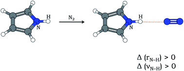
In most cases of X–H⋯Y hydrogen bonding (X is an electronegative atom and Y is any electron-rich center), the X–H bond gets elongated and the stretching frequency of X–H (νX–H) gets red-shifted. Relatively fewer cases of blue-shifting H-bonding are also known, where the X–H bond contracts leading to an increase in νX–H. Here is presented the first experimental report of blue-shifting N–H⋯N H-bonding that accompanies an increase in N–H bond length as evident from quantum chemistry computations. Pyrrole and N2 have been shown to form N–H⋯N hydrogen bond in an ‘annealed’ N2 matrix, where both doped (pyrrole) and dopant (nitrogen) molecules had scope to align themselves and form stable H-bonded clusters. The infrared spectrum of the annealed matrix shows a blue shift of νN–H by 7 cm−1. Computation of anharmonic frequencies at the MP2/6-31+G(d) level of theory reflects the observed blue shift despite N–H bond lengthening after H-bond formation.
The model system is the N–H⋯N hydrogen bonded 1:
1 cluster between pyrrole and N2. Geometry optimization of this cluster by various levels of quantum chemistry computations shows a linear geometry of N–H⋯N
N moiety (Fig. 1). The bonding between NH (of pyrrole) and N2 is further ascertained by the existence of bond critical point between the two moieties using AIM theory.33 Topological diagram citing bond critical points is provided in ESI.†
The advantages of using this H-bonding prototype are the following. (a) The N–H⋯NN geometry is linear (see Fig. 1) which minimizes the extent of dispersion interaction from remote parts of pyrrole and N2 moieties. Thus the intermolecular interaction would exclusively be H-bonding. (b) Vibrational energy distribution over internal coordinates, calculated by the method of Pulay and Torok,34 of pyrrole shows that the N–H stretching vibration is exclusively a local mode (contribution of νN–H to the normal mode energy is ∼100%). Thus, the intermode couplings are negligible and the diagonal anharmonicity (anharmonicity along normal mode vector) accounts for the correct PES of this normal mode. For the pyrrole⋯N2 cluster, the second largest contributor to the N–H stretching normal mode is νH⋯N (hydrogen bond stretching) which contributes only ∼0.5% to the normal mode energy. Also, the beauty of the system is that both the N–H and H⋯N stretches are collinear. Thus, the diagonal anharmonicity of the N–H stretching mode incorporates the effect due to coupled H⋯N stretch. Other coupling terms are very small and can be comfortably neglected. Such simplification, intrinsic to the system, improves accuracy in prediction of νN–H as additional approximations are required to incorporate intermode couplings.
All quantum chemistry computations have been carried out by GAMESS [Version: 5 Dec 2014 (R1)] program package.35 As the H-bonding interaction between pyrrole and N2 is expected to be at the weaker end, and consequently the spectral shift (ΔνN–H) to be small, a careful selection of theoretical level is necessary to depict the real picture of the system. In the present study, the widely accepted Møller–Plesset second order perturbation (MP2) theory has been used to model the N–H stretching frequency. It is a known issue that the accuracy in prediction depends on the basis set selected, and there is no any unanimous rule (as will be evident very shortly) that a higher basis set must produce more accurate results. Thus to ascertain the accuracy in theoretical prediction the following procedure is adopted. First, the diagonal anharmonicity of the N–H stretching mode of pyrrole is computed over 64 grid points using a series of different basis sets and compared with experimental data in gas phase. The following Pople type basis sets: 6-31G(d,p), 6-31+G(d), 6-31+G(d,p), 6-311++G(d,p), and also the aug-cc-pVDZ basis set have been used for computations. Results obtained from the said computations are summarized in ESI.† The best match of the computed anharmonic frequency of N–H stretching mode is found for the MP2/6-31+G(d) level of theory which predicts the νN–H of pyrrole as 3532 cm−1, while the reported experimental value in gas phase is 3531 cm−1.36 Notably, the higher basis sets, viz., 6-31+G(d,p), 6-311++G(d,p) and aug-cc-pVDZ are found to be worse in predicting the νN–H value of pyrrole. Reliability of MP2/6-31+G(d) theoretical level and the adopted approach to compute anharmonic frequency have further been checked for some H-bonded clusters of pyrrole. Obviously, the systems are so chosen that the intermode couplings with νN–H are negligibly small. As experimental data for H-bonded pyrrole⋯N2 cluster in gas phase is currently not available in literature, a structurally similar N–H⋯N hydrogen bonded system, pyrrole⋯acetonitrile (Fig. 1), has been used for the purpose. While the experimental value of νN–H of pyrrole⋯acetonitrile is 3437 cm−1 in gas phase,37 the computed anharmonic frequency for the same at MP2/6-31+G(d) theoretical level is 3439 cm−1. The same theoretical approach to pyrrole homo-dimer predicts νN–H values as 3520 and 3444 cm−1 for free and H-bonded (N–H⋯π) N–H moieties, respectively, while the reported gas-phase values36 are 3522 and 3444 cm−1. All these data for νN–H are summarized in Table 1 for a quick comparison between experimental and computed values.
Molecule/cluster | Predicted νN–H (cm−1) | Experimental νN–Ha (cm−1) |
---|---|---|
a Values taken from ref. 36 and 37. | ||
Pyrrole | 3532 | 3531 |
Pyrrole⋯acetonitrile | 3439 (N–H⋯N bonded) | 3437 |
Pyrrole⋯pyrrole | 3520 (Free N–H) | 3522 |
Pyrrole⋯pyrrole | 3444 (N–H⋯π bonded) | 3444 |
Pyrrole⋯N2 | 3540 (N–H⋯N bonded) | No previous data |
Such close agreements between experimental observations and theory are very encouraging and indicate the acceptability of MP2/6-31+G(d) level of theory for the present work. The same theoretical level was also previously used for prediction of blue-shifting hydrogen bonds in many other systems.5,8,29 Importantly, for pyrrole⋯N2 cluster, the MP2/6-31+G(d) computational level predicts a blue shift of νN–H by 8 cm−1 (see Table 1). At the same time it predicts a lengthening of N–H bond by 0.0007 Å. Similar bond (N–H) lengthening is predicted by all other theoretical levels (see ESI†). Thus, the claim of blue-shifted νN–H, in spite of lengthening of N–H bond, gets strong support from quantum chemistry computations at MP2/6-31+G(d) level of theory for hydrogen bonded cluster between pyrrole and N2. For deeper scrutiny of the said observations, a scan of H⋯N bond length is done, and at each scan point all the geometrical parameters, except the fixed H⋯N bond length, are allowed to optimize. This treatment shows an energy minimum (corresponding to equilibrium geometry) at H-bond length of 2.37 Å (Fig. 2). As the H-bond length decreases, the N–H bond increases in length and never goes through any minimum (see Fig. 2). Such behavior was earlier ascribed to ‘proper’ H-bonding, where a red shift of νX–H was expected to occur because of elongated X–H bond in equilibrium geometry.13 As the spectral shift in the present case is opposite (blue shift), a further scrutiny of the H⋯N bond energy is done. Energy decomposition analysis (EDA), as implemented in GAMESS, is carried out to evaluate H⋯N bond energy at each point of previous scan. The same EDA method was earlier used to obtain reasonable results for several H-bonded clusters.38 Interestingly, in the present study, the outcomes of EDA over the entire scan range of H⋯N bond length vary in a systematic manner and reveal that the H-bond is strongest when it is little longer (2.52 Å) than that at equilibrium geometry (see Fig. 2). Now, the stretching modes of N–H and H⋯N are coupled. An extra potential exerted upon N–H stretching mode by the adjacent hydrogen bonded part at equilibrium geometry lifts the value of νN–H. Eventually, the hydrogen bond, H⋯N, becomes ‘blue-shifting’ in spite of elongated N–H bond.
The infrared spectra of pyrrole-doped N2 matrix have been recorded at a temperature range of 8–28 K (Fig. 3). The binding energy of the pyrrole⋯N2 cluster being very small, only ∼1 kcal mol−1 (BSSE corrected) according to the calculation at MP2/6-31+G(d) level, its natural abundance in a static gas cell at room temperature must be extremely small. However, in the present study, it is shown that the same H-bonded cluster can be generated in relatively large abundance within a cold N2 matrix, and studied by infrared spectroscopy. For preparation of the seeded matrix, a mixture of pyrrole vapor and nitrogen gas at a ratio of 1:
1000 at room temperature (22 °C) was diffused through a needle and deposited on the cold (8 K) KBr window in the matrix isolation chamber under high vacuum (∼10−6 mbar). The νN–H band of the initially deposited matrix appears as multiplet centered at ∼3512 cm−1, and the overall width of the multiplet is ∼7 cm−1 (spectrum ‘a’, Fig. 3). This multiplet corresponds to the νN–H of pyrrole monomer trapped in different matrix sites. Appearance of such site splitting in matrices is common and cited earlier.39 Notably, the solvation shift (by N2 matrix) of νN–H from reported gas-phase data (and prediction at MP2/6-31+G(d) level of theory) is ∼20 cm−1 (compare with Table 1). There is no any significant IR band(s) except for pyrrole monomer in the spectrum ‘a’ of Fig. 3. However, dramatic changes occur when the matrix is annealed by heating it slowly up to 28 K and then cooling back to 8 K (a–c, Fig. 3). Spectrum ‘c’ in Fig. 3 corresponds to the resulting annealed matrix. Two stable site bands, centered at 3509 and 3514 cm−1 still persist in annealed matrix, and several new features develop. The overall spectrum of annealed matrix, keeping in mind the solvation shift by ∼20 cm−1, looks quite similar to the reported gas phase spectra of pyrrole monomer and its higher clusters,36 except for some extra peaks in the present case. Thus by a comparison with gas phase spectra, the peak at 3424 cm−1 in annealed matrix can be assigned to the pyrrole dimer, and the broad feature near 3380 cm−1 can be assigned to higher homo-clusters of pyrrole. An interesting new feature observed in the present study is a distinct blue-shifted peak at 3519 cm−1 which is practically absent in the spectrum of freshly prepared matrix, but appears after heating the matrix to a higher temperature (a–b, Fig. 3). Possibility of any blue-shifted peak due to homo-clusters of pyrrole can be ruled out from previous reports in gas phase36 as well as Ar and Xe matrices.39 Thus, the 3519 cm−1 peak must arise out of some specific interaction between the guest pyrrole molecules and the matrix molecules, i.e., N2. In freshly prepared matrix, the relative orientations of pyrrole and N2 molecules were quite random. Rigidity of the matrix opposed any reorientation of pyrrole and N2 molecules that could establish required linearity for effective N–H⋯N
N hydrogen bonding. When the temperature of the matrix is raised, the matrix softens and a number of pyrrole and N2 molecules reorient to form N–H⋯N
N hydrogen bond as shown in Fig. 1. Thus, the 3519 cm−1 peak (spectra ‘b’ & ‘c’, Fig. 3) has been assigned to the νN–H of 1
:
1 cluster between pyrrole and N2. The observed blue shift (∼7 cm−1) is again in good agreement with the prediction (+8 cm−1) by MP2/6-31+G(d) level of theory. The bottom panel of Fig. 3 depicts the computed spectral positions (with 20 cm−1 red shift for solvation effect) of pyrrole monomer, its homo-dimer as well as pyrrole⋯N2 cluster. The match of computed spectral positions with those observed in annealed matrix is outstanding.
In conclusion, this is the first report of infrared spectroscopic detection of blue-shifting NH⋯N bonding between pyrrole and N2, and the finding is well supported by quantum chemistry computation at MP2/6-31+G(d) theoretical level. The study shows that the shortening of equilibrium bond length of H-bond donor (X–H) is not a mandatory criterion for occurrence of blue-shifting H-bonding, especially when the binding energy is very small. The importance of anharmonic correction in frequency is evident for accurate prediction of spectral shifts for hydrogen bonded clusters. Another important outcome of this study is about the acceptability of theoretical level for correct prediction of spectral shifting. This study signifies the use of an optimum basis set for a particular system. Any increase in the size of basis set does not guarantee, or might not be necessary for, accurate prediction.
Footnote |
† Electronic supplementary information (ESI) available: Theoretical predictions for optimized geometrical parameters of pyrrole⋯N2 cluster, N–H stretching frequency of pyrrole, changes in N–H bond length upon H-bond formation and pictorial presentation of bond critical points. See DOI: 10.1039/c5ra17309d |
This journal is © The Royal Society of Chemistry 2015 |