DOI:
10.1039/C5RA17206C
(Paper)
RSC Adv., 2015,
5, 88456-88463
Preparation of anti-fouling silicone elastomers by covalent immobilization of carboxybetaine
Received
25th August 2015
, Accepted 10th October 2015
First published on 13th October 2015
Abstract
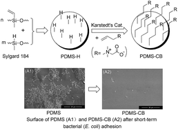
Polydimethylsiloxane (PDMS) is a widely used material for biomedical applications. In this work, a convenient method for the covalent modification of PDMS with carboxybetaine was developed and used to construct a biocompatible and anti-fouling coating. Following the preparation of a Si–H functionalized PDMS film by adjusting the molar ratio of the two components used in Sylgard 184 silicone elastomer networks, the allyl carboxybetaine (ACB) was grafted to the PDMS surface via a hydrosilylation reaction in the presence of a Karstedt's catalyst. ATR-FTIR and water contact angle measurements revealed that carboxybetaine was introduced to the PDMS surface successfully. The biocompatibilities of PDMS and carboxybetaine-modified PDMS (PDMS-CB) films were evaluated by cytotoxicity, hemocompatibility, and dynamic clotting time. The anti-fouling properties of PDMS-CB were evaluated by protein adsorption and bacterial adhesion measurements. The results showed that the carboxybetaine layer could enhance the biocompatibility of PDMS and reduce the adsorption of protein and adhesion of bacteria efficiently.
1. Introduction
Due to the unique advantages of polydimethylsiloxane (PDMS), including high gas permeability, excellent optical transparency, high chemical resistance, biological inertness, nontoxicity, ease of fabrication and low cost, PDMS has been widely used in medical implants,1,2 microengineering,3 prosthetics,4 microfluidic devices5,6 and many other areas. However, owing to its high hydrophobicity, PDMS suffers from biofouling problems such as nonspecific protein adsorption and bacterial/cell adhesion, which results in many undesirable bioreactions in biological environments.7–9 Therefore, it is highly desirable to improve the surface properties of PDMS by reducing protein adsorption and improving biocompatibility, which would significantly extend the applicability of PDMS in implantable biomaterials, biosensors, ocean engineering and membrane separation. To date, numerous reports have investigated efficient surface modification techniques to create anti-fouling coatings on PDMS.10–12 The most commonly used method for changing the surface properties of PDMS is physical modification, which includes surface adsorption of coating materials via physical interactions,13 oxidation of the PDMS surface by oxygen plasma14 and UV-ozone treatment.15 Unfortunately, the physical modification method is generally temporary, and the hydrophobicity will recover after just hours. A more effective surface modification method for PDMS is chemical modification, such as the deposition of a self-assembled monolayer (SAM) or surface grafting,16–19 and the surface modified layer is relatively stable due to the covalent bonds formed between the PDMS substrate and the coating agent.
In recent years, zwitterionic-based materials, such as sulfobetaine and carboxybetaine, have exhibited excellent non-fouling characteristics; they can greatly reduce nonspecific protein adsorption and bacterial adhesion.20–22 Thus, they are considered as ideal materials for surface modification.
In our previous work,25 carboxybetaine-functionalized polysiloxane (PDMS-g-CB) was blended with a PDMS elastomer to form a PDMS-g-CB/PDMS blended film (b-PDMS). It was found that the hydrophobic PDMS surfaces could be converted to hydrophilic surfaces with the introduction of 0.5 wt% to 2 wt% PDMS-g-CB, and the amount of adsorbed protein and adhered bacteria on b-PDMS were significantly reduced compared with untreated PDMS. Notably, most of the hydrophilic PDMS-g-CB chains were embedded in the matrix of the b-PDMS film; thus, only a small portion of the PDMS-g-CB chains were exposed on the surface of the PDMS film and contributing to the antifouling properties. Therefore, it would be more efficient to graft the carboxybetaine groups on the surface of the PDMS film. Herein, a novel zwitterionic molecule (allyl carboxybetaine, ACB) was synthesized and then employed to modify PDMS by a subsequent hydrosilylation reaction in the presence of a Karstedt's catalyst. The surface modification of PDMS was characterized by ATR-FTIR and water contact angle (WCA). The biocompatibility, including cytotoxicity, hemocompatibility, and dynamic clotting time, and the surface properties of PDMS, such as protein adsorption and bacterial adhesion, were also investigated.
2. Experimental
2.1 Materials
A PDMS precursor and curing agent (Sylgard 184) were purchased from Dow Corning (Midland, USA). N,N-Dimethylallylamine (DMAA, >99%, Haining City Huangshan Chemical Industry Co., Ltd, China) was purified by distillation before use. Sodium chloroacetate (SC) and a Karstedt's catalyst (Pt ∼ 2 wt%, platinum (0)-1,3-divinyl-1,1,3,3-tetramethyldisiloxane complex solution) were obtained from Aladdin Reagent Co., Ltd (Shanghai, China) and used as received. Escherichia coli (E. coli) and Staphylococcus aureus (S. aureus) were kindly supplied by the College of Natural Resources & Environment, South China Agricultural University. The culture medium RPMI 1640 was obtained from Invitrogen (New York, USA). Fetal bovine serum was provided by Hangzhou Sijiqing Biological Engineering Materials Co. Ltd (Hangzhou, China). MTT (3-(4,5-dimethylthiazol-2-yl)-2,5-diphenyltetrazoliumbromide) was obtained from Alfa-Aesar. All other reagents were used as received without any further purification.
2.2 Preparation of carboxybetaine-grafted PDMS films (PDMS-CB)
In this paper, carboxybetaine was grafted onto hydrophobic PDMS surfaces via a hydrosilylation reaction as shown in Schemes 1 and 2.
 |
| Scheme 1 The synthesis and structure of allyl carboxybetaine. | |
 |
| Scheme 2 The schematic of surface modification with ACB. | |
2.2.1 Synthesis of allyl carboxybetaine (ACB). ACB was synthesized via the quaternization reaction of N,N-dimethylallylamine (DMAA) with sodium chloroacetate (SC). Typically, DMAA (8.5 g, 0.1 mol) and SC (14.0 g, 0.12 mol, dissolved in 30 mL distilled water) were mixed with 50 g dry isopropanol as the solvent. Then, the mixtures were heated to 65 °C for 12 h under a nitrogen atmosphere. Afterwards, isopropanol and water were removed from the reaction vessel under reduced pressure, and the residues were redissolved in isopropanol. The insoluble impurities were separated by filtration, and the solvents were evaporated in vacuo. The residual solid was washed once again with methanol, and then the resulting product was dried in a vacuum oven at 85 °C for 3 h (87.6% yield). FT-IR (KBr, cm−1): 3010 (
CH, ν); 2967 (–CH3, ν); 2831 (–CH2, ν); 1633 (C
O, ν); 1477, 1402 (–CH3; –CH2, δas); 1338 (
CH, β); 1222, 1072, 1006 (–CH3, δ); 887, 725 (
CH, γ). 1H-NMR δ (D2O, ppm): 3.24 (s, –N–(CH3)2); 3.87 (s, –CH2–COO); 4.19 (d, –CH–CH2); 5.68–5.77 (m, CH2
CH–); 6.01–6.11 (m, CH2
CH–).
2.2.2 Preparation of Si–H functionalized polysiloxanes elastomers. The Sylgard 184 precursor (part A) was mixed thoroughly with its curing agent (part B) in 1
:
1, 2
:
1, 3
:
1, 4
:
1 and 10
:
1 ratios (by weight) and degassed under vacuum for 30 min. Then, the mixtures were cured at 80 °C for 1 h to obtain Si–H functionalized PDMS films (PDMS-H). After being cooled to room temperature, the PDMS-H films were removed from their molds and cut into small pieces, each with a diameter of approximately 10 mm. The surface structures of the resulting PDMS-H films were measured with ATR-FTIR.
2.2.3 Grafting the carboxybetaine groups on the PDMS-H surface. Typically, 20 pieces of PDMS-H film were added to a solution of ACB (10 mL, 50 wt% in dried isopropanol). Then, 200 μL of Karstedt's catalyst was added, and the mixture was stirred for 12 h at room temperature. Afterwards, the films were taken out and washed with ethanol and water 3 times and then dried with nitrogen at 55 °C for 3 h to obtain the PDMS-CB films.
2.3 Characterization
Fourier transform infrared (FT-IR) spectra were recorded with a VERTEX-70 spectrometer (BRUKER Instrument Corp., Germany) at resolution of 4 cm−1. Samples were coated on KBr plates and measured over a range of 400–4000 cm−1 at room temperature. Attenuated total reflection Fourier transform infrared (ATR-FTIR) spectra were measured on a VERTEX-70 spectrometer equipped with an ATR accessory. 64 scans were collected for each sample with a resolution of 4 cm−1 in the 4000–650 cm−1 region.
1H-NMR spectra were recorded on a Bruker ADVANCE III-400 (400 MHz) Fourier digital NMR spectrometer (BRUKER Instrument Corp., Germany) at room temperature using D2O as the solvent.
X-ray photoelectron spectroscopy (XPS) analysis was performed with a PHI X-tool XPS System (Ulvac-PHI Inc., Japan), using a non-monochromatized Al Ka X-ray source operating at 15 kV and 20 W. The binding energy scale is referenced by setting the peak maximum in the C 1s spectrum to 284.5 eV. Survey scans were performed from 1000 to 0 eV and high-resolution C 1s spectra were obtained at 45° takeoff angles.
The static water contact angles of native PDMS and PDMS-CB surfaces were measured with a contact angle meter (DropMeter A-100, MAIST Vision Inspection & Measurement Co. Ltd, China) at ambient temperatures. One drop of water (10 μL) was placed onto the surface and observed with an optical microscope. The WCA values were calculated from three measurements at different positions.
2.4 Biocompatibility evaluation
2.4.1 Cytotoxicity test (MTT method). The cytotoxicity tests were carried out using MTT method and direct contact method, according to ISO 10993-5: 2002.For MTT method, the PDMS or PDMS-CB patches (1 cm × 1 cm × 0.1 cm) were first sterilized for 3 h with ultraviolet light and then soaked in 10 mL of RPMI 1640 culture medium (the film/solution ratio was 1/10 (cm2 mL−1)) for 24 h to obtain an extract solution of the PDMS film. For comparison, the extract solution of LDPE film (negative control) and extract solution of organotin-stabilized PVC film (positive control) were also prepared.
L929 cells (murine aneuploid fibrosarcoma cells) were cultured in RPMI 1640 supplemented with 10% FBS in 96-well plates (100 μL medium per well) at a density of 1.0 × 10⁵ cells per mL. The cells were cultured overnight at 37 °C in a humidified 5% CO2 incubator after inoculation. After culturing the cells for 24, 48, and 72 h, a 5.0 mg mL−1 MTT solution (in PBS) was added to the wells, and the wells were incubated for another 4 h at 37 °C. The growth medium was subsequently removed, and 150 μL of DMSO was added, followed by vigorous shaking to dissolve the purple formazan crystals that had formed. The absorbance was measured with a Thermo MK-III MicroplateReader (Thermo, USA) at a wavelength of 570 nm.
2.4.2 Cytotoxicity test (direct contact method). L929 cell (2 mL) suspension was added to the vessels and incubated at 37 °C in a humidified 5% CO2 incubator. Next, each vessel was filled with fresh RPMI 1640 culture medium. The sterilized PDMS or PDMS-CB film patches (1 cm × 1 cm × 0.1 cm) were gently placed on the layer of cells in the center of each of the replicate vessels. One-tenth of the cell layer surface was covered by the test sample. Finally, the vessels were incubated in the same environment as described above. For comparison, an LDPE film (negative control) and an organotin-stabilized PVC film (positive control) were also prepared.
2.4.3 Hemolytic activity. The hemocompatibility of the PDMS film was tested by direct contact methods, according to ISO 10993-5: 2002 and GB/T 16175-2008. Fresh blood was obtained from an albino rabbit of the New Zealand strain (weight: 2.5 kg). The blood was then diluted with saline water (0.9% wt NaCl) to a volume ratio of 1/1.25, i.e., each sample contained 4 mL of fresh blood and 5 mL of saline water.Briefly, 5 g of sterilized PDMS film patches (5 mm × 20 mm × 1 mm) and 10 mL of normal saline were added to the diluted blood. For comparison, positive and negative controls were prepared using 10 mL of distilled water and 10 mL of saline water, respectively. After incubation for 30 min at 37 °C, 0.2 mL of diluted blood was added, and the samples were incubated for 1 h. The solution was then centrifuged at 3750 rpm (×800g) for 5 min, and the optical density (OD) of the clear supernatant fluid was measured at 545 nm (UV2300, Techcomp Co. Ltd, China). The hemolysis ratio (HR) was calculated using the following equation:
|
 | (1) |
where OD
test sample, OD
(−)control, and OD
(+)control are the OD values of the PDMS film sample, the positive control, and the negative control, respectively.
2.4.4 Dynamic clotting time test. In a dynamic clotting time experiment, 0.1 mL of rabbit blood, which was adulterated with an anticoagulant of Alsevre's solution, was first dripped onto the sample surface in a 37 °C atmosphere; second, 0.2 mol L−1 CaCl2 solution was added to initiate blood clotting. After 10, 20, 30, 40, 50 and 60 min, each sample was transferred into a beaker containing 50 mL of distilled water and then rinsed gently, and the absorbance of the supernatant liquid was measured at a wavelength of 540 nm by UV-vis.28 For each sample, an average optical density was obtained from three measurements.
2.5 Protein adsorption
Bovine serum albumin (BSA) was used as the model protein for evaluation of protein adsorption. To measure the amount of protein adsorbed onto PDMS and PDMS-CB surfaces, the films were immersed in 10 mL protein solutions (BSA, 4 mg mL−1) and incubated at 37 °C for 60 min; then the films were removed from the BSA solution and rinsed with PBS three times. Subsequently, the films were sonicated for 120 min in PBS to detach any protein adsorbed onto the native PDMS and b-PDMS surfaces. Then, the solution was collected, and the amount of protein was determined by the Micro BCA (bicinchoninic acid) Protocol (Micro BCA Protein Assay Kit, Sangon Biotech Co., Ltd, Shanghai, China).26,27 Afterwards, the amount of adsorbed protein was calculated according to the standard protein curve, which was measured with different concentrations of a protein standard.
2.6 Bacterial adhesion
Two typical gram-negative and gram-positive bacteria, i.e., Escherichia coli (E. coli) and Staphylococcus aureus (S. aureus), were used to investigate the bacterial adhesion behavior on the surface of native PDMS and PDMS-CB films. Before the bacterial adhesion experiment, E. coli and S. aureus were incubated in a broth medium (containing 3.0 mg mL−1 beef extract, 10.0 mg mL−1 peptone and 5.0 mg mL−1 sodium chloride) at 37 °C for 24 h. The bacterial concentration was measured by spectrophotometer at 540 nm (OD540) with the assumption that the OD540 = 1.0 corresponds to a bacterial concentration of approximately 109 colony-forming units (CFU) per milliliter. For the bacterial adhesion assay, the E. coli or S. aureus containing broth was washed and resuspended in PBS to obtain a bacterial suspension, which had a concentration of 108 CFU mL−1. The native PDMS and PDMS-CB films were sterilized with ultraviolet rays (30 min on each side) and immersed in the bacterial suspension in a sterile flask, then cultured at 37 °C for 20 h. Afterwards, the films were removed and rinsed three times in sterile PBS, and any bacteria which adhered on the surface were immobilized with 2.5% glutaraldehyde solution for 6 h at 4 °C. Then, the films were washed with sterile PBS three times and dehydrated stepwise in a series of ethanol/water mixtures (30%, 50%, 70%, 80%, 95% and 100% by volume, 20 min in each mixture). Finally, the dried films were coated with gold and observed with a scanning electron microscope (Zeiss EVO 18, Germany).
3. Results and discussion
3.1 Synthesis of allyl carboxybetaine (ACB)
Allyl carboxybetaine was synthesized by a quaternization reaction of 1,1-dimethylallylamine (DMAA) and sodium chloroacetate (SC) (Scheme 1). The chemical structure of the obtained product was characterized by FT-IR and 1H-NMR spectroscopy. On the basis of the FT-IR spectrum of sodium chloroacetate (Fig. 1(a)), the peak at 765 cm−1 was attributed to C–Cl stretching vibrations. After the quaternization reaction, this characteristic peak disappeared. Another change in the FT-IR spectrum after quaternization is the appearance of
CH stretching vibrations at 3010 cm−1. Those changes indicated that DMAA and SC had undergone quaternization. Fig. 2 displays the 1H-NMR spectrum of the obtained product; the signals at 5.68–5.78 ppm and 6.01–6.11 ppm were assigned to the protons of C
C, whilst the signals at 3.23 ppm and 3.86 ppm were attributed to the protons –N(CH3)2 and –CH2COO, respectively. Overall, the data from the 1H-NMR spectrum of the obtained product indicated that the target product was obtained successfully.
 |
| Fig. 1 The FT-IR spectra of (a) sodium chloroacetate (SC) and (b) allyl carboxybetaine (ACB). | |
 |
| Fig. 2 The 1H-NMR spectrum of allyl carboxybetaine (ACB). | |
3.2 Preparation of Si–H functionalized PDMS film
According to the instructions provided by the supplier, the two components of Sylgard 184 (Part A and Part B) are Si–H containing silicone and vinyl-containing silicone, as shown in Fig. 3. The two components are generally mixed at a weight ratio of 10/1 to prepare PDMS elastomer networks. Using that weight ratio, the mole ratio of Si–H groups to vinyl groups is approximately 1
:
1; so the curing reaction will finish completely and no more Si–H functional groups will remain. If the weight ratio of part A to part B is less than 10
:
1, there will be excess Si–H containing silicone. Therefore, Si–H groups would remain on the surface of PDMS after the network is completely cured.23
 |
| Fig. 3 FT-IR spectra of Part A and B of the Sylgard 184 silicone elastomer. | |
Fig. 4 shows the ATR-FTIR spectra of PDMS prepared by Sylgard 184 at different A/B ratios. When the weight ratio was less than 10
:
1, there were sharp absorption bands at 2163 cm−1 and 912 cm−1 attributed to the stretching and bending vibrations of Si–H. The appearance of Si–H characteristic bands revealed that Si–H groups could be introduced to the PDMS surface by changing the weight ratio of the two components of Sylgard 184, and the content of Si–H could be adjusted conveniently. Whereas the reaction efficiency and the mechanical performance of the PDMS-H film, the PDMS-H film with A/B = 1/1 (wt/wt) was chosen for the following grafting reaction, for the high density of Si–H groups on the surface.
 |
| Fig. 4 The ATR-FTIR spectra of PDMS-H elastomers with the A/B ratio of (a) 10/1, (b) 4/1, (c) 3/1, (d) 2/1 and (e) 1/1. | |
3.3 Preparation of carboxybetaine-grafted PDMS films (PDMS-CB)
PDMS-CB films were prepared by the hydrosilylation reaction of PDMS-H films (Sylgard 184, A/B = 1/1, wt/wt) and allyl carboxybetaine using the Karstedt's catalyst. The ATR-FTIR spectra of carboxybetaine-modified PDMS (PDMS-CB, freshly prepared and after 3 months in dry air) and Si–H functionalized PDMS (PDMS-H) are shown in Fig. 5. The most prominent differences between PDMS-H and PDMS-CB are the disappearance of the strong adsorption band for Si–H stretching vibrations at 2163 cm−1, and the appearance of a C
O characteristic adsorption band for carboxybetaine groups at 1633 cm−1;24 both of the changes indicate that allyl carboxybetaine was covalent grafted onto the PDMS surface successfully. The same evidence could be found on the XPS spectra of PDMS and PDMS-CB, as shown in Fig. 6. Fig. 6(A) shows a typical C 1s spectrum of the PDMS-H substrate, consisting of only one component centered at binding energy of 283.5 eV, corresponding to C–Si group; while in Fig. 6(B), the peaks centered at binding energy of 283.5, 284.5, 285.3, 286.9 and 288.2 eV, were assigned to C–Si, C–C, C–N, C–O and C
O, respectively, which confirmed the grafting of carboxybetaine group on the surface of PDMS.29
 |
| Fig. 5 The ATR-FTIR spectra of (a) Si–H functionalized PDMS, (b) freshly prepared carboxybetaine-modified PDMS (PDMS-CB), and (c) PDMS-CB, after 3 months in dry air. | |
 |
| Fig. 6 The XPS spectra of (a) PDMS-H and (b) PDMS-CB in C 1s region. | |
3.4 Water contact angle analyses
The hydrophilicity of the native PDMS, PDMS-H and PDMS-CB films was evaluated by water contact angle measurements (as shown in Fig. 7). The native PDMS and PDMS-H film show relatively high hydrophobicity (the water contact angles are approximately 103°). After the introduction of carboxybetaine via the hydrosilylation reaction, the hydrophilicity of PDMS significantly improved. The water contact angle of freshly prepared PDMS-CB is approximately 46°, and the average water contact angle of PDMS-CB is no more than 48° even after 3 months. These results suggest that carboxybetaine was stably grafted onto the PDMS surface, which increased the hydrophilicity of the PDMS film.
 |
| Fig. 7 Water contact angle of native PDMS, PDMS-H, and PDMS-CB (freshly prepared and after 3 months). | |
3.5 Evaluation of biocompatibility
PDMS has been widely used as an inactive surface biomaterial; thus the biocompatibility of carboxybetaine-modified PDMS films, i.e., PDMS-CB, was evaluated by cytotoxicity, hemocompatibility, and dynamic clotting time.
3.5.1 Cytotoxicity. In this study, the cytotoxicity of PDMS and PDMS-CB film toward L929 fibroblast cells was assessed by MTT (3-(4,5-dimethylthiazol-2-yl)-2,5-diphenyltetrazoliumbromide) assays and direct contact tests. For the MTT assays, the original and diluted extracts were prepared and immersed in RPMI 1640 culture medium for 24 h. L929 cells were cultured for 72 h in the negative control (extract solution of LDPE films), the positive control (extract solution of organotin-stabilized PVC films), PDMS film extract or PDMS-CB before adding MTT. The L929 cells cultured in both PDMS film extract and PDMS-CB film extract grew well, whereas in the positive control, the majority of the cells adopted a bloom form. The cell viability in the PDMS film extract and PDMS-CB film extract were similar to that in the negative control, as determined by MTT assay and shown in Fig. 8.
 |
| Fig. 8 Comparison of the cytotoxicity of the PDMS film extract, PDMS-CB film extract, the negative control (extract solution of LDPE films) and the positive control (extract solution of organotin-stabilized PVC films) toward L929 cells (n = 5). | |
In addition, the direct contact method was used to evaluate in vitro cytotoxicity. The morphologies of L929 cells incubated on the PDMS film, the PDMS-CB film, an LDPE film (negative control), and an organotin-stabilized PVC film (positive control) for 24 h are shown in Fig. 9. In agreement with the results of the MTT assay, the cell viability on the PDMS film and PDMS-CB film was similar to that on the negative control (LDPE film), and almost all of the cells adopted a bloom form in the positive control. Taken together, these results demonstrate the nontoxicity of PDMS-CB toward L929 cells.
 |
| Fig. 9 Comparison of cytotoxicity toward L929 cells in direct contact tests. (A) LDPE film (negative control), (B) organotin-stabilized PVC film (positive control), (C) PDMS film, and (D) PDMS-CB film. | |
3.5.2 Hemocompatibility. The hemocompatibility of the PDMS and PDMS-CB films was assessed by measuring hydrolysis and dynamic clotting time. The hemolytic activity was determined by hemoglobin release under the test conditions. For hemocompatibility, the hemolysis ratio must be less than 5%. The hemolysis ratios for heparinized blood mixed with extracts of either a PDMS or PDMS-CB film patch for 24, 48, 72, and 96 h were 1.12 ± 0.22%, 1.34 ± 0.27%, 1.49 ± 0.19%, and 2.96 ± 0.32% for PDMS films, and 0.92 ± 0.15%, 0.98 ± 0.27%, 1.23 ± 0.32%, and 2.13 ± 0.55% for PDMS-CB films, respectively. These values are within acceptable limits, indicating the hemocompatibility of both the PDMS and PDMS-CB films.Fig. 10 shows the clotting time curves of PDMS and PDMS-CB films. The clotting time measurement tests the activated degree of intrinsic coagulation factors. The slower the optical density value decreases with time, the longer the clotting time is.28 The curve of the PDMS film decreases more quickly in the first 20 min; therefore, the blood coagulation of the PDMS film is greater than that of the PDMS-CB film. Additionally, the optical densities are always bigger for the PDMS-CB film regardless of how many times the blood was in contact with the material, which indicates that dynamic clotting time is longer for the carboxybetaine-grafted surface. Therefore, the carboxybetaine-grafted surface can improve the blood biocompatibility of a PDMS film.
 |
| Fig. 10 Curves of dynamic clotting time for PDMS and PDMS-CB films. | |
3.6 Protein adsorption study
It is believed that most undesirable bio-adsorption and bio-fouling on the surfaces of materials are promoted by protein adsorption. Therefore, protein resistance is the primary target for construction of low-fouling or non-fouling surfaces. Herein, we chose bovine serum albumin (BSA), which exists extensively in blood and plasma, as the model protein for protein adsorption testing, and the protein adsorption on various PDMS substrates was measured by the micro-BCA method. Fig. 11 records the amount of BSA adsorbed onto the surfaces of native PDMS, PDMS-H and PDMS-CB film. There were no significant differences in the levels of BSA adsorption on the surfaces of native PDMS and PDMS-H. In contrast, the amount of BSA adsorbed onto the carboxybetaine-modified PDMS surface was reduced to 1.75 μg cm−2, a reduction of more than 75%. As PDMS-CB film containing zwitterionic groups (carboxybetaine), the surface based on charge interactions could resist nonspecific protein adsorption via a hydration layer bound through solvation of the charged zwitterionic groups in addition to hydrogen bonding.30–32
 |
| Fig. 11 Amount of BSA adsorbed onto the surface of native PDMS, Si–H functionalized PDMS and carboxybetaine-modified PDMS (freshly prepared and after 3 months). | |
3.7 Bacterial adhesion study
The adhesion of bacteria on a material is an essential factor leading to biofilm formation and infection. To construct a low-fouling surface, it is necessary to reduce or inhibit the adhesion of bacteria. To evaluate the anti-bacterial adhesion properties of the modified PDMS films, the short-term (24 h) attachment of two typical Gram-negative and Gram-positive bacteria, i.e., Escherichia coli (E. coli) and Staphylococcus aureus (S. aureus), was investigated. Fig. 12 illustrates representative SEM images of adherent bacteria on the surfaces of native PDMS, PDMS-H and PDMS-CB films (freshly prepared and after 3 months). In Fig. 12A1 and B1, numerous bacteria (E. coli or S. aureus) can be observed on the surface of native PDMS. The adhesion of both E. coli and S. aureus is significantly decreased on the carboxybetaine-modified surface (Fig. 12A2 and B2) compared with native PDMS, even after 3 months (Fig. 12A3 and B3). After counting the exact number of bacteria present on 10 regions of SEM images for each sample, the average number of adsorbed bacteria was determined, as shown in Fig. 13. The bacterial adhesion test demonstrates the excellent anti-adhesive behavior of the carboxybetaine-grafted PDMS surface, which is highly desirable for the construction of anti-fouling surfaces. It is believed that the ability of carboxybetaine modified surfaces to inhibit bacterial adhesion can be attributed to their hydration ability.22
 |
| Fig. 12 SEM images of the surfaces of (A1 and B1) native PDMS, (A2 and B2) PDMS-CB (freshly prepared), and (A3 and B3) PDMS-CB (after 3 months) after short-term bacterial adhesion (A1 to A3: E. coli; B1 to B3: S. aureus). | |
 |
| Fig. 13 The average amount of bacteria adsorbed on the sample surface (calculated from SEM images). | |
4. Conclusions
Si–H functionalized PDMS films were easily prepared by adjusting the mole ratio of the two components in Sylgard 184. Unsaturated carboxybetaine, which was synthesized via a quaternization reaction, was grafted onto the surface of Si–H functionalized PDMS films through the hydrosilylation reaction in the presence of Karstedt's catalyst. ATR-FTIR and water contact angle measurements confirmed the presence of carboxybetaine groups on the surface of PDMS film. Protein adsorption, bacterial adhesion tests, and dynamic clotting time tests suggested that carboxybetaine modified PDMS surfaces could significantly improve the blood biocompatibility, and reduce the adsorption of BSA, as well as bacteria adhesion compared with untreated PDMS surface.
Acknowledgements
The authors acknowledge financial support from the National Natural Science Foundation of China (grants 31201552 and 51473051), the Specialized Research Fund for the Doctoral Program of Higher Education (grant 20124404120025), the Fundamental Research Funds for the Central Universities (grant D2153830) and Innovation and Entrepreneurship Training Program for College Students in Guangdong Province (grant 201410564059).
References
- F. Abbasi, H. Mirzadeh and M. Simjoo, J. Biomater. Sci., Polym. Ed., 2006, 17, 341 CrossRef CAS PubMed.
- F. Abbasi, H. Mirzadeh and A. Katbab, Polym. Int., 2001, 50, 1279 CrossRef CAS PubMed.
- A. Colas and J. Curtis, Silicone biomaterials: history and chemistry & medical application of silicone, Academic Pressed, Elsevier, The Netherlands, 2004, p. 864 Search PubMed.
- P. N. Eleni, I. Katsavou, M. K. Krokida, G. L. Polyzois and L. Gettleman, Dent. Mater., 2009, 25, 1493 CrossRef CAS PubMed.
- S. L. Peterson, A. McDonald, P. L. Gourley and D. Y. Sasaki, J. Biomed. Mater. Res., Part A, 2005, 72, 10 CrossRef PubMed.
- I. Wong and C. M. Ho, Microfluid. Nanofluid., 2009, 7, 291 CrossRef CAS PubMed.
- A. Mata, A. J. Fleischman and S. Roy, Biomed. Microdevices, 2005, 7, 281 CrossRef CAS PubMed.
- D. Wu, B. Zhao, Z. Dai, J. Qin and B. Lin, Lab Chip, 2006, 6, 942 RSC.
- J. M. Anderson, N. P. Ziats, A. Azeez, M. R. Brunstedt, S. Stack and T. L. Bonfield, J. Biomater. Sci., Polym. Ed., 1995, 7, 159 CrossRef CAS PubMed.
- J. Zhou, H. Yan, K. Ren, W. Dai and H. Wu, Anal. Chem., 2009, 81, 6627 CrossRef CAS PubMed.
- Z. Zhang, J. Wang, Q. Tu, N. Nie, J. Sha, W. Liu, R. Liu, Y. Zhang and J. Wang, Colloids Surf., B, 2011, 88, 85 CrossRef CAS PubMed.
- K. A. Heyries, C. A. Marquette and L. J. Blum, Langmuir, 2007, 23, 4523 CrossRef CAS PubMed.
- S. Lee and J. Vörös, Langmuir, 2005, 21, 11957 CrossRef CAS PubMed.
- H. M. L. Tan, H. Fukuda, T. Akagi and T. Ichiki, Thin Solid Films, 2007, 515, 5172 CrossRef CAS PubMed.
- K. Efimenko, W. E. Wallace and J. Genzer, J. Colloid Interface Sci., 2002, 254, 306 CrossRef CAS.
- H. Makamba, J. H. Kim, K. Lim, N. Park and J. H. Hahn, Electrophoresis, 2003, 24, 3607 CrossRef CAS PubMed.
- J. Zhou, J. Yuan, X. Zang, J. Shen and S. Lin, Colloids Surf., B, 2005, 41, 55 CrossRef CAS PubMed.
- J. Xu, Y. Yuan, B. Shan, J. Shen and S. Lin, Colloids Surf., B, 2003, 30, 215–223 CrossRef CAS.
- H. Chen, Z. Zhang, Y. Chen, M. A. Brook and H. Sheardown, Biomaterials, 2005, 26, 2391 CrossRef CAS PubMed.
- A. J. Keefe, N. D. Brault and S. Jiang, Biomacromolecules, 2012, 13, 1683 CrossRef CAS PubMed.
- W. Yang, S. Chen, G. Cheng, H. Vaisocherova, H. Xue, W. Li, J. Zhang and S. Jiang, Langmuir, 2008, 24, 9211 CrossRef CAS PubMed.
- G. Cheng, Z. Zhang, S. Chen, J. D. Bryers and S. Jiang, Biomaterials, 2007, 28, 4192 CrossRef CAS PubMed.
- D. Guo, H. Han, J. Wang, S. Xiao and Z. Dai, Colloids Surf., A, 2007, 308, 129 CrossRef CAS PubMed.
- S. A. Snow, W. N. Fenton and M. J. Owen, Langmuir, 1991, 7, 868 CrossRef CAS.
- L. Cheng, Q. Liu, Y. Lei, Y. Lin and A. Zhang, RSC Adv., 2014, 97, 54372 RSC.
- K. Ishihara, A. Fujiike, Y. Iwasaki, K. Kurita and N. Nakabayashi, J. Polym. Sci., Part A: Polym. Chem., 1996, 34, 199 CrossRef CAS.
- S. Fujishita, C. Inaba, S. Tada, H. Kitano, M. Gemmei-Ide and Y. Saruwatari, Biol. Pharm. Bull., 2008, 31, 2309 CAS.
- J. Liu, D. Yang, F. Shi and Y. Cai, Thin Solid Films, 2003, 429, 225 CrossRef CAS.
- S. Pintoa, P. Alvesa, C. M. Matosb, A. C. Santosb, L. R. Rodriguesc, J. A. Teixeirac and M. H. Gil, Colloids Surf., B, 2010, 81, 20 CrossRef PubMed.
- S. Chen, J. Zheng, L. Li and S. Jiang, J. Am. Chem. Soc., 2005, 127, 14473 CrossRef CAS PubMed.
- J. Zheng, L. Li, H. Tsao, Y. Sheng, S. Chen and S. Jiang, Biophys. J., 2005, 89, 158 CrossRef CAS PubMed.
- S. Chen, F. Yu, Q. Yu, Y. He and S. Jiang, Langmuir, 2006, 22, 8186 CrossRef CAS PubMed.
|
This journal is © The Royal Society of Chemistry 2015 |
Click here to see how this site uses Cookies. View our privacy policy here.