Zaheer Khan*a,
Shaeel Ahmed AL-Thabaitia and
Shokit Hussainb
aDepartment of Chemistry, Faculty of Science, King Abdulaziz University, P.O. Box 80203, Jeddah, 21589, Saudi Arabia. E-mail: drkhanchem@yahoo.co.in
bNanoScience Research Laboratory, Department of Chemistry, Jamia Millia Islamia (Central University), New Delhi-110025, India
First published on 11th December 2015
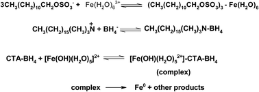
Self-assembled water soluble sheet-like zero-valent iron nano-composites have been prepared using a simple one-pot chemical reduction method using an aqueous solution of FeCl3 and NaBH4 both with and without cetyltrimethylammonium bromide (CTAB) and sodium dodecyl sulphate (SDS). It was demonstrated that dark brownish precipitates were formed immediately after the addition of NaBH4 into the ferric chloride solution. In the presence of CTAB and SDS, stable perfectly transparent brown colored iron sols were formed. UV-visible spectra show that both of the stabilizers (CTAB and SDS) formed a stable complex with Fe3+ ions. The synthesized Fe-nanoparticles were characterized using dynamic light scattering (DLS), energy dispersion X-ray spectroscopy (EDX), transmission electron microscopy (TEM), Fourier transform infrared (FT-IR) spectroscopy, ultraviolet-visible spectroscopy, and X-ray diffraction (XRD). The TEM results show that the average size of a zero-valent iron nano-sheet is about 144 to 625 nm in diameter. The mean particle size was estimated to be 203 nm with a standard deviation of 67 nm, which translated to a surface area of Fe-nanoparticles of ca. 2.0 m2 g−1. The role of surfactants and the mechanisms of nucleation and self-aggregation processes have been discussed for the first time.
Chemical reduction methods have been widely used to produce nano-structured materials because of their straightforward natures and potential to produce large quantities of the final product with and without a large number of stabilizers. Generally, a stabilizing agent is required to prevent the agglomeration or further growth of the nanoparticles in the chemical reduction methods, and some reducing agents also act as stabilizers and/or capping agents. A large number of stabilizers (carbohydrates, dendrimers, lipids, oligonucleotides, organic solvents, proteins, peptides, polymers, plants extract, phospholipids, and surfactants) have been used to obtain the desired shapes of advanced nano-materials of noble metals.25–30 Out of these, a cationic surfactant, namely, cetyltrimethylammonium bromide acts as a shape-directing agent and is also responsible for anisotropic growth. Teng and Yang explained the effects of surfactants (oleic and stearic acids) and synthetic conditions on the morphology of self-assembled monodisperse iron oxide nanoparticles with diameters of 3, 5, 10, 16 and 25 nm from the thermal decomposition of iron carbonyl.30 Lin and his co-workers used reverse micelle synthesis (cetyltrimethylammonium bromide, 1-butanol and octane) for the synthesis of gold-coated iron nanoparticles.31 The co-precipitation method for the synthesis of CTAB-capped magnetic nanoparticles (Fe3O4) was also used and explained that CTAB does not alter the super-paramagnetic properties of particles.32 Zhang and his co-workers33,34 synthesized Fe-nanoparticles (reduction of FeCl3·6H2O solution; 1.0 mol dm−3 with the drop wise addition of NaBH4; 1.6 mol dm−3), Fe-nanoparticles coated with a thin layer of palladium, and Fe-silver nanoparticles and used these (Fe-, Fe/Pd-, and Fe/Ag-nanoparticles) for the rapid and complete de-chlorination of chlorinated aromatics and polychlorinated biphenyls. He and Zhao used water-soluble starch as a potential nanoparticle stabilizer for the preparation of Fe0-based nanoparticles by reducing Fe2+ or Fe3+ in aqueous solution using NaBH4.35
Recently, we have reported the morphology of Ag- and Au-nanoparticles with surfactants, starch, and polymers, using a variety of reducing agents.28,29,36–38 Our goal in this study was to establish the roles of CTAB and SDS in the morphology of water soluble zero-valent Fe-nanoparticles synthesized from the FeCl3 (reduction potential of Fe3+/Fe0 couple = −0.04 V) and NaBH4 (versatile strong reducing agent, E0 = −1.33 V versus NHE) redox reaction at room temperature. To the best of our knowledge, the use of CTAB and SDS as a stabilizing and/or capping agent was reported for the first time on the Fe3+ ions and NaBH4 system.
![]() | (1) |
It was observed that the pH of the freshly prepared NaBH4 solution increased with ageing time (from 8.0, 8.3, 9.0 and 9.3 for time = just after preparation, 30 min, 1 h and 2 h, respectively) and finally became constant after 24 h (pH = 9.8), which might be due to the increasing sodium hydroxide concentration.40,41 Therefore, the ageing time of NaBH4 solutions and [NaBH4] are the crucial problem that we address first. In order to determine the exact ratio of Fe3+ to NaBH4 and to establish a relationship between the preparation time of NaBH4 solutions, a series of experiments were carried out under different conditions (Table 1). The reaction between FeCl3 and NaBH4 occurs within a few minutes at room temperature and is indicated by color changes from pale yellow to dark greenish/black with the formation of iron nanoparticles.
103[Fe3+] (mol dm−3) | 103[NaBH4] (mol dm−3) | 103[SDS] (mol dm−3) | Color/stability | λmax (nm) | |||
---|---|---|---|---|---|---|---|
1.0 | 0.0 | 0.0 | Orange transparent/stable | 220 | 298 | 480 | |
1.0 | 2.5 | 0.0 | Yellow transparent/stable | 220 | 346 | 480 | |
1.0 | 5.0 | 0.0 | Yellow/orange/stable | — | 355 | 480 | |
1.0 | 9.0 | 0.0 | Brown-black/stable | — | 355 | 480 | |
2.0 | 2.5 | 0.0 | Yellow-brown | 220 | 346 | 480 | |
2.0 | 5.0 | 0.0 | Brown-black ppt; 5 min | ||||
2.0 | 9.0 | 0.0 | Brown-black ppt; 4 min | ||||
1.0 | 0.0 | 2.0 | Yellow/turbid | ||||
1.0 | 0.0 | 6.0 | Yellow/stable | 238 | 330 | 480 | |
1.0 | 0.0 | 9.0 | Yellow/stable | 238 | 330 | 480 | |
1.0 | 6.0 | 6.0 | Yellow/unstable | 238 | 311 | 486 | |
1.0 | 6.0 | 8.0 | Yellow/unstable | 238 | 311 | 486 | |
![]() |
|||||||
104[CTAB] | |||||||
1.0 | 0.0 | 5.0 | Yellow/stable | 238 | 290 | 375 | 480 |
1.0 | 0.0 | 10.0 | Yellow/stable | 238 | 290 | 375 | 480 |
1.0 | 0.0 | 20.0 | Yellow/stable | 238 | 290 | 375 | 480 |
2.0 | 6.0 | 5.0 | Brown-yellow stable | ||||
2.0 | 6.0 | 8.0 | Black; ppt after 25 min | 238 | 290 | 375 | 480 |
2.0 | 6.0 | 10.0 | Black; ppt after 20 min | 238 | 290 | 375 | 480 |
2.0 | 6.0 | 12.0 | Black; ppt after 15 min | 238 | 290 | 375 | 480 |
2.0 | 6.0 | 16.0 | Black; ppt after 12 min | 238 | 290 | 375 | 480 |
2.0 | 6.0 | 20.0 | Dark yellowish | 238 | 290 | 375 | 480 |
2.0 | 6.0 | 30.0 | Yellowish transparent | 238 | 290 | 375 | 480 |
2.0 | 6.0 | 40.0 | Yellowish transparent | 238 | 290 | 375 | 480 |
Henglein et al. monitored the stepwise growth of silver clusters in aqueous solution by spectroscopic methods and suggested that the small metal particles in solution have been found to be advantageous over the water-insoluble forms because a UV-visible spectrophotometer can be used to monitor the optical changes that accompany the surface reactions of metal and semiconductor nanoparticles with plasmon resonance lines in the visible range (solutions of nanometer large particles are transparent and the scattering of light can be neglected).42,43 UV-visible spectroscopy is one of the widely used techniques for the characterization of metal nanoparticles. The shape of the spectra gives preliminary information about the size and the size distribution of the nanoparticles.44 The UV-visible spectrum of Fe3+ ions in aqueous solution is shown in Fig. 1, which shows three absorption peaks; two well defined and one very weak shoulder at wavelengths of 220 nm, 298 nm and 480 nm, respectively. The two sharp peaks at 220 nm and 298 nm correspond to Fe(H2O)63+ and Fe(OH)(H2O)52+ species present in the aqueous solution of FeCl3. UV-vis spectrophotometry measures the electronic transitions between the orbitals of soluble molecules and ions and provides information about the electronic structure of the complex. The intensity of electronic transitions (molar absorbance) occurring in the 200–500 nm region for the FeCl3 system indicates that the bands originate from ligand to metal (L → Fe3+) charge transfer transitions, although in the higher energy UV region there may also be contributions from charge transfer transitions to the solvent.45 On the other hand, the surface plasmon peak of FeNPs has been reported to appear at ca. 390 nm.44 Upon the addition of [NaBH4] = 2.5 × 10⁻³ mol dm−3 to the Fe3+ ion aqueous solution, the shape of the spectra changed drastically. The intensity of the first peak (220 nm) decreased. The second peak (298 nm) disappeared completely and a broad shoulder began to develop at ca. 346 nm indicating the formation of Fe-nanoparticles.46 The reaction between FeCl3 and NaBH4 was instantaneous and the color of the reaction mixture changed from yellow to dark brown (Table 1). Interestingly, it was observed that the position of the shoulder shifted with [NaBH4] (red-shift of total 9 nm) and a broad shoulder developed at 355 nm (the shape of the absorption band became constant with increasing [NaBH4] (Fig. 1C and D)). Surprisingly, the intensity of the weak shoulder which appeared at 480 nm also increased with [NaBH4] (Fig. 1). This phenotypic change correlated with the absorption spectra data, such that the absorption peak at 220 nm and 298 nm was shifted to 346–355 nm after the addition of the NaBH4. Interestingly, our spectra of the resulting Fe-sol showed one broad band covering the whole visible region of the spectrum. According to Rayleigh’s law, a linear relationship between −log(absorbance) and log(wavelength) is expected if the soluble iron species is present in the form of colloidal particles.47 The plot of log(absorbance) versus log(wavelength) is linear with slope = −4.0 (Fig. 2 (■)). Thus, it can be seen that the available data are consistent with the formation of soluble (colloidal) iron nano-composites as the observed product during the NaBH4 oxidation of FeCl3 under the present experimental conditions. It is well known that if the yellow color is due to the formation of colloidal iron, the spectrum will be mainly due to the scattering of light (Rayleigh’s law). The pH values of the working solutions were also found to have increased with [NaBH4] (pH = 2.0, 5.0, 7.5 and 8.5 for [NaBH4] = 0.0, 2.5, 5.0 and 9.0 × 10⁻³ mol dm−3), which is due to the hydrolysis of NaBH4 with water. Hydrolysis rates slow down with time because of the increasing concentration of the sodium hydroxide. Under our experimental conditions, ferric iron is reduced by the borohydride according to the reactions in Scheme 1.
In aqueous solution Fe3+ ions coordinate with water molecules to form octahedral Fe(H2O)63+ (water molecules coordinate the central metal ion in a pseudo octahedral arrangement) and participate in the acid–base equilibria and different hydro-oxo-aquo species of iron exist in solution, with the concentrations of these species depending on the pH.48 Under our experimental conditions (pH = 2.0 to 8.5), [Fe(OH)(H2O)6]2+ species form a complex with BH4− (Scheme 1, eqn (3)). The next reaction is a rate determining oxidation–reduction which leads to the formation of metallic Fe, an iron–borohydride complex, and [BH3] as intermediates. Ultimately, an intermediate gets converted into the stable products (eqn (5) and (6)). Nuclei, Fe0 obtained by reduction (eqn (4)) can react either with water molecules to form Fe2O3 or with other nuclei to give pure Fe metallic nanoparticles. Table 1 also shows that the Fe-nanoparticles precipitated, and the precipitation time strongly depends on the NaBH4–Fe3+ ratios. The very fast nucleation by the precipitation of Fe-nanoparticles is the reason why nanosized, but agglomerated, particles form. Nuclei cannot grow when there is no material left. However, long term Ostwald ripening, the growth of larger particles by dissolution of smaller ones which finally vanish due to different, size dependant chemical potentials as a driving force, cannot be excluded. Thus, stabilising the particles, e.g. with a suitable stabilizer, is necessary to suppress particle growth.
Fig. 3 shows TEM images of dark yellow colored iron sols which are composed of typical morphologies (nano-rods, chains, and irregular nano-layers aggregated into chain-like iron) of FeNPs. The sizes measured from the micrograph range from 20 to 40 nm, and the mean grain size is 30 nm. The resultant particles do not appear as discrete nanoscale particles, but form much bulkier dendritic flocs with varying density. This type of aggregation was attributed to the magnetic forces among the FeNPs. Therefore, a greater surface area and reactivity are expected. Our TEM images (chain-like structures) are in accordance to the results of Zhang and Manthiram with the formation of chains composed of nanometer size Fe alloy particles with magnetic metals and a nonmagnetic metal. The magnetic interaction between the adjacent particles plays a determining role in the chain structure formation (chain length = 30 nm). Boron has an indirect role in the formation of the chain structures.49 Visual observations also suggested the evolution of hydrogen gas in the form of bubbles from the water solutions of NaBH4. The pH of the freshly prepared NaBH4 solution was found to increase with ageing time (from 8.0, 8.3 9.0 and 9.3 for time = just after preparation, 30 min, 1 h and 2 h, respectively) and finally became constant after 24 h (pH = 9.8)), which is due to the increasing sodium hydroxide concentration. Sodium borohydride also provides an inert atmosphere automatically due to the formation of hydrogen gas, protecting the FeNPs from oxidation (molecular hydrogen can react with many elements and compounds, but at room temperature the reaction rates are usually so low as to be negligible. This apparent inertness is in part related to the very high dissociation energy of the molecule). However, the intervention of classical electrochemical/corrosion reactions by which iron is oxidized from exposure to oxygen and water cannot be ruled out completely because metallic or zero-valent iron (Fe0) is a moderate reducing reagent, which can react with dissolved oxygen and to some extent with water.50
![]() | ||
Fig. 3 TEM images showing the self-association of Fe-nanoparticles. Reaction conditions: [Fe3+] = 1.0 × 10⁻³ mol dm−3, and [NaBH4] = 9.0 × 10⁻³ mol dm−3. |
In Scheme 2, eqn (7) represents the complete dissociation of SDS into CH3(CH2)10OSO3− and Na+ because at lower concentrations, surfactants simply behave as electrolytes. The next step is the fast complex formation between CH3(CH2)10OSO3− and Fe3+ (eqn (8)). The formation of the SDS–Fe3+ complex may therefore be explained by the formation of ionic bonds between the sulfate groups of SDS and the Fe3+ ions, similar to the results and explanation that described the formation of SDS, alkylthiol and alkylsilane coatings on iron, silica and gold substrates, respectively.16,51,52
The spectra of the Fe3+ ion and CTAB aqueous solution showed 4 peaks (two peaks at 238 nm, and 290 nm and two weak shoulders at 375 nm and 480 nm) instead of three peaks of Fe3+ ions only (Fig. 4). The intensity of all peaks increases and they shift to a higher wavelength (red-shift) in the presence of CTAB. The increase in absorbance might be attributed to the solubilisation and/or incorporation of Fe3+ ions into the Stern layer of CTAB micelles. As a result, Fe3+ ions come close together in the small volume of micelles, which in turn increases the absorbance. Inspection of Table 1 and Fig. 4 data clearly indicates that the pre- and post-micellar effects on complexation formation can be rationalized by considering the distribution and/or solubilization of the Fe3+ ions among the different pseudo phases present in the reaction medium (Scheme 3).
In Scheme 3, eqn (9) represents the complete dissociation of NaBH4 into Na+ and BH4− ions in aqueous solution.6 It is well known that surfactants simply behave as an electrolyte at lower concentrations and exist with their counter ions in equilibrium. Therefore, eqn (10) shows the ionization of CTAB molecules. Finally, ionized CTA and BH4− form an ion–pair complex (CTA–BH4−).
![]() | ||
Fig. 5 Time-resolved UV-vis spectra of SDS-capped Fe-nanoparticles. Reaction conditions: [Fe3+] = 1.0 × 10⁻³ mol dm−3, [NaBH4] = 6 × 10⁻³ mol dm−3, and [SDS] = 8.0 × 10⁻³ mol dm−3. |
The size of the nanodisks varied from 187 to 625 nm and 144 to 360 nm for the two different scale bars with a range of 500 nm and 200 nm respectively. The mean particle size was calculated to be 411 nm and 240 nm with a standard deviation of 102 and 26 for the ranges of 500 and 200 nm, respectively. The observed various bands (Fig. 5) are also congruent with the optical extinction of Fe-nanodisks with anisotropic growth of FeNPs. SDS decreases the electron gaining tendency (reduction) of Fe3+ ions from the reductant but does not totally prevent it. These results and explanations are in accordance with the observations and theory reviewed by Goia and Egon Matijevic53 (complex formation reduces the value of the standard redox potential. The decrease in the potential strongly depends on the stability of the complex or the solubility product). The standard reduction potential value of Fe3+ somehow decreases after the complex formation with SDS (Fig. 4). The surface area (S) of the SDS capped FeNPs were calculated using the following relation:35
r = 3/[ρ*S] |
Visual observation showed that the typical color of the Fe3+ ions changed from pale yellow, to dark yellow, which indicated the nanoparticles morphology altering with [CTAB] at different reaction stages (Table 1). The UV-visible absorption spectrum of the resulting FeNPs prepared with [CTAB] = 20.0 × 10⁻⁴ mol dm−3 at a constant [Fe3+] = 2.0 × 10⁻³ mol dm−3 and [NaBH4] = 6.0 × 10⁻³ mol dm−3 shows a large number of peaks at around 238 nm, 290 nm, 375 nm, and 480 nm (Fig. 7). The intensity of all bands decreases with reaction time. The shape of the CTAB–Fe3+ spectra changed entirely very quickly in the presence of NaBH4 (Fig. 4). The presence of various absorption bands indicates the anisotropic growth of FeNPs of various shapes and sizes with a wide distribution.23 In order to obtain insight into the nature of the sol, Rayleigh's law (absorbance = concentration/wavelength) was also used. The plot of log(absorbance) versus log(wavelength) is linear with slope = −1.6 (Fig. 2; (●)). The available statistics are consistent with the formation of colloidal iron sols in the presence of CTAB. The mechanism for the formation of the ion–pair complex between the CTAB and BH4− is as follows: on the basis of observed results, explanations, and previous discussions, the mechanism in Scheme 4 is now proposed for the formation of FeNPs.
![]() | ||
Fig. 7 Time-resolved UV-vis spectra of CTAB-capped Fe-nanoparticles. Reaction conditions: [Fe3+] = 2.0 × 10⁻³ mol dm−3, [NaBH4] = 6.0 × 10⁻³ mol dm−3, and [CTAB] = 20.0 × 10⁻⁴ mol dm−3. |
In Scheme 4, eqn (12) shows the complex formation between CTA–BH4 and reactive species iron. In the next step, the complex undergoes the oxidation–reduction mechanism (eqn (13); rate-determining step) and forms metallic Fe and other reaction products. Eqn (14) is the fast adsorption of CTA onto the surface of the FeNPs. The rates of redox reaction are decreased with CTAB (Fig. 2 and 7) which might be due to the strong association between CTA and BH4−. As a result, the reducing nature of the complexes of borohydride ions are diminished but not totally prevented which, in turn, decreases the reaction rates.53
In order to quantify the kinetics of the Fe3+–BH4− redox system with and without CTAB, preliminary observations showed that the formation of FeNPs is very fast at room temperature. The orange color of the Fe3+ ions disappears immediately, the reaction mixture became brown-black and the rates of the reaction strongly depend on the [NaBH4]. Inspection of Fig. 1 shows that the absorbance of the Fe3+ ions at 220 nm and 298 nm decreases with [NaBH4]. Owing to the fast disappearance of the orange color, conventional kinetic experiments did not give useful information. Therefore, in order to see any changes in the reduction rates of Fe3+ ions by NaBH4, a series of experiments were carried out in the presence of CTAB concentrations varying from 4.0 × 10⁻⁴ to 20.0 × 10⁻⁴ mol dm−3. Surprisingly, the fast reaction becomes slow in the presence of CTAB. The apparent-first-order rate constants (kobs, s−1) were determined from the slopes of the initial tangents to the plots of log(absorbance) versus time (Fig. 2S; ESI†). Interestingly, it was observed that the reaction rates vary linearly with [CTAB] (104kobs = 0.4, 1.1, 1.8, 2.3, 3.2, 3.8 and 5.7 s−1 for [CTAB] = 2.0, 4.0, 6.0, 8.0, 10.0, 12.0 and 16.0 × 10⁻⁴ mol dm−3, respectively). To confirm the formation of a perfectly transparent iron sol, the fulfilment of the Beer–Lambert law using the resulting black brown-yellow solution was checked. The wavelength 480 nm was chosen by monitoring the absorbance at different time intervals for different [Fe3+] (at this wavelength, an aqueous FeCl3 solution has negligible absorbance; Fig. 4). The results are presented graphically in Fig. 3S (ESI†). The law is obeyed for the entire concentration range of Fe3+ ions used in the present investigations (for a short reaction time, the Beer–Lambert plot is linear and passes through the origin). The plot deviates from linearity with the reaction time at higher [Fe3+], which might be attributed to the aggregation of small nanoparticles. As a result, the size of the Fe-nanoparticles would be increased. Thus we may state confidently that the formation of nucleation centres is not directly proportional to the [Fe3+] (small Fe3+ ions being enough to initiate the formation of a metal nucleation centre which acts as a catalyst for the reduction of other Fe3+ ions present in solution).42 The extinction coefficient (ε) calculated from the slope of the linear region of Fig. 3S† was found to be ε480 = 5.00 × 10² mol−1 dm3 cm−1 and 8.0 × 10² mol−1 dm3 cm−1 for SDS- and CTAB-capped Fe-nanoparticles.
Fig. 8 shows TEM images of dark black-brown colored iron sols. Fig. 8A shows FeNPs composed of typical morphologies (quantum dots or tiny particles and irregular shaped iron turning-like particles). The high-resolution images obtained from such nanoparticles clearly show the aggregation of small grains or iron turning Fe-nanocrystals, which are arranged in an unsymmetrical style leading to the formation of flower and/or branched like Fe-nano leaves with many tiny particles (Fig. 8B). The resulting particles aggregated due to magnetic forces and do not appear as discrete nanoscale particles, but form much bulkier dendritic flocs with varying density. The size of some denser flocs can be much greater than 0.5 μm. Similar TEM images of discrete particles were observed by He and Zhao and Zhang and Manthiram for magnetic FeNPs and nonmagnetic CuNPs, respectively.35,49 Finally, we observed a well defined Fe-nanosheet (diameter = 0.27 μm) in the TEM images of the perfectly transparent dark black-brown color iron sols (Fig. 8C). The successful self-assembly of nanoparticle arrays depends on the magnetostatic force, which favours the formation of magnetically aligned chains of magnetic dipoles, rather than two- or three-dimensional structures. Fig. 8C also showed that the surface of the Fe-nano sheet was covered by amorphous materials. It is known that iron can be readily oxidized once it is exposed to air. The oxidation of oleic acid-stabilized FeNPs has been observed for compositions of both iron and its alloys.54 The origin of these materials might be due to the oxidation by oxygen dissolved in the reaction medium or chemisorbed surfactant(s) (CTAB or SDS), and some oxidation may also occur when the samples for analysis are prepared in air. Metallic iron is a moderate reducing reagent, which can react with dissolved oxygen and to some extent with water. In light of this, oxidation of FeNPs takes place by molecular oxygen (Scheme 5).
![]() | ||
Fig. 8 TEM images of CTAB-capped Fe-nanoparticles. Reaction conditions: [Fe3+] = 1.0 × 10⁻³ mol dm−3, [NaBH4] = 6.0 × 10⁻³ mol dm−3, and [CTAB] = 20.0 × 10⁻⁴ mol dm−3. |
In Scheme 5, eqn (15) shows the formation of Fe3+ ions which ultimately convert to iron oxide nanoparticles (Fe2O3; eqn (16)).
To confirm the formation of oxide nanoparticles, XRD patterns of the as prepared SDS stabilized FeNPs were also recorded (Fig. 9). It is well established that Fe2O3 (cubic maghemite) and Fe3O4 (magnetite) have an inverse spinel structure. Their XRD patterns differ only in the relative intensities of given crystalline planes. The diffraction peaks at 30.1, 35.1, 43.2, 57.2, and 63.0° 2θ can be indexed to the Miller indices as (220), (311), (400), (511), and (440) planes. In the FT-IR spectra of FeNPs, the major peaks of CTAB shifted (little blue shift; Fig. 10A) from 2919 cm−1, 2854 cm−1 (CH2 stretching), 1491 cm−1 (alkyl chains deformation), to 2916 cm−1, 2849 cm−1, and 1406 cm−1, respectively (Fig. 10B), and peak intensity also decreased. Interestingly, two new peaks appear at 1635 cm−1 and 3420 cm−1 due to hydroxyl groups attached to the iron nanoparticle surface, as water molecules chemically adsorbed onto the magnetic particle surface.55 FT-IR spectra of pure SDS is shown in Fig. 11A. The peaks at 2840 cm−1 and 2921 cm−1 are due to the aliphatic –C–H stretching frequency. The bands at 1460 cm−1 are due to the bending –C–H of methyl and methylene groups. The band at 1050 cm−1 represents the characteristic frequency of the SO42− group and the 3432 cm−1 peak corresponds to H–OH stretching.56 In FeNPs, the major peaks of SDS shifted (little blue shift) from 2921 cm−1, 2840 cm−1 (CH2 stretching), 1460 cm−1 (alkyl chains deformation), and the 3432 cm−1 peak corresponding to H–OH stretching, to 2915 cm−1, 2835 cm−1, 1455 cm−1, and 3420 cm−1 respectively (Fig. 11B), and the peak intensity also decreased. Comparison of the FT-IR spectra clearly shows that the FeNPs are capped and/or stabilized by both CTAB as well as SDS. This is not very surprising judging from the fact that both surfactants adsorbed onto the surface of the resulting FeNPs through various interactions, which protected the nanoparticles from the dissolved molecular oxygen present in the reaction mixture.
![]() | ||
Fig. 9 X-ray diffraction patterns of CTAB-capped Fe-nanoparticles. Reaction conditions: [Fe 3+] = 2.0 × 10⁻³ mol dm−3, [CTAB] = 20.0 × 10⁻⁴ mol dm−3, and [NaBH4] = 6.0 × 10⁻³ mol dm−3. |
![]() | ||
Fig. 10 FT-IR spectra of only CTAB (A), and CTAB-capped Fe-nanoparticles (B). Reaction conditions: [Fe 3+] = 2.0 × 10⁻³ mol dm−3, [CTAB] = 20.0 × 10⁻⁴ mol dm−3, and [NaBH4] = 6.0 × 10⁻³ mol dm−3. |
![]() | ||
Fig. 11 FT-IR spectra of only SDS (A), and SDS-capped Fe-nanoparticles (B). Reaction conditions: [Fe3+] = 2.0 × 10⁻³ mol dm−3, [SDS] = 8.0 × 10⁻³ mol dm−3, and [NaBH4] = 6.0 × 10⁻³ mol dm−3. |
DLS measurements were also performed to determine the size and the size distribution of the SDS- and CTAB-capped Fe-nanoparticles. The figure collects the hydrodynamic radii of the nanoparticles, taken from the position of the peaks of the intensity distribution function. The hydrodynamic size measured by DLS is the size of a hypothetical hard sphere that diffuses in the same fashion as that of the particle being measured. Hydrodynamic effective diameters of the particles are summarized in Fig. 12. As expected, the SDS-capped Fe-nanoparticles have a smaller radius (ca. 200 nm) in comparison to the CTAB-capped nanoparticles (ca. 800 nm), which is rationalized in terms of the strong interactions between the Fe-nanoparticles and the negative head group of SDS. On the other hand, the hydrodynamic radius is slightly larger than the particle size determined by the TEM (Fig. 6 and 8), indicative of a surface layer of the stabilizing agent and/or the hydration sphere. The DLS effective diameter ranges reported above may be indicative of some occurrence of particle aggregation in aqueous suspension (Fig. 12). The larger values of the hydrodynamic radii using DLS compared to that of using TEM are expected because DLS measures the structure along with its hydration. In order to obtain more information about the composition and elements present in the nanoparticles, the intensity of the transmission electron signal was measured using detectors (energy-dispersive X-ray analysis). EDX spectra of (a) CTAB–FeNPs and (b) SDS–FeNPs are depicted in Fig. 13, which showed the presence of Fe by the appearance of Fe peaks at 1.0, 6.6 and 7.1 keV. Both EDX spectra do not contain any peaks of Na, Br, Cl, B, N, and O, suggesting that the reaction product was composed of high purity Fe-nanoparticles (Fe = 99.33 and 79.05% for CTAB and SDS, respectively). Interestingly, the peak for sulphur also appeared in the elemental EDX (Fig. 13B), which could confirm that SDS chelating/capping and/or stabilizing agents have remained in the system. The absence of an O peak in the EDX confirmed the formation of pure Fe-nanoparticles with no oxide, which might be due the strong capping actions of SDS and CTAB.
Footnote |
† Electronic supplementary information (ESI) available. See DOI: 10.1039/c5ra17061c |
This journal is © The Royal Society of Chemistry 2016 |