
Engineered regenerated bacterial cellulose scaffolds for application in in vitro tissue regeneration
Shaukat Khana,
Mazhar Ul-Islamab,
Muhammad Wajid Ullaha,
Muhammad Ikramc,
Fazli Subhanc,
Yeji Kima,
Jae Hyun Janga,
Sik Yoonc and
Joong Kon Park*a
aDepartment of Chemical Engineering, Kyungpook National University, Daegu, Republic of Korea. E-mail: parkjk@knu.ac.kr; Fax: +82 539506615; Tel: +82 539505621
bDepartment of Chemical Engineering, College of Engineering, Dhofar University, Salalah, Sultanate of Oman
cDepartment of Anatomy, Pusan National University School of Medicine, Yangsan, Gyeongsangnam-do, Korea
First published on 30th September 2015
Abstract
In this study, we sought to synthesize regenerated bacterial cellulose (rBC) scaffolds for application in in vitro tissue regeneration. Bacterial cellulose (BC) was dissolved in N-methyl morpholine-N-oxide (NMMO), and salt crystals were added as porogens, followed by casting in molds and incubation in water. The synthesized scaffolds were characterized using Fourier transform infrared (FT-IR) spectroscopy and field-emission scanning electron microscopy (FE-SEM). The FT-IR spectra exhibited bands characteristic for BC in rBC scaffolds, indicating no alteration in chemical structure, while FE-SEM revealed a porous structure of the rBC scaffold. The scaffolds exhibited very high swelling ratio, indicating enhanced water absorption and nutrient exchange capacity. The in vitro biocompatibility of the rBC scaffolds was tested based on the adhesion, growth, and proliferation of animal fibroblasts (NIH 3T3), and the osteogenesis of animal osteoblasts (MC3T3-E1). Results indicated good cell adhesion, penetration, and proliferation. Alkaline phosphatase (ALP) activity and Alizarin red staining (ARS) revealed osteogenic differentiation of animal osteoblasts on the scaffolds. These results demonstrate that the rBC scaffolds are potential candidates for future tissue engineering applications.
Introduction
Tissue engineering offers new perspectives in medical treatment through the reconstruction of damaged and diseased organs. This approach involves a three dimensional (3-D) matrix serving as a template to support the infiltration, growth, differentiation, and proliferation of cells to form the targeted tissue or organ.1 This 3-D matrix, known as a scaffold, must be nontoxic and biocompatible, with the appropriate surface chemistry for easy cell adhesion, differentiation, and proliferation; the scaffold morphology should resemble the micro scale architecture of the native extracellular matrix (ECM).2 Also, the pores within the 3-D matrix should be uniform and interconnected to facilitate cellular infiltration, vascularization, exchange of nutrients and metabolic wastes, and should also have adequate mechanical properties to facilitate tissue formation with structural integrity.3,4The 3-D scaffolds are commonly fabricated using natural and synthetic polymers, such as cellulose, gelatin, chitosan, collagen, polyglycolic acid (PGA), and polycaprolactone (PCL).5 Cellulose is the most common and most important biopolymer that is naturally available. It is considered an unlimited raw material, meeting the growing demand for green and biocompatible products as it is safe, stable, biodegradable, nontoxic, inexpensive, and renewable.6 In particular, bacterial cellulose (BC) is a highly pure biopolymer, produced by a group of acetic acid bacteria that does not contain biogenic impurities like hemicelluloses, lignin, and pectin.7 The biosynthesis of BC is a complex process, resulting in a 3-D gelatinous nanofibrillar hydrogel on the surface of the growth medium. The cellulose chain molecules are connected to each other primarily via strong hydrogen bonding and van der Waals forces, resulting in elementary fibrils that combine to make larger microfibrils, which in turn assemble into fibers.8 The microstructures formed by the ultrafine microfibrils of BC are 1 to 9 μm in length, resulting in a dense, reticulated structure that is stabilized by strong hydrogen bonds. These networks exhibit high crystallinity (around 60%) and a higher degree of polymerization (16![[thin space (1/6-em)]](https://www.rsc.org/images/entities/char_2009.gif) 000 or 20000) compared to the plant cellulose.9 The regular crystalline arrangement of the glucan molecules results in the distinct diffraction pattern, reactivity, and swelling of BC.9
000 or 20000) compared to the plant cellulose.9 The regular crystalline arrangement of the glucan molecules results in the distinct diffraction pattern, reactivity, and swelling of BC.9
BC has found various applications in food, paper and electronic industries. BC combines unique mechanical and structural properties with high porosity, high water holding capacity, in situ moldability, permeability for gas and fluid exchange, biocompatibility, and nontoxicity, elevating the demand for BC applications, especially in biomedical fields.10 BC has been patented for use as artificial blood vessels and temporary skin substitutes (Biofill and BASYC). Additional potential biomedical applications of BC include vascular grafts, artificial skin, bone regeneration, cartilage replacement, artificial cornea, and as tissue engineering scaffolds.11 BC has recently had great success in tissue regeneration studies in vitro and in vivo.
The surface properties of scaffolds, including morphology, wettability, chemistry, surface charge, and presence of hydrophobic and hydrophilic functional groups, play an important role in cell–material interactions.12 Surface modification of BC has led to considerable improvement in its biocompatibility. BC with its surface modified by treatment with plasma containing nitrogen exhibited greater cell adhesion properties when compared to pure BC.13 In another study, the BC surface was modified by the introduction of cationic and anionic charges. Improved adhesion of MG63 cells was observed on the cationic BC, compared to the native and anionic counterparts.14 A BC surface coated with ECM proteins resulted in increased adhesion of mesenchymal and neuronal cells.15 Improved adhesion of fibroblast cells was observed on a BC surface coated with a cellulose-binding matrix fused with Arg–Gly–Asp.16 Scientists have also augmented the microporosity of BC for better cell infiltration. For example, in situ production of BC on a honeycombed-patterned agarose film resulted in the fabrication of BC with a regular, porous structure.16 Similarly, BC scaffolds with high porosity and interconnectivity were fabricated by adding paraffin and starch microparticles to BC harvesting media.17 However, it is difficult to control the BC to microparticle ratio, as well as the shape of the scaffold produced by this method.
A porogen/particulate leaching method has also been applied by several researchers in the fabrication of scaffolds using biopolymers. Porogens are either placed in a mold followed by polymer casting, or suspended in the polymer solution and then casted in a mold. This step is followed by porogen leaching and solvent removal, leaving behind the porous polymer scaffold. Porogen materials normally used include salt,18 paraffin,19 ice,20 gelatin,21 and sugar.22 The primary concern with this fabrication method is the removal of porogen particles from the casted scaffolds. Therefore, this method is only considered suitable for the fabrication of very thin 3-D membranes. Besides its limitation to very thin scaffolds, its disadvantages also include the use of toxic solvents, the long time required for solvent removal (up to weeks), and the residual porogen particles in fabricated scaffolds.23
In the present study, we report a simple approach for the preparation of 3-D rBC scaffolds through a combined solvent casting and particulate leaching method, applying NMMO as a solvent and salt crystals as porogens. This fabrication route not only enables us to control the BC to porogen ratio and the shape of the rBC scaffold, but also ensures the quick and complete removal of solvent and porogen when incubated in water. The resulting scaffolds are highly porous and exhibited an excellent swelling ratio in water. Our in vitro assessment of their biocompatibility with animal cells had promising results.
Materials and methods
Materials
Glucose, sodium hydroxide (NaOH), phosphate buffered saline (PBS), glutaraldehyde, acetic acid, and succinic acid were purchased from Sigma-Aldrich (St. Louis, MO, USA). Yeast extract and peptone were purchased from Becton, Dickinson and Company (Le Pont de Claix, France). The reagents were used without any additional processing.Preparation of BC sheets and synthesis of 3-D rBC scaffolds
Gluconacetobacter hansenii PJK (KCTC 10505BP) was cultured using a basal medium, while the BC sheets were prepared as previously reported by our group.10 The concentration of glucose, yeast extract, peptone, acetic acid and succinic acid in the basal medium was 10 g L−1, 10 g L−1, 7 g L−1, 1.5 mL L−1 and 0.2 g L−1, respectively. 1 M NaOH solution was used to adjust the pH of the medium to 5.0. The media was then sterilized at 121 °C for 15 min. G. hansenii PJK colonies were inoculated in the medium and incubated at 30 °C and 150 rpm for 24 h. Sterilized rectangular container (30 cm × 30 cm × 10 cm) was used for the preparation of BC sheets. The basal medium was inoculated with 5% pre-culture and incubated under static conditions at 30 °C for 10 days. The prepared BC sheets were treated with 0.5 M NaOH at 121 °C for 15 min, followed by washing with distilled water. The 3-D rBC scaffolds were synthesized through a solution casting method using sodium chloride (NaCl) crystals as porogens. Briefly, BC was powdered and then dissolved in NMMO (4% by weight). NaCl was also powdered and sieved (crystal size = 200–400 μm) before adding to the BC solution (BC to salt ratio was 1:3). The BC–salt mixture was casted into a mold and allowed to solidify, followed by salt leaching and solvent removal by incubating in water at room temperature. A BC solution casted without porogen served as a control (Fig. 1b). The scaffolds were freeze-dried after salt leaching and solvent removal was complete.
 | ||
| Fig. 1 Images of freeze dried simple and 3-D rBC scaffolds with cm scale in background (a). Surface and cross-sectional morphology of the scaffolds (b). | ||
Characterization
Fourier transform infrared (FT-IR) spectra of the freeze-dried rBC and 3-D rBC scaffolds were recorded using a Spectrum GX & Auto Image. FT-IR spectrophotometer [spectral range: 4000–400 cm−1; beam splitter: Ge coated on KBr; detector: deuterated triglycine sulfate; resolution: 0.25 cm−1 (step selectable); PerkinElmer, Melville, NY, USA]. The samples were mixed with KBr and formed into pellets prior to FT-IR analysis (IR grade; Merck, Darmstadt, Germany). Field-emission scanning electron microscopy (FE-SEM) images of the freeze-dried rBC and 3-D rBC scaffolds were obtained using Hitachi S-4800 (Tokyo, Japan) and Horiba EDX-350 instruments (Tokyo, Japan). Briefly, samples were fixed on a brass holder and then coated with osmium tetroxide using a VD HPC-ISW osmium coater (Tokyo, Japan) prior to FE-SEM observations. Scaffold photographs were obtained using an Olympus digital camera (Model 5050, Olympus, Tokyo, Japan).Swelling behavior and mechanical properties of the scaffolds
The swelling behavior of the scaffolds was examined to determine their water absorption as previously described.24 The known dry weight of each scaffold (wd) was soaked in water at room temperature, and weighed (ww) after soaking for a determined length of time. The swelling ratio (SR) was calculated as:| SR = (ww − wd)/wd × 100 |
The mechanical properties of the simple rBC and 3-D rBC scaffolds were measured using an Instron Universal Testing machine (Model 4465, USA) according to the procedure described by the American Society for Testing and Materials (ASTM D 882). Briefly, two metal clamps were placed at either end of each 10 cm × 1 cm rectangular strip of dried sample with a thickness of 1 mm. The clamps were then mounted on an Instron 4465 that measured both elongation and maximum tensile load before fracture. The average values were taken from at least triplicate experiments.
Cell culture
Animal fibroblasts (NIH 3T3) were cultured in a 5% CO2, humid atmosphere at 37 °C in minimum essential medium (MEM) having fetal bovine serum (FBS) (10%), penicillin (100 U mL−1) and streptomycin (100 U mL−1). The in vitro biocompatibility assessment was carried out by cutting the scaffolds into round pieces (diameter = 20 mm and thickness = 1 mm) and autoclaved at 121 °C for 15 min for sterilization. Afterward, the samples were incubated in cell culture medium for 24 h. NIH 3T3 (2.7 × 10⁴ cells per sample) were cultured on the scaffolds, followed by cell adhesion, proliferation, penetration, and cytotoxicity assessments.In vitro cell viability and spreading characterization
The attachment and spreading of NIH 3T3 cells on the simple rBC and 3-D rBC scaffolds were evaluated by FE-SEM. Cell morphology was observed after 1 day, 1 week, and 3 weeks of culture. The cell cultured scaffolds were washed thrice with PBS and fixed with 2.5% glutaraldehyde for 10 min. The scaffolds were again washed repeatedly with PBS, followed by drying prior to FE-SEM observations.Confocal laser scanning microscopy
Confocal microscopy analysis was performed as previously reported.25 Briefly, animal fibroblasts (NIH 3T3) were seeded on 3-D rBC fibrous scaffolds at 1 × 10⁴ cells per scaffold, and cultured for 7 days with occasionally changing media. The cultured cells were washed with PBS, and fixed with 4% paraformaldehyde for 15 min at 4 °C. Subsequently, the fixative was removed with cold PBS, followed by permeabilization with 0.1% Triton X-100 in PBS for 5 min. The cells were again washed with cold PBS followed by incubation with 1% bovine serum albumin (BSA, Sigma-Aldrich) for 60 min at ambient temperature. Excess solution was shaken off, and the cells were incubated for 1 h at room temperature with fluorescein isothiocyanate (FITC)–phalloidin (diluted 1:100; Promega, Madison, WI, USA). The cells were then rinsed in cold PBS and mounted on glass slides using Vectashield® containing 4,6-diamidino-2-phenylindole (DAPI) (Vector Laboratories, Burlingame, CA, USA). Fluorescence of cells was observed using a confocal laser scanning microscope (Olympus FV1000-IX81).
Confocal image acquisition
Confocal microscope (Olympus FV1000-IX81) equipped with a 40× oil-immersion objective was used for image recording. The excitation and emission parameters wavelengths for Alexa 488 were fixed as: 488 nm and 500–540 nm, respectively, and for DAPI as 358 nm and 461 nm, respectively. Gains were adjusted to avoid saturation in pixel intensity.3-D reconstruction
3-D reconstruction involves the creation of a 3-D model from a set of recorded 2-D images. When multiple images are available, then the position of a 3-D reconstruction point is the intersection of the respective projection rays. The relations between multiple images show that corresponding sets of points contain a structure which is related to the calibration and poses of the camera. The respective 3-D reconstruction image of the 3-D rBC scaffold was recorded as; stacks of confocal images were collected through the scaffold with step size of 5 μm between adjacent optical planes, starting from one pole of the scaffold. After thresholding, this stack was used to generate a 3-D animation sequence by using the 3-D projection.Alkaline phosphatase activity
The differentiation of osteoblast cells was evaluated by the expression of alkaline phosphatase (ALP) activity as reported previously.26 Briefly, after 5 and 10 days culture of animal osteoblast (MC3T3-E1) cells on rBC and 3-D rBC scaffolds, ALP activity was determined using an ALP assay kit according to the manufacturer's instructions (Sigma-Aldrich). The cells were washed twice with PBS, and subsequently lysed through sonication using cell lysis buffer [10 mM Tris–HCl, containing 2 mM MgCl2 and 0.05% Triton X-100 (pH 8.2)] at 4 °C. Cell lysates were then centrifuged at 10000 rpm for 10 min at 4 °C, and the supernatant was utilized for the assays. Lysates were incubated in ALP detection buffer for 30 min at 37 °C. Then, 0.1 M NaOH was added to stop the reaction, and the samples were monitored at 405 nm. Total protein content was measured spectrophotometrically using a Micro-BCA protein assay kit (Pierce, Rockford, IL, USA) and read at 562 nm. The enzymatic activity of ALP was normalized to the total protein content of the sample (405/562 nm).
Alizarin red staining (ARS) of mineralized osteoblast cultures
This assay was performed using a previously reported method.26 Briefly, cell cultures were washed two times with PBS, and then fixed with 2.5% glutaraldehyde for 15 min. After fixation, cells were washed with water and stained with 0.2% Alizarin red (Sigma-Aldrich) in 2% ethanol for 15 min. Cells were then washed four times with water, and dried on 37 °C.Cell toxicity
A 3-dimethylthiazol-2,5-diphenyltetrazolium bromide (MTT) colorimetric assay was performed to find out the ratio of viable cells during 1, 3, 5 and 7 days of cell culture on simple rBC and 3-D rBC scaffolds.27 Briefly, cultures on the simple rBC and the 3-D rBC scaffolds were rinsed with PBS, followed by addition of MTT solution (0.5 mg mL−1 in Dulbecco's medium without phenol red) and incubated at 37 °C for 4 h. Later, the supernatant was discarded and 200 μL of DMSO was added for 30 min at 37 °C to dissolve the formazan crystals. The optical density of the formazan solution was recorded at 540 nm through a microplate reader. The cells cultured on the simple rBC served as a control.Statistical analysis
All quantitative data were expressed as the mean ± standard deviation. Statistical comparisons were performed using one-way ANOVA with SPSS 13.0 for Windows software (SPSS Inc., Chicago, IL, USA). P values less than 0.05 were considered statistically significant.Results and discussion
Synthesis and macro- and microstructural characteristics of the scaffolds
Highly porous rBC scaffolds were fabricated through a solvent casting and particulate leaching method using NMMO as solvent and salt as porogen materials. The influence of the BC concentration, the size of the salt crystals, and the BC to salt ratio on the final porous structure of the scaffold was preliminarily assessed: the optimal conditions in terms of structural homogeneity were obtained with 4% BC in NMMO, 200–400 μm salt crystals, and a BC to salt ratio of 1:3. Characterization of the 3-D rBC scaffolds was carried out as a direct comparison with simple rBC fabricated with the same BC concentration without porogens. Fig. 1a depicts the macroscopic images of the freeze-dried simple and the 3-D rBC scaffolds. The images highlight the macroscopic morphology of the prepared scaffolds, indicating the greater porosity of 3-D rBC compared to that of simple BC. The porosity of both samples was further confirmed through SEM micrographs. The SEM images in Fig. 1b depict the surface and cross-sectional morphology of the fabricated scaffolds. The images confirm the compact structure of simple rBC, due to the absence of porogens in the fabrication process. In comparison, leaching out of porogens from the 3-D rBC scaffolds left behind large interconnected pores. These channels extended throughout the scaffold, as indicated by the surface and cross-section SEM images (Fig. 1b).
The microstructure design, including pore size and porosity, are crucial for the determination of cellular activities leading to tissue regeneration. Uniform and interconnected pores facilitate the diffusion of cells, nutrients, and wastes, and oxygen exchange, resulting in enhanced cellular ingrowth. Scaffold porosity and pore size have been reported to have profound effects on cell proliferation.28 Therefore, controlled porosity and pore size are important factors in scaffold fabrication for successful tissue regeneration applications. Among the scaffold fabrication techniques, electrospinning is of particular interest as it is an easy and suitable method for continuous and large scale preparation of micro- and nanofibers. Micro- and nanofibers of infinite length are produced from polymer solutions under the action of electric field. This fabrication process is simple, low cost and offers the possibility to fabricate any soluble polymer into ultrathin fibers.29 However, the required high voltage and poor cost to yield efficiency limits the applications of this process. Another feasible approach for microfibers fabrication is the centrifugal spinning. This approach applies the centrifugal force to shear the polymer solution into fibers. However, fiber uniformity, quality and productivity are the major issues with this method.30 Another technique involving production of polymer fibers is the solution or melts blowing method. It involves the extrusion of polymer solution or melts through an orifice under high air pressure. The air pressure causes the polymer solution or melts to elongate into microfibers. Gyration, the combination of centrifugal spinning and solution blowing, offers not only the manipulation of the fiber diameter but also fiber diameter distribution and fiber length.30,31 However, both the electrospinning and gyration methods involve processing of polymer solution at room temperature. BC/NMMO solution is stable around 50 °C and solidifies very quickly room temperature. Therefore, it is very hard to fabricate rBC scaffolds through electrospinning and gyration methods.
Our method involves a solvent casting/particulate leaching method for rBC scaffold fabrication. The most common problem with solvent-assisted particulate leaching methods is the difficulty in complete removal of the porogen due to encapsulation by the hydrophobic polymer. However, in our case, the hydrophilic nature of BC allows the easy and complete leaching of NMMO and porogen microparticles in water, leaving behind a pure and highly porous rBC scaffold. The macroscopic and SEM images confirm the successful fabrication of highly porous rBC scaffolds through our proposed solvent casting/particulate leaching (NMMO/salt) method. This fabrication technique is environmentally friendly, cost effective, simple, and adaptable to different shapes and geometries.
The FT-IR spectra of native BC, simple rBC, and 3-D rBC are depicted in Fig. 2. In these spectra, the stretching of –OH groups, including –CH2–OH and –CH–OH, are observed around 3400 cm−1, while that of aliphatic saturated –C–H groups was assigned to the peak around 2900 cm−1.32 The stretching band of H–O–H adsorbed on cellulose is observed around 1650 cm−1, while the absorption bands for C–H2 symmetric bending and wagging, –OH wagging, and C–O–C pyranose ring skeletal vibration are observed around 1417 and 1312, 1165, and 1060 cm−1, respectively.33 The FT-IR spectra of the simple rBC, and 3-D rBC showed the characteristic absorption bands of cellulose II. The band for C–H2 symmetric bending and wagging observed at 1417 and 1312, respectively in BC spectra shifted to lower wave numbers in both the simple and 3-D rBC films. These results suggest that the intermolecular hydrogen bonding involving the exocyclic OH groups is destructed during the regeneration process. Also, the intensity of the characteristic band for the amorphous region of BC at 895 cm−1 was increased in simple and 3-D rBC films compared to native BC as a result of regeneration. These results proved the transformation of cellulose I to cellulose II during the regeneration process.34 However, it is evident from the figure that the FT-IR spectra of native BC and simple and 3-D rBC films show absorption bands for the same functional groups indicating no alterations in the chemical structure of BC during regeneration. This indicates that only physical changes occur during the dissolution and regeneration process.
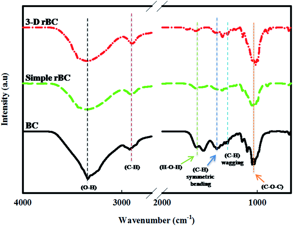 | ||
| Fig. 2 FT-IR spectroscopy of native BC, simple rBC and 3-D rBC. | ||
Swelling studies and tensile properties
The time dependent percent swelling of simple rBC and 3-D rBC scaffolds is depicted in Fig. 3a. The swelling ratio of the simple rBC increased to 169%, 251%, 272% and 423% at 10, 20, 30 and 60 min, respectively, and reached a saturation of 461% at 90 min. This gradual increase in the swelling ratio is likely attributed to the compact nature of simple rBC, allowing the water to penetrate very slowly. In comparison, the swelling ratio of the 3-D rBC scaffold increased rapidly to 833% in the initial 10 min, and reached a saturation of 1314% in only 30 min. The significant swelling of the 3-D rBC scaffolds indicates the hydrophilic character of BC and the porous structure of the sample accommodating free standing water, while the rapid saturation is attained due to its interconnected porous structure. BC is known for its high water absorption capacity. It physically entraps the water molecules on its surface, as well inside the porous fibrillar network.35 This property contributes to its potential numerous applications in the biomedical field, including wound healing, the treatment of skin burns, and tissue regeneration.36 The high water holding capacity of our scaffolds is highly favorable for biomedical applications, allowing an easy and continuous transfer of nutrients to cells throughout the tissue regeneration period. This would also be helpful in absorbing the wound exudates that help in quick healing. As the swelling behavior is dependent on the porosity of rBC films, it can be easily controlled by maintaining the pore size for particular application. | ||
| Fig. 3 Swelling ratio profiles of simple rBC and 3-D rBC during incubation in water at room temperature (a), mechanical properties including tensile strength, % strain and Young's modulus of Simple rBC and 3-D rBC scaffolds (b). | ||
Fig. 3b shows the tensile strength results of the simple and 3-D rBC scaffolds. The maximum tensile strength at the breaking point for simple rBC was higher than for the 3-D rBC. The tensile strength for Simple rBC was found to be 4.39 MPa, while that of 3-D rBC was 3.9 MPa as shown in Fig. 2. Similarly, Young's modulus and average % strain for simple rBC was 0.85 GPa and 2.1% while that of 3-D rBC was 0.74 GPa and 1.92%, respectively.
Mechanical properties such as tensile strength, Young's modulus and % strain at break are particularly important for scaffolds used in tissue engineering fields, because they determine the shape-retaining ability of the scaffolds during practical applications. Fig. 3b shows a decrease in the mechanical properties of 3-D compared to the simple 2-dimensional (2-D) rBC scaffolds. This decrease can be attributed to the high porosity of 3-D rBC scaffolds compared to the simple rBC scaffolds. These results are in complete agreement with previous studies of Zhang and coworkers who constructed poly(lactic-co-glycolic acid) (PLGA) 3-D scaffolds and determined their mechanical properties. They also found that the Young's modulus decreased as the pore size of scaffold increased and attributed this trend to the slight reduction in the scaffolding materials when the pore size was increased.37
Cell adhesion, growth, and proliferation on scaffolds
Porosity and pore size play an important role in cell attachment and proliferation in 3-D porous scaffolds. Cell adhesion, growth, and proliferation on the simple and 3-D rBC scaffolds were assessed by SEM (Fig. 4). The images revealed strong cell adhesion on the surface of simple rBC after one day, while cells were attached inside the pores within the 3-D rBC scaffold after one day. Prolonged incubation of one and three weeks resulted in filopodia formation in both the samples. The cells presented 2-D spreading on the surface of simple rBC, while the cells occupied the pore walls to form ECM in the porous 3-D rBC scaffolds. A clear difference was observed between cell growth patterns on the simple rBC and the 3-D rBC samples presenting multilayers and a much extended network of filopodia on the later samples. | ||
| Fig. 4 SEM images indicating cell adhesion, growth and proliferation on the simple and 3-D rBC scaffolds during 1 day, 1 week and 3 weeks of incubation period. | ||
The biocompatibility of simple rBC films is well known. Previously, we reported the excellent biocompatible behavior of simple rBC toward animal osteoblast and fibroblast cells.10,33 Studies have also demonstrated the superiority of 3-D over 2-D cell cultures. The differentiation of fibroblast cells and formation of ECM was comparatively evaluated in 2-D and 3-D cell culture models. It was concluded that the 3-D culture environment enhances cell proliferation and differentiation compared to their 2-D counterparts.38 In the case of BC, the small pore size of native BC and simple rBC films restrict cell penetration and, in turn, its application as a 3-D scaffold for tissue regeneration applications. Therefore, we intended to enhance the porosity and pore size of BC by fabrication of 3-D scaffolds with a solvent casting/porogen leaching method. Our results revealed the successful penetration of cells into the 3-D rBC scaffolds, indicating their potential application in the tissue regeneration field.
Morphological and immunocytological behavior
Morphological and immunocytological observation was carried out using confocal microscopy to analyze cell morphology and infiltration into the 3-D rBC matrix. Fig. 5a depicts the differential interference contrast (DIC), DAPI, F-actin and DAPI/F-actin merged images of stained fibroblasts cultured for three weeks, indicating the growth and proliferation pattern in the 3-D rBC scaffold. The cells exhibited excellent adhesion, growth, and proliferation, as well as infiltration, inside the 3-D scaffold. These results are in complete agreement with the SEM images obtained after the same incubation time (3 weeks). Optical slicing of the sample in the Z-direction through Z-stack confocal microscopy allowed us to observe the penetration of cells into the 3-D rBC scaffold. Fig. 5b and c contain the Z-stack images of optical slices from top to bottom with a 5 μm slice thickness and the reconstructed 3-D projection image of the scaffold. These images clearly demonstrate the successful growth, proliferation, and infiltration (upto 100 μm) of cells inside the 3-D rBC scaffolds, indicating their suitability for tissue regeneration applications. | ||
| Fig. 5 Confocal images of DAPI and F-actin stained fibroblast cells cultured on 3-D rBC scaffolds for 3 weeks (a). Z-stack images obtained from surface to bottom of the scaffold with 5 μm plane thickness are shown (b). Reconstructed 3-D projection image of the scaffold (c). The scale bar represents 100 μm. | ||
Osteogenesis
ALP activity was used as an indicator of osteoblastic differentiation of MC3T3-E1 cultured on simple and 3-D rBC scaffolds. As shown in Fig. 6a, the ALP activity of the cells on scaffolds increased with incubation time. The relative ALP activity of simple rBC increased from 100% to 146% ± 12.6%, while that of 3-D rBC scaffolds increased from 153% ± 10.4% to 195% ± 15.1% when incubated for one week and two weeks, respectively. The degree of ALP activity expressed by the 3-D rBC scaffold was significantly higher compared to that expressed by simple rBC after two weeks of incubation. We also investigated the mineralization process using an ARS assay. Fig. 6b, depicts the Alizarin staining results observed after one and two weeks of culture in both simple and 3-D RBC scaffolds. Mineralization increased with incubation time, and both samples exhibited an increased level of mineralization at two weeks. ARS levels were significantly greater with the 3-D rBC scaffolds compared to the simple rBC (Fig. 6b). In the 3-D samples, osteoblasts exhibited significant calcium production and deposition onto the scaffolds. This data demonstrates the osteogenic nature of the 3-D rBC scaffolds. | ||
| Fig. 6 Determination of osteogenesis and mineralization through ALP activity and ARS, respectively. Relative ALP activity of simple rBC and 3-D RBC scaffolds (a), ARS of the scaffolds (b). Graph shows mean value ± SD (P < 0.05). | ||
ALP is an enzyme widely recognized as an early marker of osteogenic activity.39 During bone formation, ALP is responsible for the generation of inorganic phosphate, which is required for hydroxyapatite crystallization. ALP also facilitates mineral (calcium) precipitation through the hydrolysis of pyrophosphate, a mineralization inhibitor.40 BC has been previously investigated for its osteogenic properties, including ALP activity and mineralization tests. Fang et al.41 investigated the proliferation and osteoblastic differentiation of human bone marrow stromal cells (hBMSC) on BC. Significantly high ALP activity was recorded for hBMSCs cultured on BC. Saska et al.42 evaluated BC for in vitro bone regeneration, and reported significantly high values of ALP activity for cell cultures grown on pure BC. The same study also reported a significant level of mineralization for BC using an ARS assay.42 It can be inferred from our results that BC scaffolds can support the proliferation of osteoblasts, as well as the expression of genes important for osteogenesis such as ALP, osteocalcin, collagen I, and osteopontin.43 We conclude from our ALP activity and ARS assay results that 3-D rBC scaffolds can support the expression of genes important for osteogenesis.
Cell viability
Proliferation of NIH 3T3 cells on simple rBC and 3-D rBC scaffolds was evaluated using a MTT assay (Fig. 7). The results indicate an increase in cell numbers for both the samples with respect to time. There were no significant differences in cell viability on simple rBC or 3-D rBC scaffolds on day one and day three. This signifies that during the initial three days, cell proliferation was unaffected by the morphology differences of rBC and 3-D rBC. However, on day five and day seven, a significant increase in cell proliferation on 3-D rBC scaffolds was recorded compared to simple rBC. This is attributed to the availability of a larger space for cellular growth and proliferation in the highly porous 3-D rBC scaffolds compared to simple rBC. Similar results were obtained by Alves et al.44 when they compared 2-D collagen scaffolds with their 3-D counterparts for fibroblast proliferation. Rothan et al.45 evaluated the differentiation of fibroblasts and the formation of ECM in both 2-D and 3-D culture systems consisting of polycaprolactonetriol–citrate scaffolds. They reported a significant increase in cell proliferation and differentiation in 3-D culture systems compared to their 2-D counterparts. These results suggest improved cell viability in 3-D rBC scaffolds compared to simple rBC, attributed to the 3-D porous structure of the former.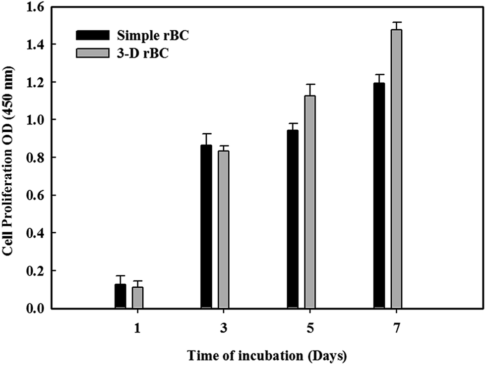 | ||
| Fig. 7 Cell proliferation on simple and 3-D rBC scaffolds determined through MTT assay after 1, 3, 5 and 7 days of cell incubation. Graph shows mean value ± SD (P < 0.05). | ||
Conclusion
Highly porous 3-D rBC scaffolds were fabricated using an easy and robust solvent casting/particulate leaching method. Scaffold indicated porous geometry along with transformation from cellulose I to cellulose II. High swelling ratio (liquid absorbing capabilities) and reasonable mechanical features encouraged the applications of synthesized 3-D rBC scaffold in biomedical fields. The biocompatibility tests conducted with animal cells indicated strong adhesion, penetration, and proliferation in the 3-D scaffolds. Additionally, the ALP activity and ARS revealed the osteogenic differentiation and successful mineralization on the scaffolds. The cell growth in 3-D scaffold, their penetration and viability was a bright sign for practical applications in tissue regeneration and wound healing. Furthermore, this study could be extended for developing composite 3-D scaffolds with biocompatible materials to further improve the applicability in biomedical field.Acknowledgements
This research was supported by the Basic Science Research Program through the National Research Foundation (NRF) funded by the Ministry of Education, Science and Technology, Korea (NRF-2014-R1A1A2055756) and by the BK21 plus (2014–2019), Korea (21A.2013-1800001).References
- L. Ma, C. Y. Gao, Z. W. Mao, J. Zhou, J. C. Shen, X. Q. Hu and C. M. Han, Biomaterials, 2003, 24, 4833–4841 CrossRef CAS
.
- M. Kim and G. H. Kim, RSC Adv., 2015, 5, 26954–26964 RSC
- X. H. Wu, Z. Y. Wu, J. Qian, Y. G. Yan, J. Wei, H. Lic and J. C. Su, RSC Adv., 2015, 5, 36007–36014 RSC
- N. Banerjee and J. Park, Korean J. Chem. Eng., 2015, 32, 1207–1217 CrossRef CAS
- S. Jebahi, H. Oudadesse, G. B. Saleh, M. Saoudi, S. Mesadhi, T. Rebai, H. Keskes, A. Feki and H. Feki, Korean J. Chem. Eng., 2014, 31, 1616–1623 CrossRef CAS
- L. Y. Zhu, X. Q. Yan, H. M. Zhang, S. J. Yao and L. Jiang, Korean J. Chem. Eng., 2015, 32, 369–372 CrossRef CAS
- W. Shao, H. Liu, X. Liu, S. Wanga and R. Zhang, RSC Adv., 2015, 5, 4795–4803 RSC
- R. M. Dominques, M. E. Gomes and R. L. Reis, Biomacromolecules, 2014, 15, 2327–2346 CrossRef PubMed
- P. C. Tischer, M. R. Sierakowski, H. Westfahl Jr and C. A. Tischer, Biomacromolecules, 2010, 11, 1217–1224 CrossRef CAS PubMed
- S. Khan, M. Ul-Islam, W. A. Khattak, M. W. Ullah and J. K. Park, Cellulose, 2015, 22, 565–579 CrossRef CAS
- S. Khan, M. Ul-Islam, W. A. Khattak, M. W. Ullah and J. K. Park, Carbohydr. Polym., 2015, 127, 86–93 CrossRef CAS PubMed
- A. Vesel, I. Junkar, U. Cvelbar, J. Kovac and M. Mozetic, Surf. Interface Anal., 2008, 40(11), 1444–1453 CrossRef CAS PubMed
- R. A. N. Pertile, F. K. Andrade, C. Alves and M. Gama, Carbohydr. Polym., 2010, 82, 692–698 CrossRef CAS PubMed
- J. Courtenay, Surface Modified Cellulose Scaffolds for Tissue Engineering, 3rd South West Regional Regenerative Medicine Meeting, 2015 Search PubMed
- R. Pértile, S. Moreira, F. Andrade, L. Domingues and M. Gama, Biotechnol. Prog., 2012, 28, 526–532 CrossRef PubMed
- F. K. Andrade, S. M. Moreira, L. Domingues and F. M. Gama, J. Biomed. Mater. Res., Part A, 2010, 92, 9–17 CrossRef CAS PubMed
- H. Backdahl, M. Esguerra, D. Delbro, B. Risberg and P. Gatenholm, J. Tissue Eng. Regener. Med., 2008, 2, 320–330 CrossRef PubMed
- A. G. Mikos, A. J. Thorsen, L. A. Czerwonka, Y. Bao and R. Langer, Polymer, 1994, 35, 1068–1077 CrossRef CAS
- P. X. Ma and J. W. Choi, Tissue Eng., 2001, 7, 23–33 CrossRef CAS PubMed
- G. Chen, T. Ushida and T. Tateishi, Mater. Sci. Eng., C, 2001, 17, 63–69 CrossRef
- Q. Zhou, Y. Gong and C. Gao, J. Appl. Polym. Sci., 2005, 98, 1373–1379 CrossRef CAS PubMed
- J. S. Capes, H. Y. Ando and R. E. Cameron, J. Mater. Sci.: Mater. Med., 2005, 16, 1069–1075 CrossRef CAS PubMed
- D. W. Hutmacher, Biomaterials, 2000, 21, 2529–2543 CrossRef CAS
- G. Turco, E. Marsich, F. Bellomo, S. Semeraro, I. Donati, F. Brun, M. Grandolfo, A. Accardo and S. Paoletti, Biomacromolecules, 2009, 10, 1575–1583 CrossRef CAS PubMed
- D. J. Choi, S. M. Choi, H. Y. Kang, H. J. Min, R. Lee, M. Ikram, F. Subhan, S. W. Jin, Y. H. Jeong, J. Y. Kwak and S. Yoon, J. Biotechnol., 2015, 205, 47–58 CrossRef CAS PubMed
- N. Su, Q. Sun, C. Li, X. Lu, H. Qi, S. Chen, J. Yang, X. Du, L. Zhao, Q. He, M. Jin, Y. Shen, D. Chen and L. Chen, Hum. Mol. Genet., 2010, 19, 1199–1210 CrossRef CAS PubMed
- H. Wang, C. Chu, R. Cai, S. Jiang, L. Zhai, J. Lu, X. Li and S. Jiangbc, RSC Adv., 2015, 5, 53550–53558 RSC
- V. Karageorgiou and D. Kaplan, Biomaterials, 2005, 26, 5474–5491 CrossRef CAS PubMed
- O. A. Inozemtseva, Y. E. Salkovskiy, A. N. Severyukhina, I. V. Vidyasheva, N. V. Petrova, H. A. Metwally, I. Y. Stetciura and D. A. Gorin, Russ. Chem. Rev., 2015, 84, 251–274 CrossRef CAS PubMed
- S. Mahalingam and M. Edirisinghe, Macromol. Rapid Commun., 2013, 34, 1134–1139 CrossRef CAS PubMed
- S. Zhang, B. T. Karaca, S. K. van Oosten, E. Yuca, S. Mahalingam, M. Edirisinghe and C. Tamerler, Macromol. Rapid Commun., 2015, 36, 1322–1328 CrossRef CAS PubMed
- M. Ul-Islam, N. Shah, J. H. Ha and J. K. Park, Korean J. Chem. Eng., 2011, 28, 1025–1103 Search PubMed
- S. Khan, M. Ul-Islam, W. A. Khattak, M. W. Ullah and J. K. Park, Carbohydr. Polym., 2015, 127, 86–93 CrossRef CAS PubMed
- Y. O. Sang, I. Y. Dong, S. Younsook, C. K. Hwan, Y. K. Hak, S. C. Yong, H. P. Won and H. Y. Ji, Carbohydr. Res., 2005, 340, 2376–2391 CrossRef PubMed
- M. Ul-Islam, T. Khan, W. A. Khattak and J. K. Park, Cellulose, 2013, 20, 589–596 CrossRef CAS
- M. Ul-Islam, W. A. Khattak, M. Kang, S. M. Kim, T. Khan and J. K. Park, Cellulose, 2013, 28, 253–263 CrossRef
- Y. S. Zhang, K. P. Regan and Y. Xia, Macromol. Rapid Commun., 2013, 34, 485–491 CrossRef CAS PubMed
- E. Cukierman, R. Pankov, D. R. Stevens and K. M. Yamada, Science, 2001, 294, 1708–1712 CrossRef CAS PubMed
- E. Bonucci, Biological Calcification: Normal and Pathological Processes in the Early Stages, Springer, Berlin-Heidelberg, 2nd edn, 2007, pp. 491–506 Search PubMed
- C. D. Hoemann, H. El-Gabalawy and M. D. McKee, Pathol. Biol., 2009, 57, 318 CrossRef CAS PubMed
- B. Fang, Y. Z. Wan, T. T. Tang, C. Gao and K. R. Dai, Tissue Eng., 2009, 15, 1091–1098 CrossRef CAS PubMed
- S. Saska, L. N. Teixeira, P. T. de-Oliveira, A. M. M. Gaspar, S. J. L. Ribeiro, Y. Messaddeqa and R. Marchettoa, J. Mater. Chem., 2012, 22, 22102–22112 RSC
- J. J. Kim, R. K. Singh, S. J. Seo, T. H. Kim, J. H. Kim, E. J. Leeab and H. W. Kim, RSC Adv., 2014, 4, 17325–17336 RSC
- L. B. Alves, V. C. Mariguela, M. F. Grisi, S. L. Souza, A. B. Novaes, M. Taba, P. T. Oliveira and D. B. Palioto, J. Appl. Oral Sci., 2015, 23, 206–214 CrossRef PubMed
- H. A. Rothan, I. Djordjevic, H. Bahrani, M. Paydar, F. Ibrahim, N. Abd Rahmanh and R. Yusof, Int. J. Med. Sci., 2014, 11, 1029–1038 CrossRef CAS PubMed
| This journal is © The Royal Society of Chemistry 2015 |