O. E. Meirona,
L. Houbenb and
M. Bar-Sadan
*a
aBen-Gurion University of The Negev, Department of Chemistry, Beer Sheva, Israel. E-mail: barsadan@bgu.ac.il
bChemical Research Support, Weizmann Institute of Science, Rehovot, Israel
First published on 12th October 2015
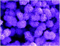
Nanoflowers of layered materials are promising building blocks for photocatalysis due to their unique morphology and exposed edges. Colloidal synthesis of MoS2, MoSe2 and their alloys was used to produce fine nanoflowers with tunable composition. The alloys follow Vegard's law, showing homogeneous composition. They exhibit fully tunable optical properties, where the exciton positions change with alloy composition. Samples annealed under vacuum retain their fine morphology and their composition, which closely follows the feed ratio. Time series analysis was used to investigate the formation mechanism. The results support a growth mechanism of fast-precipitating amorphous material, followed by crystallization of a few layers of small sheets, which curl and tangle around themselves. Electron tomography of the time series reveals a perforated structure without a dense core, supporting the suggested growth mechanism. These structures show multiple exposed edge sites, making them promising materials for applications such as photocatalysis.
One of the most promising morphologies for catalysis is nanoflowers,11 which exhibit a relatively large ratio between exposed edges and basal planes. These materials are usually produced via a hydrothermal synthesis in an autoclave.12 Electrodes synthesized using the above-mentioned technique were successfully used to reduce polysulfides in quantum dot sensitized solar cells.11 Although these materials are very promising, closer inspection reveals that the petaled edges are more than 10 nm thick. Having more than 15 layers prevents the utilization of quantum size effects for improving the activity of the exposed layer edges.13 In addition, hydrothermal synthesis requires high pressure in order to produce supercritical water, which makes it only suitable for small scale production. By switching to colloidal synthesis, a larger number of parameters can be controlled, ultimately leading to better morphology control. Recently, MoSe2 nanoflowers were synthesized via colloidal synthesis by using MoO2(acac)2 and dibenzyldiselenide.14 The resulting ultrathin structures resembled the petaled particles. However, they were an order of magnitude smaller, and had only 2–5 layers at the edges. Their exact growth mechanism is still unclear. These fine petaled structures were selenium-deficient, and demonstrated better efficiency for hydrogen evolution.
Morphology control is not the only approach to modifying TMD properties. Doping and alloying arise as promising methods for chemically modifying TMD properties. First principles studies have shown that atom dopants can induce changes in physical properties such as conductivity and magnetism in TMDs.15,16 Among other qualities, the semiconducting nature of TMDs was switched from p- to n-type and vice versa using different dopants.16–19 Alloys are also used in order to tune the electronic structure of TMDs, specifically the materials' bandgap and band alignment. The two most researched alloys are MoxW1−xS2 (ref. 20) and Mo(SxSe1−x)2.21–23 Both are produced primarily using solid state chemistry techniques such as chemical vapor deposition (CVD) and chemical vapor transport (CVT). Early density function theory research predicted that Mo(SxSe1−x)2 should be stable at room temperature,24 making it one of the most researched alloys of MoS2. Recent work explored the catalytic activity of Mo(SxSe1−x)2 alloys grown by CVT.25 The alloys are more effective cathode materials than either pure MoS2 or MoSe2 towards HER.14
Even though alloying and doping play a crucial role in fine tuning the electronic properties of TMDs, only a handful of papers have been published so far on these materials. The biggest challenge is to reproducibly produce alloys with a specific composition in large quantities. Using CVD or CVT, only partial control is achieved over the alloys' compositions. A previous study showed that by varying the substrate position inside the CVD furnace relative to the precursors, the alloy composition can be controlled.22 However, it causes a composition gradient on the substrate, making it impractical for large scale applications. In order to resolve these issues, a different chemical approach has to be explored.
A recent report describes the colloidal synthesis of Mo(SxSe1−x)2 in an oleylamine and octylamine solution, using MoCl5 and elemental S and Se.26 The alloyed nanoflakes show improved performance of hydrogen production compared to pure MoS2 and MoSe2. However, one major problem that occurs when preparing HER electrodes is the loss of the exposed edges due to aggregation of the layers. In flower-like particles, more active edges are exposed, which will possibly lead to better performance.
In this work, we synthesized alloys of Mo(SxSe1−x)2 nanoflowers using a colloidal process. Control over the composition of the alloys was achieved using a bottom-up strategy at relatively low temperatures (≤300 °C). Chalcogenide precursors with similar reactivities were used in order to form the desired homogeneous alloy for each Se/S composition. The produced alloys exhibited tunable band gaps as a function of composition. The mechanism of formation was evaluated through a time series experiment and electron tomography of the particles, revealing that the particles formed from an amorphous material, and that there was no dense core at the center of the particles. This finding confirms a previously suggested formation mechanism, where separated sheets fold onto each other to form the nanoflower structures.
The desired molar ratios of MoO2(acac)2 and chalcogenide precursors were degassed in a three-necked flask with 10 ml OA under vacuum at 110 °C for 15–30 min. The flask was backfilled with N2, and heated to 300 °C for 20 min. After the flask was cooled to room temperature, the product was precipitated by adding excess 1-butanol. The crude product was collected by centrifugation. The product was washed with the following solutions: 50
:
50 (toluene
:
ethanol), 70
:
30 (toluene
:
ethanol), pure toluene, twice with 70
:
30 (ethanol
:
DW), and once with acetone. It was then dried overnight under vacuum. The materials produced using this method were amorphous. In order to improve their crystallinity, the products were sealed in a quartz ampule under vacuum and annealed at 873 °C in a tube furnace for 16–24 h.
![]() | ||
| Fig. 1 TEM images of Mo(SxSe1−x)2 alloys. The left panel shows the as-synthesized materials. The right panel shows the products after annealing in vacuum. The scale bar in all the images is 200 nm. | ||
HR-TEM images revealed that the edges were constructed of 3–5 layers, and that the number of layers is retained after annealing, as can be seen in Fig. 2. In agreement with the literature, the crystallinity of TMDs produced at a low temperature is poor. These materials are often described as amorphous MoSx or MoS3.30,31 Therefore, the syntheses products were annealed at above the growth temperature of MoS2 (873 °C). During the annealing process, the materials are sensitive to oxidation,32 so they require an inert atmosphere (N2) or a reducing environment (such as a mixture of 5% H2 in Ar). Alternatively, the annealing can be performed under vacuum conditions in a sealed quartz ampule, preventing the loss of the more volatile S during the annealing stage. Annealing the samples under vacuum in a sealed quartz ampule retains the size and morphology of the delicate flowers without thickness or stoichiometry change, as can be seen in Fig. 1 and 2 and Table 1. The annealed samples were used for further characterization.
:
Se ratio calculated from ICP-MS results
| Feed ratio | Experimental value | ||
|---|---|---|---|
| S | Se | S | Se |
| 0.0 | 1.0 | 0.00 | 1.00 ± 0.03 |
| 0.2 | 0.8 | 0.23 | 0.77 ± 0.03 |
| 0.5 | 0.5 | 0.51 | 0.49 ± 0.03 |
| 0.8 | 0.2 | 0.80 | 0.20 ± 0.03 |
| 1.0 | 0.0 | 1.00 | 0.00 ± 0.03 |
In order to determine the alloys' compositions more accurately, ICP-MS spectrometry was used. Table 1 summarizes the results of the different alloys. Measuring the concentration of S using ICP-MS introduces substantial errors: it has many known interferences, it is only about 15% ionized in the Ar plasma, and all isotopes are subject to severe overlapping from mainly O- and N-based signals. Therefore, only the Mo
:
Se ratio was calculated, and S content was determined based on the Se ratios. We used the synthesis of pure MoSe2 to determine the Mo
:
Se ratio of 1
:
1.62, in accordance with previously reported Mo-rich MoSe2−x nanoflowers.14 The results for the ternary compounds were normalized to the MoSe2 values, and showed a very good fit of the measured S
:
Se with the feed ratio.
Pure MoS2 and MoSe2 have hexagonal structures of the P63/mmc space group (PDF-37-1492 for MoS2 and 77-1715 for MoSe2). Fig. 3a shows XRD data of pure and alloyed materials. There is a shift towards higher diffraction angles with increasing S content, in accordance with a reduction in cell parameters as the larger Se atoms are replaced by smaller S atoms. Dashed lines were added to assist in tracking the transition. Cell parameters of the different alloys were calculated, and the a lattice parameter was plotted against the Se content (Fig. 3b). The plot has good linear correlation, which is consistent with Vegard's law. It suggests that S and Se are homogeneously distributed in the lattice. The UV-Vis spectra of the pure and alloyed materials contain the two excitons of MoS2 and MoSe2, as can be seen in Fig. 4a. Both excitons shift gradually to the lower wavelengths as the S content increases, further confirming the ternary mixture's homogeneity. In accordance, the Raman spectra presented in Fig. 4b show a gradual increase in the MoS2 E12g and A1g modes with increasing S content, as previously reported.22 As the Se content fraction decreases, there is a dampening and decrease in the MoSe2 and A1g modes.
In order to investigate the sensitivity of the alloyed nanoflowers to alternative synthetic protocols, different precursors were used for the synthesis. We found that replacing MoO2(acac)2 with different Mo precursors, e.g. MoCl5, MoO3, Na2MoO4·2H2O or (NH4)2MoO4, prevents the formation of the nanoflower structures and produces other morphologies (see Fig. S1 in the ESI†). Changing the S or Se precursor to elemental S and Se also resulted in loss of the flower-like morphology. The combination of MoO2(acac)2 and DBSe/DBS proved to be unique in achieving the nanoflowers in oleylamine.
Previous reports provide two growth schemes: either the initial structures are amorphous sheets that curl around themselves to produce flowers,14 or the flower-like structure is the nucleus of the growth, followed by an outward growth of the thin petals.33 These two mechanisms have different reaction rates: a faster reaction yields the sheets morphology, and a slower reaction produces the flower morphology, which is then extended by growth.
To better understand the formation mechanism, we performed a time series analysis. The synthesis procedure was slightly modified as follows: the molybdenum and selenium precursors were degassed in two separate flasks. The molybdenum solution was heated to 300 °C, and the selenium solution was then injected rapidly to start the reaction. Aliquots of 1 ml were quenched in 1-butanol at four different time points during the reaction, and analyzed. Fig. 5a–d presents TEM images of the aliquots at times 1, 3, 5 and 10 min, respectively. In addition, electron tomography was used to understand the 3D structure of the particles from the time series and to determine the growth mechanism. Using electron tomography, it is possible to isolate a slice of the analyzed volume without the superimposed information from the top and bottom parts of the sample. Fig. 6 presents the 2D images from the time series at times 1, 3 and 5 minutes, alongside the corresponding slices from the 3D reconstructions.
![]() | ||
| Fig. 5 Time series analysis of MoSe2 particles. TEM images of MoSe2 nanoflowers taken at times: (a) 1 min. (b) 3 min. (c) 5 min. (d) 10 min. | ||
Fig. 5a and 6a show that after 1 minute, small ∼50 nm amorphous clusters form in the solution, that later serve as a reservoir of material for the crystallization stages. After 3 minutes, the amorphous material is consumed to form curled and tangled sheets that crystallize within the amorphous clusters. Voids are also created in this process, since the crystallized form is denser than the amorphous cluster. Adjacent clusters also merge, as shown in Fig. 5b and 6b. There is no preferred growth direction of the sheets, and they appear to favor random lateral growth. It is important to note that no dense core is visualized, ruling out a radial growth mechanism where a crystallized seed serves as the substrate for further growth. The amount of amorphous material decreases with time, with a slight increase in the number of crystalline layers (from 1–3 after 3 minutes to 3–5 after 5 minutes). The major change seen in the particles over time is the crystallization of the amorphous material, with a relatively minor change in lateral size after 5 minutes. We therefore conclude that the growth relies on the reservoir of material produced by the initial cluster formation. Our findings support the formation mechanism described in ref. 14 and in contrast to the one described in ref. 33.
A consequence of this reaction mechanism is the control of the homogeneity of the final product, since the efficient formation of an amorphous material reservoir allows the incorporation of both S and Se during crystallization to form homogeneous solid solutions, without relying on their reactivity and diffusion in the solution. In order to confirm that the same mechanism governs the growth of the ternary compounds, tomography of the Mo(S0.51Se0.49)2 sample was performed. Fig. 7 presents three slices of the tomogram from a central section of the particle. The slices in Fig. 7b–d show three cut planes in different directions from the tomogram. They show that there are voids at the core of the particle, in accordance with the growth mechanism described above. The particle seems perforated, with delicate petals extruding from the center, like in the pure material's structure. An additional 3D visualization is shown in Fig. 7a, featuring the sponge-like structure which lacks a dense center. Additional movies of the tilt series, a scroll through the tomogram slices and a rotation of the 3D visualization is available in the ESI.†
Footnote |
| † Electronic supplementary information (ESI) available: Movies of the electron tomography tilt series, a scroll through the tomogram slices and a rotation of the 3D visualization is available in the ESI. See DOI: 10.1039/c5ra16853h |
| This journal is © The Royal Society of Chemistry 2015 |