Utilizing an aggregate forming microenvironment sensitive coumarin–cholesterol conjugate as a sensor of pluronic organization and micro-polarity

Ivy Sarkara, H. Surya Prakash Rao*b, Avinash Desaib and Ashok Kumar Mishra*a
aDepartment of Chemistry, Indian Institute of Technology Madras, Chennai-600 036, India. E-mail: mishra@iitm.ac.in
bDepartment of Chemistry, Pondicherry University, Pondicherry-605 014, India

Received 17th August 2015 , Accepted 2nd November 2015

First published on 4th November 2015


Abstract

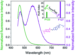
A new fluorescent cholesterol molecular probe, 3-acetyl-7-(diethylamino)-2H-chromen-2-one attached cholesterol (Cum–Chl), has recently been introduced as a reporter of micro-heterogeneous media. The H-aggregate forming ability of Cum–Chl provides a useful fluorescence parameter, Imonomer/Iaggregate to understand the micro-polarity of anisotropic media. Pluronics are surfactant based polymeric anisotropic media having important applications in therapeutics. Being a sensitive indicator of pluronics micro-polarity Imonomer/Iaggregate can help in selecting pluronics for medicinal purposes. Additionally, temperature and concentration induced sol–gel transition of pluronics (P123, F127) have been successfully investigated by Cum–Chl using its Imonomer/Iaggregate value, along with other conventional fluorescence parameters. Finally, this molecular probe, Cum–Chl emerges as a good sensor of progressive polymeric association and micro-polarity of pluronics.


Introduction

Pluronics are water soluble tri-block copolymers composed of hydrophilic poly(ethyleneoxide) (PEO) and hydrophobic poly(propylene oxide) (PPO) units.1 They are often abbreviated as PEOx–PPOy–PEOx, where x and y denote the number of the respective units. Modulation in the behavior of PEO and PPO units as a function of physical and chemical parameters has increased the industrial applications of pluronics over conventional surfactants.2 A more pronounced temperature effect on PPO solubility over PEO imparts amphiphilicity to pluronics.3,4 Thus, these block polymers form micelle followed by gelation both as a function of temperature and concentration, unlike conventional surfactants. Anhydrous hydrophobic core region of the spherical micelle consists of PPO units helps in solubilization of water insoluble drug molecules.5–7 Highly hydrated hydrophilic outer region, corona consists of PEO units keeps the micelle dispersed in water.5,8,9 Thus the effective volume of the micelle increases to double than its desired volume.10 Pluronics are largely used in drug therapy due to its water solubility and ability to intake drug molecules inside the hydrophobic core.11–13 Above CMC (critical micelle concentration) or CMT (critical micelle temperature) pluronics remain as liquid crystalline phase composed of several lypotropic structures (e.g. cubic, hexagonal, lamellar) followed by the formation of gel at higher concentration or temperature.10,11,14,15 The complex phase diagram of pluronics are well studied using different experimental techniques.10,16–19 It should be noted that, CMC and CMT values of pluronics differ from batch to batch due to the change in poly dispersity index.11,20,21 In this work, concentrations and temperatures are chosen such that pluronics remain above the CMC/CMT but below the transparent hydrogel.11 Below CMC/CMT pluronics remain as monomers which have effective biological activity and translocation ability through the cell membranes.8 So, pluronics are used above CMC in drug composition so that in blood it dilutes back to the monomer.8 In the present study, we have chosen two pluronics: hydrophobic P123 and hydrophilic F127. Few important parameters of these two available in literature are provided in ESI, Table S1 for better understanding of their properties.12,22

Conjugation of small fluorophores with different molecules of interest has increased the application of conventionally used fluorophores. Conjugated probes are important because of their low perturbation and site specificity.23 A coumarin–cholesterol conjugate, Cum–Chl (7-((diethylamino)-3-(3-cholesteryl)propanoyl)-2H-chromen-2-one) has recently been introduced by us as a new fluorescent molecular probe for micro-heterogeneous media.24 Conjugated probes are important for different domains of interest like biochemistry, biophysics, cell biology, drug delivery, imaging, sensing, analytical purposes etc.23–30 As a result, conjugated probes have become a point of interest for multi-disciplinary and inter-disciplinary researchers. Fig. 1 shows the molecular structure of parent fluorophore, Cum and its cholesterol-conjugate, Cum–Chl. Along with the monomer emission at 470 nm, Cum–Chl has a fairly red shifted H-aggregate emission at 566 nm in aqueous medium. For this molecule, it was established that the monomer to aggregate fluorescence intensity ratio, Imonomer/Iaggregate can be used as a faithful parameter in addition to the usual unimolecular photophysical fluorescence sensitivity parameters (e.g. intensity, shift of emission maxima, anisotropy, lifetime etc.) in micro-heterogeneous lipid bilayer and bile salt media.24 The immense industrial application of pluronics and the modulation in the behavior of PPO and PEO units with temperature have increased its popularity in scientific research. Pluronics with medium PPO block length and reasonably hydrophobic structure are found to be suitable for drug delivery.8 This needs a proper choice of pluronics on basis of their micro-polarity. In this article, we have used characteristic fluorescence feature of Cum–Chl, Imonomer/Iaggregate to monitor the micro-polarity of different pluronics. Moreover, micro-environmental changes of pluronics as a function of temperature and concentration have also been looked into.


image file: c5ra16549k-f1.tif
Fig. 1 Molecular structure of 3-acetyl-7-(diethylamino)-2H-chromen-2-one (Cum) and its cholesterol conjugate, 7-((diethylamino)-3-(3-cholesteryl)propanoyl)-2H-chromen-2-one, (Cum–Chl).

The main objective of this paper is to monitor the micro-polarity difference of two pluronics (P123, F127) using Imonomer/Iaggregate parameter of Cum–Chl. The progressive polymeric association of pluronics as a function of temperature and concentration has also been monitored by the Imonomer/Iaggregate value along with other fluorescence parameters. Non-interfering small parent molecular probe, Cum has been considered as a reference for this purpose.

Materials and methods

Materials

Pluronic P123 and F127 were purchased from Sigma Chemical Co. (Bangalore, India). Same batch of pluronics have been used throughout. Triple distilled water used for all experiments was prepared by using KMnO4 and NaOH. Spectroscopic grade ethanol was used for making stock solution of probes.

Preparation of pluronic solutions

10% (w/v) solution of pluronic P123 and F127 in triple distilled water were used for the temperature dependent experiments. Temperature was varied from 5–24 °C and 13–34 °C, respectively, for P123 and F127 solutions. For concentration dependent experiments stock solutions of 30% (w/v) pluronics were prepared. Then stock polymeric solutions were diluted with triple distilled water to get solutions with 0–24% (w/v) concentration. Concentration dependent experiments were carried out at 10 °C and 35 °C for both P123 and F127 solutions. As pluronics are soluble in cold water only, polymeric solutions were kept in fridge to ensure complete dissolution. Then probe solution was added with the prepared polymeric solutions and kept over-night to ensure homogeneity.

Physical measurements

DSC measurements of pluronic solutions have been performed with TA-DSC Q-200 instrument.

Fluorescence measurements were carried out with Fluoromax 4 (Horiba Jobin Yvon) spectrofluorimeter having 150 W xenon lamp as source of excitation. 2/2 slit width has been used for all experiments. Steady state fluorescence anisotropy (rSS) was determined using the following equation.31

image file: c5ra16549k-t1.tif
here, IVV and IVH are fluorescence intensities and subscripts V (vertical) and H (horizontal) signify orientation of excitation and emission polarizer. G is the instrument correction factor.

Data acquisition was done by Horiba Jobin Yvon TCSPC lifetime instrument in time-correlated single-photon counting arrangement. Nano-LED of 444 nm was used as excitation source. The pulse repetition rate was set at 1 MHz. Instrumental full width at half-maxima of the 444 nm LED, including the detector response was measured to be ∼1.2 ns. The instrument response function was collected using scattered medium, LUDOX AS40 colloidal silica. IBH software was used for the decay analysis. Decays were fitted to get a symmetric distribution keeping χ2 value in the range of 0.99 ≤ χ2 ≤ 1.4. Average fluorescence lifetime (τaverage) was calculated using the following equation31 where τi is the lifetime of i-th component with amplitude βi, where n signifies number of component present.

image file: c5ra16549k-t2.tif

Ethanolic stock solution of Cum and Cum–Chl were used for all experiments and finally diluted with water. Ethanol contamination was kept less than 1% to avoid interference. Final probe concentration was maintained at 2.5 μM for all experiments. The desired temperature was controlled using water circulation through jacketed cuvette holder from a refrigerated bath (Julabo, Germany).

Results and discussions

Temperature dependent sol–gel transition study of pluronics (P123, F127) by Differential Scanning Calorimetric (DSC) analysis

DSC is a convenient thermo-analytical tool to monitor phase transition of several substances. DSC analyses of 10% polymeric solutions have been carried out to obtain the sol–gel transition temperature. Fig. 2a and b show the thermograms of 10% P123 and F127, respectively, both in presence and absence of the probe (Cum–Chl). The sol–gel transition temperatures of 10% P123 and F127 are found to be 15 °C and 21 °C, respectively. Additionally, it doesn't change in presence of the probe (Cum–Chl) as the structures of polymeric association remain unaltered.32
image file: c5ra16549k-f2.tif
Fig. 2 Differential scanning calorimetric thermograms of pluronics, (a) 10% P123 and (b) 10% F127 in absence and presence of Cum–Chl.

Temperature induced sol–gel transition study of pluronics by fluorescence spectroscopy

Temperature induced sol–gel transition is the unique property of polymeric surfactants. This increases hydrophobicity of the pluronic micro-environment as a function of temperature. Here, 10% solution of P123 and F127 have been analyzed by using fluorescence across their sol–gel transition temperature using Cum–Chl and Cum.15,33 Fig. 3a and b depict the fluorescence spectra of Cum–Chl, in 10% P123 and F127 media, respectively, as a function of temperature, at λex 440 nm. It shows fluorescence response of Cum–Chl towards two pluronics of different hydrophobicity is markedly different from each other. Fluorescence intensity of Cum–Chl monomer (∼470 nm) is much lower in magnitude in 10% F127 media (Fig. 3b) as compared to that of P123 media (Fig. 3a). Also, with increasing temperature monomer intensity in 10% F127 has been found to increase only by two fold, whereas, it is six fold in 10% P123 media. A significant presence of H-aggregate (∼566 nm) in F127 media (Fig. 3b) as compared to P123 media (Fig. 3a) is also evident. This distinctive behavior of Cum–Chl in P123 and F127 media is due to the hydrophobicity difference of these two. With a larger PEO unit F127 is more hydrophilic than P123. As the breaking of Cum–Chl H aggregate is a function of medium polarity F127 disaggregates H-aggregate less efficiently as compared to P123. Thus higher H-aggregate population of Cum–Chl in F127 media is found (Fig. 3a and b). This has been reflected in the Imonomer/Iaggregate value, given in Fig. 3c and d. Almost 30 fold higher value of Imonomer/Iaggregate in P123 than F127 signifies higher partitioning of the monomer into P123 micelles than F127. Thus the value of Imonomer/Iaggregate reports the micro-polarity of pluronics media qualitatively. Similar observation has also been found previously for bile salts with different hydrophobicity.24 Sodium cholate (NaC) with three hydroxyl groups is more hydrophilic as compared to sodium deoxycholate (NaDC) with two hydroxyl groups. As a result, Imonomer/Iaggregate value in NaDC media was found to be 4 times higher as compared to the NaC media.24 Here, in P123 media re-distribution in the population of monomer and aggregate form is also evident by an iso-emissive point ∼560 nm (Fig. 3a). The H-aggregate emission band intensity has been found to decrease with increasing temperature (ESI Fig. S1a and b) in both pluronics.
image file: c5ra16549k-f3.tif
Fig. 3 Fluorescence spectra of Cum–Chl in presence of 10% (a) P123 and (b) F127 as a function of temperature; at λex 440 nm. Variation of Imonomer/Iaggregate in presence of 10% (c) P123 and (d) F127 with increasing temperature, inset shows its derivative plot.

Fig. 3c and d show the variation of Imonomer/Iaggregate with sol–gel transition in two pluronics media. It shows marked change during sol–gel transition (Fig. 3c and d) which is 15 °C and 21 °C for 10% P123 and F127 media, respectively. This shows Imonomer/Iaggregate value of Cum–Chl is sensitive towards the sol–gel transition of pluronics. Along with that, during sol–gel transition monomer emission of Cum–Chl in 10% P123 has been found to undergo a hypsochromic shift of 10 nm (ESI Fig. S1a). During sol–gel transition, with increasing temperature highly hydrated pluronic micelles are known to undergo progressive dehydration.34–36 This enhances hydrophobicity of pluronic micro-environment with increasing temperature, resulting in the blue shift of emission maxima. As a result, increase in the Imonomer/Iaggregate value (Fig. 3c and d) also signifies the progressive dehydration of pluronics. Steady state fluorescence anisotropy (rSS) of Cum–Chl monomer has also been found to increase with increasing temperature in 10% P123 media (ESI Fig. S2). The sharp increase of fluorescence anisotropy up to the sol–gel transition temperature followed by labeling effect originally reflects the increase in micro-viscosity of P123 media with increasing temperature. The slower solvation dynamics in interfacial region than in the bulk water also supports this observation.37

Along with the change in polarity during sol–gel transition of pluronics viscosity of the medium also change appreciably.1–4 So, it is necessary to verify the effect of medium viscosity on the fluorescence properties. Glycerol and ethylene glycol have been chosen which are known to undergo change in viscosity with change in temperature to verify the viscosity effect on fluorescence. As for example, viscosity of glycerol changes from 12[thin space (1/6-em)]070 cP (0 °C) to 612 cP (30 °C) with increasing temperature with no change in polarity.38 Fig. 4a and b show the fluorescence spectra of Cum–Chl in glycerol and ethylene glycol, respectively, at different temperatures. The characteristic aggregate emission at 566 nm does not appear with increase in viscosity. Thus it appears that the formation of aggregates in water is essentially polarity induced. The only observation of the increase in monomeric fluorescence at 485 nm with increasing viscosity is likely due to the suppression of the non-radiative decay rates as expected.


image file: c5ra16549k-f4.tif
Fig. 4 Fluorescence spectra of Cum–Chl in (a) glycerol and (b) ethylene glycol at two different temperatures; at λex 440 nm.

Fig. 5a and b give the excitation spectra of Cum–Chl at λem 566 nm in 10% P123 and F127 media, respectively, with increasing temperature. It shows in polymeric media of P123, H-aggregate band at ∼370 nm decreases significantly as compared to that in water (inset of Fig. 5a). This is because of the redistribution of monomeric and aggregate form in this media. But, in 10% F127 media there is hardly any change in the aggregate band intensity (∼370 nm) (Fig. 5b). This also justifies our previously described model.


image file: c5ra16549k-f5.tif
Fig. 5 Excitation spectra of Cum–Chl in presence of 10% (a) P123 and (b) F127 with increasing temperature, inset shows excitation spectra of Cum–Chl in water; at λem 566 nm.

Fluorescence lifetime of Cum–Chl has been found to change in 10% P123 media but remain unaltered in 10% F127 media. Fig. 6a and b show the fluorescence lifetime decay profiles of Cum–Chl monomer and aggregate form, respectively, in 10% P123 as a function of temperature. Inset of Fig. 6a and b show the variation of respective τaverage values with increasing temperature. The onset of the increase in monomer lifetime and decrease in aggregate lifetime (inset of Fig. 6a and b) follow the sol–gel transition of 10% P123. Table S2 (ESI) summarizes the fluorescence lifetime data of Cum–Chl monomer and aggregate in 10% P123 media with increasing temperature. Residue distribution plots of the same have been given in ESI Fig. S3.


image file: c5ra16549k-f6.tif
Fig. 6 Fluorescence lifetime decay profiles of Cum–Chl (a) monomer (λem = 470 nm) and (b) aggregate (λem = 566 nm) in presence of 10% P123 with increasing temperature, inset shows variation of average fluorescence lifetime; at λex 444 nm.

The above mentioned distinct fluorescence behavior of Cum–Chl in different pluronics is not observed with the parent fluorophore, Cum. Fig. 7a and b depict the fluorescence spectra of Cum in 10% P123 and F127, respectively, as a function of temperature, at λex 440 nm. For both the pluronics fluorescence intensity has been found to increase in similar order of magnitude along with ∼20 nm blue shift of emission maxima (normalized spectra, ESI Fig. S4). Fig. 7c and d show the intensity variation of Cum in 10% P123 and F127 media, respectively, as a function of temperature where the onsets have been found at the sol–gel transition temperature of the respective pluronics. Fig. 7e and f show fluorescence lifetime decay profiles of Cum in 10% P123 and F127 media, respectively, with temperature. Average fluorescence lifetime values have been found to increase during sol–gel transition. The onsets of the increase in the average fluorescence lifetimes of Cum follow the sol–gel transition of 10% P123 and F127 media, inset of Fig. 7e and f, respectively. The increase in the steady state fluorescence intensity has correspondence with increase in average fluorescence lifetime (τaverage) value. Tables S3 and S4 (ESI) summarize the fluorescence lifetime data of Cum in 10% P123 and F127 media, respectively, with increasing temperature. Residue distribution plots of the same have been given in ESI Fig. S5 and S6, respectively.


image file: c5ra16549k-f7.tif
Fig. 7 Fluorescence spectra of Cum in presence of 10% (a) P123 and (b) F127 as a function of temperature; at λex 440 nm. Variation of fluorescence intensity in presence of 10% (c) P123 and (d) F127 with increasing temperature, inset shows derivative plot. Fluorescence lifetime decay profiles of Cum in presence of 10% (e) P123 and (f) F127 with increasing temperature, inset shows variation of average fluorescence lifetime and its derivative plot.

Being a small molecule, Cum is expected to behave like a distributive probe located randomly inside the pluronic micelles.14 Unlike Cum, Cum–Chl is a highly non-polar molecule with a large steroidal cholesterol moiety. As a result, it is located preferentially inside the hydrophobic core of spherical micelle. This specific location of Cum–Chl can be supported from the study by George et al. where they have shown the presence of HUF, a long tailed molecule, inside the core region.14 Likewise, we can assume the hydrophobic cholesterol moiety of Cum–Chl to be buried inside the hydrophobic core region keeping the fluorophore, coumarin near the PPO/PEO interface.14,32 Additionally, it has been confirmed that the intrinsic fluorescence of P123 and F127 don't contaminate the fluorescence of the probe molecules (ESI, Fig. S7). Moreover, the intrinsic fluorescence of pluronics does not change with increasing temperature, unlike the probes in pluronic media.34

Concentration dependent sol–gel transition study of pluronics by fluorescence spectroscopy

After exploring the temperature induced sol–gel transition of pluronics, concentration induced changes have also been studied. This is particularly important from the drug delivery point of view. Here, 0 to 24% of pluronic solutions have been used for studying the concentration induced changes at two different temperatures. All the used P123 solutions remain as flowing liquid throughout the concentration and temperature ranges. But, 20% and 24% solutions of F127 were found to be thick non-flowing gel at 35 °C. Fig. 8a and b show the concentration dependent spectral change of Cum–Chl in P123 media at, 10 °C and 35 °C, respectively. And Fig. 8c and d show the concentration dependent spectral change of Cum–Chl in F127 media at, 10 °C and 35 °C, respectively. As described in the temperature study section, F127 cannot disaggregate Cum–Chl H-aggregates efficiently, unlike P123. Similar observations have also been made in this section. Marginal increase in monomer intensity and appreciable presence of aggregate form in F127 give its evidence. In P123 media with increasing concentration of the polymer, monomeric fluorescence intensity increases along with the concomitant decrease in the aggregate fluorescence intensity (Fig. 8a and b) with an iso-emissive point at ∼560 nm. This signifies the redistribution of monomeric and aggregate form in presence of P123. In both P123 and F127 media with increasing concentration of the polymers, H-aggregate fluorescence of Cum–Chl has been found to decrease as evident from the normalized spectra, ESI Fig. S8. Fig. 8e and f show the comparative variations of Imonomer/Iaggregate at two different temperatures in P123 and F127 media, respectively. Imonomer/Iaggregate value in F127 media (Fig. 8f) is smaller in magnitude than the corresponding values in P123 media (Fig. 8e), which supports our previously described model. At higher temperature, the value of Imonomer/Iaggregate is higher as compared to that at lower temperature in both the media. This indicates higher partitioning of Cum–Chl monomer inside the micelle at higher temperature. Concentration dependent lifetime analysis has not been performed because of the increasing scattering.39
image file: c5ra16549k-f8.tif
Fig. 8 Fluorescence spectra of Cum–Chl, with increasing % of P123 at (a) 10 °C and (b) 35 °C and with increasing % of F127 at (c) 10 °C and (d) 35 °C. Comparative variation of Imonomer/Iaggregate at two different temperatures, with increasing % of (e) P123 and (f) F127; at λex 440 nm.

Poor disaggregation of Cum–Chl H-aggregates with increasing concentration of F127 is also evident from the excitation spectra (ESI Fig. S9a and b). In P123 media, there is clear cross-over of two bands (370 nm band for H-aggregates and 440 nm band for monomer) with increasing concentration (inset of ESI Fig. S9a). But, in F127 media these two bands are almost overlapping with each other although the concentration has been varied from 0% to 24% (inset of ESI Fig. S9b). In addition, nonbonding association of Cum–Chl H-aggregates with F127 is evident from the broadening of excitation spectra (ESI Fig. S9c), at lower temperature. But this broadening effect is not found at higher temperature (ESI Fig. S9d) possibly due to the breaking of this weak association.

As seen in the temperature study section, Cum unlike Cum–Chl is not capable of distinguishing the pluronics on basis of their HLB index. Fig. 9a and b show the concentration dependent spectral change of Cum in P123 media at, 10 °C and 35 °C, respectively. And Fig. 9c and d show the concentration dependent spectral change of Cum in F127 media at, 10 °C and 35 °C, respectively. Form these two sets of spectra it is evident that there is almost similar kind of intensity enhancement with increasing concentration of two polymers. A hypsochromic shift of ∼20 nm has been found at the higher temperature but not at lower temperature (ESI Fig. S10). Fig. 9e and f show the comparative intensity variation of Cum at two different temperatures, in P123 and F127 media, respectively. At lower temperature (10 °C) fluorescence intensity increases as a function of polymer concentration, but no labeling effect has been found. But, at higher temperature (35 °C) intensity increase is much higher in order followed by saturating effect (Fig. 9e and f). This indicates better partitioning of the probe into the micelle at higher temperature.11 The onsets of fluorescence intensity at 1% P123 and F127 indicate the concentration induced phase transition of pluronics at that particular temperature (35 °C).


image file: c5ra16549k-f9.tif
Fig. 9 Fluorescence spectra of Cum, with increasing % of P123 at (a) 10 °C and (b) 35 °C and with increasing % of F127 at (c) 10 °C and (d) 35 °C. Comparative variation of fluorescence intensity at two different temperatures, with increasing % of (e) P123 and (f) F127; at λex 440 nm.

Cum–Chl has been used to monitor both the temperature and concentration induced changes of pluronics. In both the studies, Imonomer/Iaggregate indicates differentiating effect towards pluronics of different micro-polarity. Polarity sensing ability of Imonomer/Iaggregate makes Cum–Chl a better reporter of micro-heterogeneous media than Cum. Increase in the Imonomer/Iaggregate value during sol–gel transition also indicates progressive dehydration as removal of water increase non-polarity of micelles. Moreover, temperature and concentration induced modulation of pluronics have been monitored by both Cum–Chl and Cum.

Conclusions

Aggregate forming new fluorescent cholesterol molecular probe, Cum–Chl has been employed for exploring the micro-heterogeneous polymeric media, pluronics. With more number of PEO units F127 is more hydrophilic than P123. As a result, the ability of F127 in disaggregating the H-aggregates of Cum–Chl is lower than that of P123. The remarkably low value of Imonomer/Iaggregate in F127 media than P123 both as a function of temperature and concentration supports this model. This helps us to distinguish anisotropic media on basis of their micro-polarity in a simple way. This also demonstrates the superiority of derivatized probe, Cum–Chl over small distributive probe, Cum. Temperature and concentration induced sol–gel transition of pluoronics have also been monitored by the monomer-to-aggregate fluorescence intensity ratio, Imonomer/Iaggregate along with other conventionally used fluorescence parameters. All the fluorescence parameters (Imonomer/Iaggregate, emission maxima shift, anisotropy, lifetime) show marked change during sol–gel transition of pluronics (P123 and F127) both as a function of temperature and concentration. Breaking of aggregate form also indicates the dehydration of pluronics micelles during sol–gel transition. Finally, this study provides a well-to-do fluorescent molecular probe, Cum–Chl, to report both the micro-polarity and sol–gel transition of pluronics.

Acknowledgements

A. K. M. thanks DST, Government of India for financial assistance in the form of major project. I. S. thanks CSIR, New Delhi, India for her research fellowship.

References

  1. P. Alexandridis and T. A. Hatton, Colloids Surf., A, 1995, 96, 1–46 CrossRef CAS.
  2. K. Nakashima and P. Bahadur, Adv. Colloid Interface Sci., 2006, 123–126, 75–96 CrossRef CAS PubMed.
  3. K. S. Mali and G. B. Dutt, J. Chem. Sci., 2007, 119(2), 147–159 CrossRef CAS.
  4. R. Ganguly, V. K. Aswal, P. A. Hassan, I. K. Gopalakrishnan and S. K. Kulshreshtha, J. Phys. Chem. B, 2006, 110, 9843–9849 CrossRef CAS PubMed.
  5. I. Goldmints, F. K. von Gottberg, K. A. Smith and T. A. Hatton, Langmuir, 1997, 13, 3659–3664 CrossRef CAS.
  6. A. V. Kabanov, I. R. Nazarova, I. V. Astafieva, E. V. Batrakova, V. Y. Alakhov, A. A. Yaroslavov and V. A. Kabanov, Macromolecules, 1995, 28, 2303–2314 CrossRef CAS.
  7. K. T. Oh, T. K. Bronich and A. V. Kabanov, J. Controlled Release, 2004, 94, 411–422 CrossRef CAS PubMed.
  8. E. V. Batrakova and A. V. Kabanov, J. Controlled Release, 2008, 130(2), 98–106 CrossRef CAS PubMed.
  9. J. Jansson, K. Schillén, G. Olofsson, R. Cardoso da Silva and W. Loh, J. Phys. Chem. B, 2004, 108, 82–92 CrossRef CAS.
  10. G. Wanka, H. Hoffmann and W. Ulbricht, Macromolecules, 1994, 27, 4145–4159 CrossRef CAS.
  11. M. E. Mohanty, V. J. Rao and A. K. Mishra, Spectrochim. Acta, Part A, 2014, 121, 330–338 CrossRef CAS PubMed.
  12. A. V. Kabanov, E. V. Batrakova and V. Y. Alakhov, J. Controlled Release, 2002, 82, 189–212 CrossRef CAS PubMed.
  13. K. S. Oh, J. Y. Song, S. H. Cho, B. S. Lee, S. Y. Kim, K. Kim, H. Jeon, I. C. Kwon and S. H. Yuk, J. Controlled Release, 2010, 148, 344–350 CrossRef CAS PubMed.
  14. S. George, M. Kumbhakar, P. K. Singh, R. Ganguly, S. Nath and H. Pal, J. Phys. Chem. B, 2009, 113, 5117–5127 CrossRef CAS PubMed.
  15. P. Alexandridis, J. F. Holzwarth and T. A. Hatton, Macromolecules, 1994, 27, 2414–2425 CrossRef CAS.
  16. K. Zhang and A. Khan, Macromolecules, 1995, 28, 3807–3812 CrossRef CAS.
  17. T. Ci, T. Li, L. Chen, G. Chang, L. Yu and J. Ding, Polym. Chem., 2013, 4, 3245–3255 RSC.
  18. C. Chaibundit, N. M. P. S. Ricardo, N. M. P. S. Ricardo, F. de M. L. L. Costa, M. G. P. Wong, D. Hermida-Merino, J. Rodriguez-Perez, I. W. Hamley, S. G. Yeates and C. Booth, Langmuir, 2008, 24, 12260–12266 CrossRef CAS PubMed.
  19. C. Chaibundit, N. M. P. S. Ricardo, F. de M. L. L. Costa, S. G. Yeates and C. Booth, Langmuir, 2007, 23, 9229–9236 CrossRef CAS PubMed.
  20. B. Chu, Langmuir, 1995, 11, 414–421 CrossRef CAS.
  21. K. Mortensen, W. Batsberg and S. Hvidt, Macromolecules, 2008, 41, 1720–1727 CrossRef CAS.
  22. R. Nagarajan, Colloids Surf., B, 1999, 16, 55–72 CrossRef CAS.
  23. P. Somerharju, Chem. Phys. Lipids, 2002, 116, 57–74 CrossRef CAS PubMed.
  24. H. S. P. Rao, A. Desai, I. Sarkar, M. Mohapatra and A. K. Mishra, Phys. Chem. Chem. Phys., 2014, 16, 1247–1256 RSC.
  25. I. Sarkar, H. Malini, T. M. Das and A. K. Mishra, RSC Adv., 2015, 5, 64604–64613 RSC.
  26. N. A. Lopez and C. J. Abelt, J. Photochem. Photobiol., A, 2012, 238, 35–40 CrossRef CAS PubMed.
  27. H. Yokota, K. Saito and T. Yanagida, Phys. Rev. Lett., 1998, 80, 4606–4609 CrossRef CAS.
  28. X. Zhao, R. Tapec-Dytioco and W. Tan, J. Am. Chem. Soc., 2003, 125, 11474–11475 CrossRef CAS PubMed.
  29. M. Becuwe, D. Landy, F. Delattre, F. Cazier and S. Fourmentin, Sensors, 2008, 8, 3689–3705 CrossRef CAS.
  30. M. Breadmore, E. Hilder and A. Kazarian, Capillary Electrophoresis of Carbohydrates, 2011, pp. 23–51 Search PubMed.
  31. J. R. Lakowicz, Principles of Fluorescence Spectroscopy, Springer, New York, 3rd edn, 2006 Search PubMed.
  32. T. Costa, J. S. de Melo, M. da G. Miguel, B. Lindman and K. Schillén, J. Phys. Chem. B, 2009, 113, 6205–6214 CrossRef CAS PubMed.
  33. P. Linse and M. Malmsten, Macromolecules, 1992, 25, 5434–5439 CrossRef CAS.
  34. J. Swain and A. K. Mishra, Phys. Chem. Chem. Phys., 2015, 17, 16752–16759 RSC.
  35. V. Singh, P. Khullar, P. N. Dave and N. Kaur, Int. J. Ind. Chem., 2013, 4, 1–18 CrossRef.
  36. J. J. Escobar-Chavez, M. Lopez-Cervantes, A. Naik, Y. N. Kalia, D. Quintanar-Guerrero and A. Ganem-Quintanar, J. Pharm. Pharm. Sci., 2006, 9, 339–358 CAS.
  37. K. Bhattacharyya, Acc. Chem. Res., 2003, 36(2), 95–101 CrossRef CAS PubMed.
  38. http://www.ips-engineering.it/biochemical-engineering/technical-data-book/105-data-book/189-viscosity-of-glycerol-water-solutions.
  39. A. C. Kumar, H. Erothu, H. B. Bohidar and A. K. Mishra, J. Phys. Chem. B, 2011, 115, 433–439 CrossRef CAS PubMed.

Footnote

Electronic supplementary information (ESI) available: Few important parameters of P123 and F127, normalized fluorescence spectra of Cum–Chl in presence of 10% (a) P123 and (b) F127 with increasing temperature; at λex 440 nm, variation of steady state fluorescence anisotropy (rSS) of Cum–Chl monomer in presence of 10% P123 with increasing temperature; at λex 440 nm, fluorescence lifetime data of Cum–Chl monomer and aggregate form in presence of 10% P123 with increasing temperature, (λex = 444 nm, λem = 470 nm for monomer and λex = 444 nm, λem = 566 nm for aggregate), residue distribution plots of Cum–Chl monomer and aggregate form in presence of 10% P123 with increasing temperature (corresponds to Table S2), normalized fluorescence spectra of Cum in presence of 10% (a) P123 and (b) F127 with increasing temperature; at λex 440 nm, fluorescence lifetime data of Cum in presence of 10% P123 with increasing temperature (λex = 444 nm, λem = 480 nm), residue distribution plots of Cum in presence of 10% P123 with increasing temperature (corresponds to Table S3), fluorescence lifetime data of Cum in presence of 10% F127 with increasing temperature (λex = 444 nm, λem = 480 nm), residue distribution plots of Cum in presence of 10% F127 with increasing temperature (corresponds to Table S4), intrinsic fluorescence of 10% P123 and F127, (a) emission spectra at λex 440 nm, (b) excitation spectra at λem 470 nm and (c) excitation spectra at λem 566 nm, normalized fluorescence spectra of Cum–Chl, with increasing % of P123 at (a) 10 °C and (b) 35 °C and with increasing % of F127 at (c) 10 °C and (d) 35 °C; at λex 440 nm, fluorescence excitation spectra of Cum–Chl (λem = 566 nm) in presence of (a) P123 and (b) F127 at 10 °C, inset shows normalized spectra, fluorescence excitation spectra of Cum–Chl (λem = 470 nm) in presence of F127 at (c) 10 °C and (d) 35 °C, normalized fluorescence spectra of Cum, with increasing % of P123 at (a) 10 °C and (b) 35 °C and with increasing % of F127 at (c) 10 °C and (d) 35 °C; at λex 440 nm. See DOI: 10.1039/c5ra16549k

This journal is © The Royal Society of Chemistry 2015
Click here to see how this site uses Cookies. View our privacy policy here.