
3D printing of an extremely tough hydrogel
Junhua Weia,
Jilong Wanga,
Siheng Sua,
Shiren Wang*b,
Jingjing Qiu*a,
Zhenhuan Zhanga,
Gordon Christophera,
Fuda Ningc and
Weilong Congc
aDepartment of Mechanical Engineering, Texas Tech University, Box 41021, Lubbock, TX 79409, USA. E-mail: jenny.qiu@ttu.edu; Fax: +1 506 742 3540; Tel: +1 806 742 3563
bDepartment of Industrial and Systems Engineering, Texas A&M University, College Station, TX 77843, USA. E-mail: s.wang@tamu.edu; Tel: +1 979 458 2357
cDepartment of Industrial Engineering, Texas Tech University, Box 43061, Lubbock, TX 79409, USA
First published on 8th September 2015
Abstract
Because of their low viscosity and large gelling temperature range, precise 3D printing of agar hydrogels has not been achieved even though the agar double network hydrogels are tough and self-recoverable. In this work, a super tough agar double network hydrogel was precisely printed by adding alginate. The addition of alginate not only increases the ink viscosity and printable period, but also improves its rheological characteristics towards precise processing control. Moreover, the entanglement of the alginate chains with the agar double network hydrogel restricts the agar helical chain bundles from pulling out under stress, which toughens the hydrogel.
1. Introduction
The meniscus is a wedge-shaped piece of cartilage that acts as a “shock absorber” in the knee.1 Every year about 1.5 million people have surgery to repair a tear in the meniscus of their knee across the United States and Europe.2,3 Although a meniscectomy is the most common surgery to relieve symptoms in the short term, follow-ups have indicated that removing part of the meniscus increases the risk of osteoarthritis.4,5 Until now, meniscus transplantation has still been the favored treatment.6,7 However, as with all other transplantations, the shortage of donors, as well as the risk of pathogen transmission, immune rejection and tissue mismatch, restricts the wide application of meniscus transplantation.8 As a result, synthesizing a substitute for the natural meniscus has been proposed. After decades of investigation by tissue engineers, all the primary results indicate that 3D printed hydrogels are the most promising solution for future meniscus substitutes based on their excellent performance as small tissue substitutes, like skin,9,10 vascular grafts,11,12 and tracheal splints.133D printing is an additive manufacturing process aimed at rapid production with high shape fidelity. Although it was first proposed by Charles W. Hull in 1986,14 the introduction of hydrogels to the 3D printing technique happened recently. In the past decade, several methods have been proposed to print hydrogels, like inkjet printing using microdrops15 and stereolithography using UV photopolymerization.16 Extrusion printing, a modified fused deposition modeling which extrudes continuous liquid inks to form a solid layered structure, is supposed to be the best strategy because it provides high cell density deposition, uses a range of materials, and balances the printer cost and printing quality.17 Although printing tissues with simple structures, like skin and vascular grafts, has been investigated and has succeeded in clinic applications,9–12 printing load-bearing tissue with precise shape requirements is still in the primary stages because the toughness requirement and fast gelling mechanism make the printing conditions complicated.
In order to limit mismatch during transplantation, a 3D printed meniscus substitute needs to have a precise shape according to a 3D model from radiographic and magnetic resonance imaging.18 As the meniscus is a tissue used to distribute loading, the ability to withstand stress is required in its substitute. Although several attempts have been made previously to produce a meniscus substitute with good geometric fidelity by using a hydrogel,19,20 they are not strong enough to substitute for its function. On the other hand, novel hydrogels have been designed with different toughening mechanisms in recent years.21–26 However, none of them can be used in extrusion printing because of the lack of a quick transition from liquid ink into a solid pattern during printing. During printing, ink in the liquid phase is extruded out of the nozzle and gelled upon the substrate. In order to have high shape fidelity, the gelation process is required to be quick enough to prevent the ink from spreading and sagging. In short, a tough hydrogel with a quick liquid–solid phase transition is hard to achieve using traditional hydrogel design.
In order to print a tough hydrogel, the emerging interpenetrating hydrogel is one of the most promising options because one fast curing network can be extruded to produce the shape of the structure while another network toughens and integrates the hydrogel. Inspired by this concept, Bakarich et al. used an ionic-covalent entanglement (ICE) gel as a 3D printing ink due to its high viscosity.27 However, its mechanical performance was still poor. In order to improve it, they used carrageenan instead of alginate.28 Although the toughness of this ICE gel is significantly improved, it is still far lower than natural cartilage. Meanwhile, they have tried to produce an epoxy/hydrogel hybrid to achieve comparable mechanical performance with cartilage.29 However, the high concentration of epoxy required for strong mechanical performance may significantly change the lubrication and friction properties of the printed hydrogel which are essential for its biomechanics as a tissue substitute. Until now, no pure hydrogel with mechanical properties comparable with cartilage has ever been printed.
Compared with the ICE gel, the agar double network hydrogel (DN gel) is far tougher. With 38 MPa compressive strength and 1 MPa tensile strength,30 it presents superb mechanical properties. Its unique thermal recovery mechanism makes it highly valuable for long-term applications. However, the low viscosity and long gelation temperature range make the printing of agar ink extremely difficult.
This paper outlines a feasible method for printing a thermally reversible hydrogel with processing control. The addition of a high viscosity polymer significantly improves the ink’s rheological characteristics. As a proof of this methodology, low viscosity agar DN gel was printed by adding alginate. The addition of alginate not only gives the ink a suitable viscosity for printing, but also ensures the printing precision. The entanglement of the alginate within the hydrogel networks further improves the modulus of the printed hydrogel. Based on this method, a super tough hydrogel with a precise structure was printed which is a huge step forward for organ and functional tissue substitutes.
2. Materials and methods
2.1. Materials
Acrylamide (AAm), N,N′-methylenebis(acrylamide) (MBAA), agar, CaCl2, and Irgacure 2959 were purchased from Sigma-Aldrich. The alginate was kindly supplied by FMC biopolymer.2.2. Hydrogel formulation
The hydrogel was prepared by dispersing 200 mg of agar, 2.84 g of AAm, 1.85 mg of MBAA, 89.7 mg of Irgacure 2959, and 200 mg of alginate in 10 ml of DI water (deionized water). In this system, the agar and alginate are the natural polymers which form networks in the hydrogel, AAm is the monomer, MBAA is the crosslinking agent, and Irgacure 2959 is the photo-initiator. The mixture was heated at 90 °C and constantly stirred to obtain a transparent solution. After 15 min of nitrogen gas bubbling, the transparent solution was injected into a steel syringe which was wrapped with a temperature controller. As shown in Table 1, two different alginate concentrations were added to test their influence on the hydrogel’s mechanical performance. The printing temperature was determined by examining the gelation point using a temperature ramp during a steady shear. After printing, the hydrogel structures were photopolymerized under UV light (375 nm) for 1 h with nitrogen gas production to fabricate the PAAm network (polyacrylamide). The resulting hydrogels are named DN gels as they have a hydrogen bonded helical agar chain30 network and a chemically crosslinked PAAm network. Because the printed hydrogels tended to stick to the glass substrate after UV irradiation, the glass slides were submerged in DI water or 0.5 M CaCl2 solution for 15 min to lubricate the hydrogels. A short period of hydrogel submerging has very limited influence on its weight or shape as the swelling ratio (additional weight after submerging over original weight) is calculated to be only ∼6%. Because the introduction of Ca2+ ions crosslinks the alginate network, the hydrogel is named TN gel (triple network hydrogel) as it has a third network: the ionic crosslinked alginate formed after submerging in CaCl2.| Sample | Agar (mg) | Alginate (mg) | Irgacure 2925 (mg) | AAm (g) | MBAA (mg) | Submerging solution |
|---|---|---|---|---|---|---|
a DN indicates the agar/PAAm double network hydrogel. DN/A indicates the double network hydrogel is prepared with uncrosslinked alginate. TN indicates the crosslinked alginate network is formed. The subscript, e.g. 1![[thin space (1/6-em)]](https://www.rsc.org/images/entities/char_2009.gif) :1, indicates the weight ratio of agar to alginate in the hydrogel. :1, indicates the weight ratio of agar to alginate in the hydrogel. |
||||||
| DN | 200 | 0 | 89.7 | 2.84 | 1.85 | Water |
| DN/A1:0.5 | 200 | 100 | 89.7 | 2.84 | 1.85 | Water |
| TN1:0.5 | 200 | 100 | 89.7 | 2.84 | 1.85 | CaCl2 |
| DN/A1:1 | 200 | 200 | 89.7 | 2.84 | 1.85 | Water |
| TN1:1 | 200 | 200 | 89.7 | 2.84 | 1.85 | CaCl2 |
2.3. Characterizations
Dogbone (ASTM D412) was printed for the tensile tests (AGS-X, SHIMADZU). 4 pieces of each sample were measured. The tensile measurement was performed at a crosshead speed of 20 mm min−1. The stress with 0 to 10% strain was used to calculate the elastic modulus (E).Rheological testing was used to establish the optimal printing conditions. The printing temperatures of the inks with and without alginate were determined in the temperature sweep described here. The printing temperature was determined as the temperature where the viscosity begins to sharply increase. The testing ink was first stirred and heated at 90 °C until a transparent solution was obtained. An AR-G2 stress controlled rheometer (TA, Instrument) with double gap cylinder geometry was used to measure the solution viscosity. A temperature ramp experiment was performed from 90 °C to 30 °C with a cooling rate of 3 °C min−1; during the experiment the complex viscosity was measured at a strain of 1% under an angular frequency of 10 rad s−1. In order to determine the printable period for the DN and DN/A inks, the inks were first heated to obtain a transparent solution, then cooled to their printing temperature before being injected into the rheometer. As the stable storage modulus indicates the stable state of the ink (the increasing of the ink viscosity is stable), the storage modulus vs. time spectrum was recorded to find the printing period of each ink.
2.4. 3D printing system
A Leapfrog Creatr 3D printer was modified to print hydrogels. The heater and nozzle on the moving platform for printing thermoplastic materials were replaced by a light weight syringe pump (NE-500 OEM, New Era), which has an infusion range from 0.73 μl h−1 to 2100 ml h−1 for a 60 cm3 syringe. In order to maintain the printing temperature, a thermal controller with a syringe heating pad (HEATER-KIT-5SP, New Era) was used to wrap the 25 ml steel syringe. Blunt tip needles with inner diameters from 18 to 26 gauge were used. The G-code for the printer was generated from commercial software: Simplify 3D. The printing setup is presented in Fig. 1(a) and (b).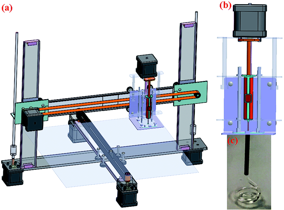 | ||
| Fig. 1 (a) The modified hydrogel 3D printer; (b) on the moving platform, a pump drives the syringe which is covered by a heating pad. (c) The long and blunt needle used to print agar gel. | ||
In order to limit the memory effect and initiate the gelation process during extrusion, a long needle (25 mm) is used. In the long needle, the hydrogel solution travels for a long time in the constricted die. The memory effect of the solution after the extrusion is released due to the long relaxation time as illustrated in Fig. 1(c). In addition, the gelation process initiates in the cold needle. Both of these effects ensure an extruded string with high shape fidelity.
2.5. Printing procedure
After adjusting the Z distance between the substrate and nozzle and injecting the ink into the syringe, the printing begins when the ink is cooled to its printing temperature. The solid structure was printed layer by layer (Fig. 2(a)) according to the G-code to produce a solid structure as shown in Fig. 2(b). The printed structure was further UV cured for 1 h under nitrogen gas protection to polymerize the PAAm network.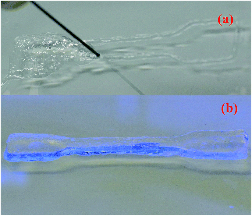 | ||
| Fig. 2 The 3D printing process for a dogbone using DN/A1:1 ink at 45 °C. | ||
3. Results and discussion
3.1. Rheological results
Previous research indicated that the minimum ink viscosity for the extrusion printer is ∼30 mPa s.17 However, higher viscosity with higher surface tension is of benefit to printing structures with precision requirements. Although the viscosity of DN ink is slightly higher than the minimum viscosity requirement, its long gelation temperature range restricts it from printing precise structures. As shown in Fig. 3(a), the complex viscosity of the DN ink increased when the temperature decreased from 90 °C. The peak viscosity appeared at around 60 °C. When the temperature continued to decrease, the viscosity decreased again. In order to have a precisely printed structure, a controllable viscosity increase is preferred because the ink can turn from liquid state to solid state when the temperature decreases slightly. However, for the DN ink, a long gelation temperature range can be found from the viscosity result with a wide viscosity peak from 55 to 70 °C. As it is hard and expensive to precisely control the temperature during printing, the agar gel can be only printed in cold liquid medium for quick gelation according to previous research.31,32 However, printing by the liquid medium method has a fatal disadvantage: low precision. When the agar ink is extruded into the cold medium, the agar forms a gelled string. However, the gelled string is easily twisted due to the medium turbulence which is caused by the movement of the nozzle. This turbulence results in the gelled strings laying down randomly on the bottom of the medium. In this work, a medium free method was used to print agar gel with highly precise control by adding alginate to improve the ink’s rheological characteristics.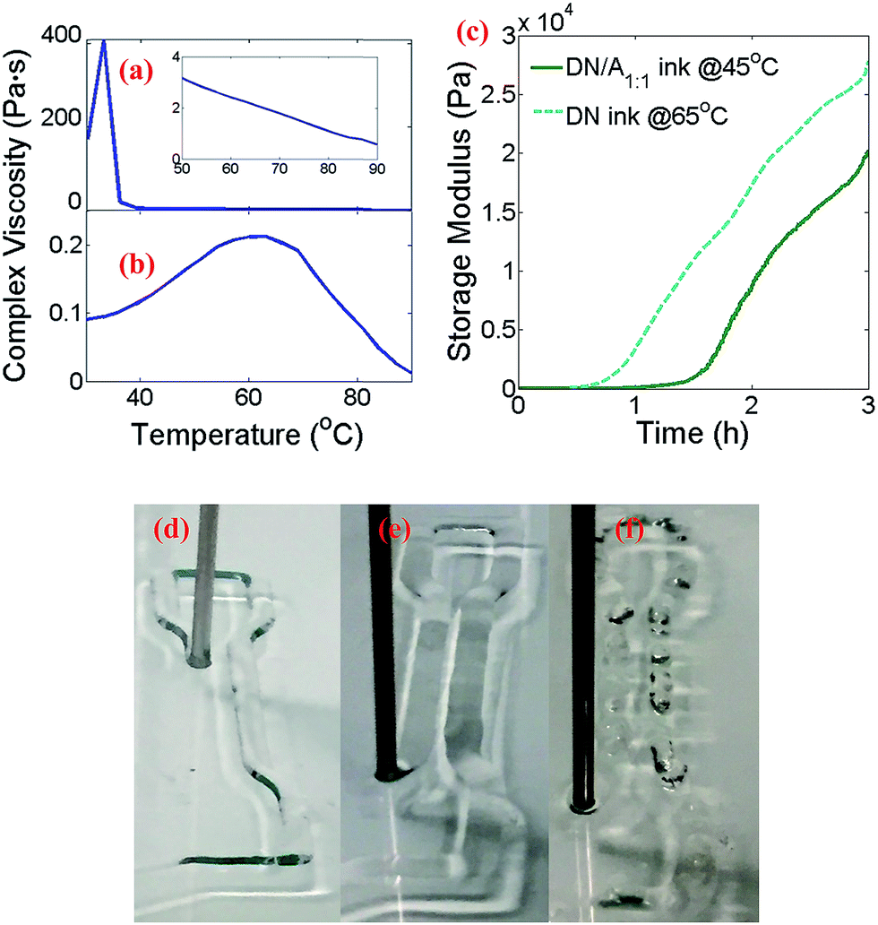 | ||
| Fig. 3 The rheological results of (a) DN ink and (b) DN/A1:1 ink. (c) The printable period of DN ink and DN/A ink. The patterns printed by DN/A1:1 at 45 °C (d), DN ink at 65 °C (e), and DN ink at 45 °C (f). | ||
After adding alginate, the viscosity of the DN/A1:1 ink significantly increased by 2000 times as shown in Fig. 3(b). A sharp viscosity peak around 40 °C is found. The agar forms a semi-gel state at ∼45 °C when its viscosity begins to increase rapidly. In the high viscosity alginate solution, the growth of the agar gel is hard because its chains are trapped within the alginate matrix. The agar is semi-gelled within the alginate matrix when the temperature is lower than ∼60 °C. This solution is feasible to be extruded because the alginate matrix restricts the agar from gelling to the whole body. When extruding, the semi-gelled agar ink further grows as the temperature further drops in the long needle. As the high viscosity of the ink provides high surface tension and agar performs a quick gelation process during printing, this method provides an innovative approach to fabricate precise structures from a thermal sol–gel reversible hydrogel. The printable periods of DN and DN/A1:1 inks at their printing temperatures were further measured by the rheometer as shown in Fig. 3(c). Due to gelation, the printable period of agar ink is limited to its stable period of the storage modulus. The storage modulus vs. time spectra indicated that the rheological characteristics of the DN ink significantly changed at 65 °C only 0.8 h after preparation, while the DN/A1:1 ink can stay as long as 1.4 h at 45 °C. The longer printable period of the DN/A1:1 is probably because the alginate solution restricts the agar gel from growing.
Fig. 3(d–f) also presents the printing process with different inks and conditions. As shown in Fig. 3(d), the pattern printed with DN/A1:1 at 45 °C shows a similar width to the nozzle and a sharp edge. Compared with that, the pattern printed with DN ink at 65 °C shows spreading and sagging which indicates it is not suitable for precise printing. One pattern was printed by DN ink at 45 °C to investigate the state of the agar at low temperature. As shown in Fig. 3(f), the semi-gelled agar was extruded. However, the surrounding low viscosity liquid agar spread and sagged while the gelled agar was randomly spread. In short, the addition of alginate plays a vital role in the precise 3D printing of agar gel.
Fig. 4 presents printed dogbone and meniscus structures from DN and DN/A1:1 inks at 65 °C and 45 °C, respectively. Sharp edges can only be printed with the DN/A1:1 ink. Although the shape can be roughly printed with the DN ink, the edges and the details of the structure are lost which is essential for the fixation of tissues during surgery. This printing methodology for semi-gelled agar within high viscosity alginate solution is proved to be feasible to print structures with a high shape fidelity requirement.
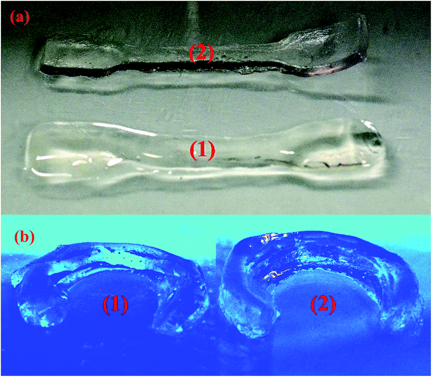 | ||
| Fig. 4 The dogbone (a) and meniscus (b) structures printed by DN (1) and DN/A1:1 (2) inks at 65 °C and 45 °C, respectively. | ||
3.2. Shape fidelity
The shape fidelity of the printed patterns from different inks was estimated by measuring the thickness and width of the dogbone neck. As all the patterns were printed using exactly the same 3D model and the same printing path, the differences in thickness and width represent the difference between the designed and printed patterns.As shown in Table 2, the addition of alginate can significantly improve the shape fidelity. Due to swelling, the printed DN patterns are lower and fatter than the design. Because the addition of alginate improved the rheological characteristics of the ink, the geometry of the TN1:0.5 becomes closer to the design. By further improving the rheological characteristics through increasing the alginate concentration, the printed pattern exhibited excellent shape fidelity which is similar to the designed shape. The slightly larger shape of TN1:1 probably comes from the shape memory of the ink.
| Sample | Thickness (mm) | Width (mm) |
|---|---|---|
| Design | 2.0 | 3.0 |
| DN | 1.705 ± 0.255 | 3.675 ± 0.045 |
| TN1:0.5 | 1.790 ± 0.010 | 3.480 ± 0.010 |
| TN1:1 | 2.097 ± 0.143 | 3.083 ± 0.057 |
3.3. Mechanical properties
Table 3 summarizes the mechanical properties of the printed patterns. Although the tensile strength and toughness increased by adding alginate, their elongation decreased. This phenomenon is the result of the higher crosslinking density produced by physical entanglement of the alginate within the DN gel. After ionic crosslinking of the alginate, the elongation further decreased while the tensile strength significantly improved again.| Sample | E (MPa) | Strength (kPa) | Elongation (%) | Toughness (kJ m−3) |
|---|---|---|---|---|
| DN | 0.07 ± 0.01 | 402.50 ± 76.38 | 1017.11 ± 84.12 | 2.18 ± 0.15 |
| DN/A1:0.5 | 0.44 ± 0.04 | 581.87 ± 118.12 | 922.94 ± 105.28 | 3.66 ± 1.04 |
| TN1:0.5 | 0.55 ± 0.07 | 662.50 ± 67.21 | 224.35 ± 50.04 | 0.95 ± 0.07 |
| DN/A1:1 | 0.81 ± 0.11 | 781.25 ± 53.9 | 691.17 ± 55.1 | 3.86 ± 0.24 |
| TN1:1 | 0.87 ± 0.11 | 1096.20 ± 46.64 | 228.06 ± 42.72 | 1.61 ± 0.15 |
The toughness describes the energy consumption of breaking. As presented in Table 3, the highest toughness of all the samples is achieved by the DN/A1:1 gel. The failure of a material generally involves two sequential processes: initial fracture formation (nucleation) and subsequent fracture propagation (growth). In the DN, DN/A1:0.5 and DN/A1:1 gels, there is no nucleation in the gels because of the agar chain pullout mechanism.33 Upon slow strains to the gels, the aggregated agar helical bundles start to separate from each other while the agar chains unzip and pull out progressively from the agar bundles as well. During this process, the agar network still remains unbroken, but the DN gel becomes soft. This mechanism allows extremely long elongation of the agar/PAA DN gel. While the addition of the alginate chains entangles and restricts the pulling of the agar bundles which concentrates the stresses, the DN/A gel tends to be broken at smaller elongation, but large elongation can still be achieved by DN/A because the pull out mechanism of the agar gel restricts nucleation, as shown in Fig. 5(c). TN1:0.5 and TN1:1, on the other hand, performed a quite different breaking process. As the alginate network is ionic crosslinked, the stress is concentrated on the connected alginate chains and consequently causes the chains to break preferentially. Although the agar network was still pulled out as shown in Fig. 5(d), the fracture grows and causes the hydrogel to break at low elongation. Due to the different breaking mechanisms, DN/A1:1 gel achieved the highest toughness at 3.86 kJ m−3 while the TN1:1 gel presented the highest tensile strength at 1.096 MPa.
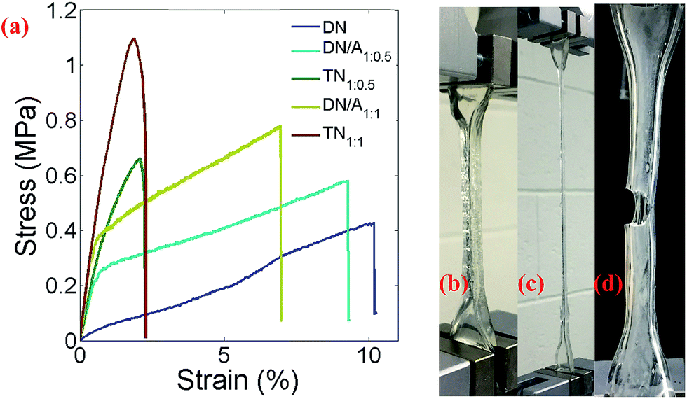 | ||
| Fig. 5 Tensile testing results for the printed hydrogels (a) and the tensile experiments of the DN/A1:1 hydrogel (b and c) and TN1:1 gel (d). | ||
Table 4 shows a comparison of the tensile properties of the 3D printed tough hydrogel in previous work with our work and natural bovine knee joint cartilage. As expected, the agar DN gel shows better mechanical properties than the ICE hydrogels.34 Moreover, the triple network hydrogel (agar/PAAm/crosslinked alginate) in this work possesses the highest tensile strength ever achieved by a printed hydrogel and the double network hydrogel (agar/PAAm) with uncrosslinked alginate chains balanced the strength and elongation to achieve extremely high toughness. The printed agar gel with a crosslinked alginate network shows comparable mechanical performance to the natural cartilage. These results indicate that this printable, precisely controllable, and super tough hydrogel is feasible for use as a cartilage substitute and can be used for future printable organs which require strong mechanical performance.
| Material | Strength/kPa | Elongation/% | Toughness/kJ m−3 | Ref. |
|---|---|---|---|---|
| PAAm/alginate | 140 | 220 | 154 | 27 |
| κ-Carrageenan/poly(oxyalkylene amine) | 600 | 350 | 1400 | 28 |
| Alginate/gelatin | 840 | 55 | 35 | |
| poly(ethylene glycol) diacrylate | ∼34 | 50 | 16 | |
| Agar/PAAm | 402.50 | 1017.11 | 2180 | This work |
| Agar/PAAm/crosslinked alginate | 1096.20 | 228.06 | 1610 | This work |
| Agar/PAAm/uncrosslinked alginate | 781.25 | 691.17 | 3860 | This work |
| Bovine knee joint cartilage1 | 530–9000 | 40–164 | 36 |
4. Conclusion
This work provides a feasible method for printing a super tough agar/PAAm DN gel with enhanced mechanical performance for precise structures. Due to the high ink viscosity after adding alginate, the agar can be extruded in the semi-gelled state which ensured its quick gelling after extrusion. Meanwhile, the entanglement of the alginate toughens the DN gel and makes it reach extremely high mechanical performance. Ionic crosslinking of the alginate has also been used to further toughen the hydrogel. However, stress concentration onto the alginate network induces the failure of the hydrogel at low elongation which decreases its toughness. This work not only provides a feasible method for achieving high precision of thermo-reversible hydrogels by precise control of the printing temperature, but also presents an easy approach to balance the toughness and tensile strength of a tough hydrogel for suitable applications. As the hydrogel printed by this method has high shape fidelity, extremely high toughness, and high strength, it fulfills the requirements of load-bearing tissue substitutes.Acknowledgements
The authors would like to acknowledge the support from NSF grant (number 1228127).References
- J. Xu, The Praeger Handbook of Acupuncture for Pain Management: A Guide to How the "Magic Needles" Work, Praeger, 2014 Search PubMed
.
- F. R. Noyes, T. P. Heckmann and S. D. Barber-Westin, J. Orthop. Sports Phys. Ther., 2012, 42, 274–290 CrossRef PubMed
- I. D. Hutchinson, C. J. Moran, H. G. Potter, R. F. Warren and S. A. Rodeo, Am. J. Sports Med., 2014, 42, 987–998 CrossRef PubMed
- R. Allaire, M. Muriuki, L. Gilbertson and C. D. Harner, J. Bone Jt. Surg., Am. Vol., 2008, 90, 1922–1931 CrossRef PubMed
- C. J. Moran, S. Atmaca, H. A. Declercq, M. J. Cornelissen and P. C. Verdonk, Int. Orthop., 2014, 38, 1937–1944 CrossRef PubMed
- R. Verdonk, K. F. Almqvist, W. Huysse and P. C. Verdonk, Sports Med. Arthrosc., 2007, 15, 121–125 CrossRef PubMed
- F. McCormick, J. D. Harris, G. D. Abrams, K. E. Hussey, H. Wilson, R. Frank, A. K. Gupta, B. R. Bach and B. J. Cole, Am. J. Sports Med., 2014, 42, 892–897 CrossRef PubMed
- P. Beaufils and R. Verdonk, The Meniscus, Springer-Verlag, Berlin Heidelberg, 2010 Search PubMed
- A. Skardal, D. Mack, E. Kapetanovic, A. Atala, J. D. Jackson, J. Yoo and S. Soker, Stem Cells Transl. Med., 2012, 1, 792–802 CrossRef CAS PubMed
- S. Michael, H. Sorg, C. T. Peck, L. Koch, A. Deiwick, B. Chichkov, P. M. Vogt and K. Reimers, PLoS One, 2013, 8, 57741 Search PubMed
- C. Norotte, F. S. Marga, L. E. Niklason and G. Forgacs, Biomaterials, 2009, 30, 5910–5917 CrossRef CAS PubMed
- M. Kamei, W. B. Saunders, K. J. Bayless, L. Dye, G. E. Davis and B. M. Weinstein, Nature, 2006, 442, 453–456 CrossRef CAS PubMed
- D. A. Zopf, S. J. Hollister, M. E. Nelson, R. G. Ohye and G. E. Green, N. Engl. J. Med., 2013, 368, 2043–2045 CrossRef CAS PubMed
- C. W. Hull, US Pat., US4575330, 1986
- K. Pataky, T. Braschler, A. Negro, P. Renaud, M. P. Lutolf and J. Brugger, Adv. Mater., 2012, 24, 391–396 CrossRef CAS PubMed
- L. A. Hockaday, K. H. Kang, N. W. Colangelo, P. Y. C. Cheung, B. Duan, E. Malone, J. Wu, L. N. Girardi, L. J. Bonassar, H. Lipson, C. C. Chu and J. T. Butcher, Biofabrication, 2012, 4, 35005 CrossRef CAS PubMed
- S. V. Murphy and A. Atala, Nat. Biotechnol., 2014, 32, 773–785 CrossRef CAS PubMed
- B. Shaffer, S. Kennedy, J. Klimkiewicz and L. Yao, Am. J. Sports Med., 2000, 28, 524–533 CAS
- D. L. Cohen, E. Malone, H. Lipson and L. J. Bonassar, Tissue Eng., 2006, 12, 1325–1335 CrossRef CAS PubMed
- J. J. Ballyns, D. L. Cohen, E. Malone, S. A. Maher, H. G. Potter, T. Wright, H. Lipson and L. J. Bonassar, Tissue Eng., Part C, 2010, 16, 693–703 CrossRef CAS PubMed
- J. Y. Sun, X. H. Zhao, W. R. K. Illeperuma, O. Chaudhuri, K. H. Oh, D. J. Mooney, J. J. Vlassak and Z. G. Suo, Nature, 2012, 489, 133–136 CrossRef CAS PubMed
- C. W. Peak, J. J. Wilker and G. Schmidt, Colloid Polym. Sci., 2013, 291, 2031–2047 CAS
- T. Nakajima, T. Kurokawa, H. Furukawa, Q. M. Yu, Y. Tanaka, Y. Osada and J. P. Gong, Chin. J. Polym. Sci., 2009, 27, 1–9 CrossRef CAS
- J. Wei, J. Wang, S. Su, M. Hasan, J. Qiu and S. Wang, New J. Chem., 2015 10.1039/c1035nj01250c
- J. L. Wang, J. H. Wei, S. H. Su, J. J. Qiu and S. R. Wang, J. Mater. Sci., 2015, 50, 5458–5465 CrossRef CAS
- J. Wei, J. Wang, S. Su, S. Wang and J. Qiu, J. Mater. Chem. B, 2015, 3, 5284–5290 RSC
- S. E. Bakarich, M. I. H. Panhuis, S. Beirne, G. G. Wallace and G. M. Spinks, J. Mater. Chem. B, 2013, 1, 4939–4946 RSC
- S. E. Bakarich, P. Balding, R. Gorkin, G. M. Spinks and M. I. H. Panhuis, RSC Adv., 2014, 4, 38088–38092 RSC
- S. E. Bakarich, R. Gorkin, M. I. H. Panhuis and G. M. Spinks, ACS Appl. Mater. Interfaces, 2014, 6, 15998–16006 CAS
- Q. Chen, L. Zhu, C. Zhao, Q. M. Wang and J. Zheng, Adv. Mater., 2013, 25, 4171–4176 CrossRef CAS PubMed
- R. Landers, A. Pfister, U. Hubner, H. John, R. Schmelzeisen and R. Mulhaupt, J. Mater. Sci., 2002, 37, 3107–3116 CrossRef CAS
- W. M. Kühtreiber, R. P. Lanza and W. L. Chick, Cell Encapsulation Technology and Therapeutics, Birkhäuser, 1999 Search PubMed
- Q. Chen, L. Zhu, L. Huang, H. Chen, K. Xu, Y. Tan, P. Wang and J. Zheng, Macromolecules, 2014, 47, 2140–2148 CrossRef CAS
- S. Naficy, H. R. Brown, J. M. Razal, G. M. Spinks and P. G. Whitten, Aust. J. Chem., 2011, 64, 1007–1025 CrossRef CAS
- B. Duan, L. A. Hockaday, K. H. Kang and J. T. Butcher, J. Biomed. Mater. Res., Part A, 2013, 101, 1255–1264 CrossRef PubMed
- A. K. Williamson, A. C. Chen, K. Masuda, E. J. M. A. Thonar and R. L. Sah, J. Orthop. Res., 2003, 21, 872–880 CrossRef CAS
| This journal is © The Royal Society of Chemistry 2015 |