Ye Won Choi,
Jae Jun Lee,
Ga Rim You,
Sun Young Lee and
Cheal Kim*
Department of Fine Chemistry and Department of Interdisciplinary Bio IT Materials, Seoul National University of Science and Technology, Seoul 139-743, Korea. E-mail: chealkim@seoultech.ac.kr; Fax: +82-2-973-9149; Tel: +82-2-970-6693
First published on 8th October 2015
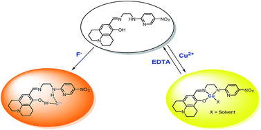
A bi-functional colorimetric chemosensor 1, based on julolidine moiety and N-(2-aminoethyl)-5-nitropyridin-2-amine, has been synthesized and characterized. The sensor 1 has proven to be highly selective and sensitive to Cu2+ with a color change from colorless to yellow in aqueous solution. The sensing mechanism of 1 for Cu2+ was proposed to be the ligand-to-metal charge-transfer (LMCT), which was explained by theoretical calculations. It was also found that the 1–Cu2+ complex could be recycled simply through treatment with an appropriate reagent such as EDTA. Importantly, the sensor 1 could be used to detect and quantify Cu2+ in water samples. Moreover, 1 showed a selective colorimetric response toward fluoride due to the increase in the intramolecular charge transfer (ICT) band by a deprotonation process without any inhibition from other anions such as CH3COO− and CN−.
Among various important anionic analytes, fluoride (F−) is of particular interest owing to its established role in dental care and clinical treatment for osteoporosis.5 However, an acute intake of a large dose or chronic ingestion of lower doses of fluoride can cause many serious disease such as gastric and kidney disorders, dental and skeletal fluorosis, and urolithiasis in humans, and even death.6 Also, they contribute significantly to environmental pollution.7 For these reasons, the improved chemosensor for the detection and sensing of F− with high selectivity is of current interest. However, many anion sensors are not capable of distinguishing fluoride effectively from anions such as CH3COO− and CN−, because they possess similar basicity to F− and easily form hydrogen bonds.8 Therefore, it is highly desirable to develop selective methods capable of discriminating fluoride, especially from competing anions (CH3COO− and CN−).
Up to now, various methods for detecting cations and anions have been developed in a variety of complex environments, such as chromatographic, fluorometric and colorimetric chemosensor, flow injection and electrochemical analysis.9 Among them, colorimetric chemosensors are the most promising in sensor field as it does not need any expensive instrumentation.10 The analyte determination can be carried out by the naked eye under visible light. Therefore, it is of considerable importance to develop probes with colorimetric sensing ability for detection of both Cu2+ and F−.
Julolidine moiety has been well known as a chromophore and chemosensors with the julolidine moiety often showed a good colorimetric response with target analytes.11 On the other hand, the presence of the electron-withdrawing nitro group on the amino-pyridyl moiety would increase not only the degree of π-conjugation but also the hydrogen bonding ability of the amino NH proton.12 Therefore, we designed and synthesized a new chemosensor 1 based on the combination of the julolidine and nitropyridine moieties, and tested its sensing properties towards various metal ions and anions.
Herein, we report a new bi-functional chemosensor 1 for Cu2+ and F−, which was synthesized in one step by condensation reaction of 8-hydroxyl-julolidine-9-carboxaldehyde and N-(2-aminoethyl)-5-nitropyridin-2-amine (Scheme 1). The sensor 1 can detect Cu2+ by color change from colorless to yellow in aqueous solution. Additionally, 1 showed a distinctly red-shifted absorption spectrum with the intense color change in the presence of F−.
:
3; v/v, 10 mM bis–tris, pH 7.0). As shown in Fig. 1, the absorption spectrum of free 1 showed a maximum peak centered at 382 nm. The addition of 12 equiv. of Cu2+ into the 1 solution resulted immediately in a significant enhancement of absorbance at 450 nm with the color change from colorless to yellow (Fig. 1b). Under the identical condition, there were no appreciable changes of the absorption in the presence of Al3+, Zn2+, Cd2+, Mg2+, Cr3+, Co2+, Ni2+, Na+, K+, Ca2+, Mn2+, Pb2+, Fe2+ and Fe3+. Hg2+, Ag+, and Au3+ produced precipitate due to their insolubility in acetonitrile/buffer solution (7
:
3). The results demonstrated that 1 was characteristic of high selectivity toward Cu2+ over other metal ions.
An UV-vis titration experiment was performed to investigate the interaction of Cu2+ and 1 (Fig. 2). Upon the gradual addition of Cu2+ to a solution of 1, the absorbance peak at 382 nm steadily decreased and two new absorption bands at 275 and 450 nm appeared concomitantly, resulting in a color change from colorless to yellow. The isosbestic point at 350 nm was clearly observed, indicating the formation of a single species between 1 and Cu2+. Moreover, the new peak at 450 nm in the visible region with molar extinction coefficient in the thousands, 7.32 × 10³ M−1 cm−1, is too large to be Cu-based d–d transitions and thus must be ligand-based transitions.13 Therefore, we proposed that the new peak at 450 nm might be attributed to a ligand-to-metal charge-transfer (LMCT).
![]() | ||
Fig. 2 Absorption spectral changes of 1 (20 μM) after addition of increasing amounts of Cu2+ in acetonitrile–water (7 : 3, v/v). Inset: absorption at 450 nm versus the number of equiv. of Cu2+ added. | ||
A Job plot analysis showed a 1
:
1 stoichiometry for the 1–Cu2+ complex (Fig. S1†), which was further confirmed by ESI-mass spectrometry analysis (Fig. S2†). The positive ion mass spectrum of ESI-mass indicated that a peak at m/z = 520.73 was assignable to [1(–H+) + Cu2+ + DMSO]+ [cald, m/z = 521.12]. Based on Job plot, ESI-mass spectrometry and the crystal structures of similar type of Cu complexes reported in the literature,14 we proposed the structure of 1
:
1 complex of 1 and Cu2+ as shown in Scheme 1.
From UV-vis titration data, the association constant for 1–Cu2+ complex was determined to be 1.5 × 10³ M−1 using Benesi–Hildebrand equation15 (Fig. S3†). This value was within the range of those (103 to 1012) reported for Cu2+ sensing chemosensors.16 The detection limit (3σ/K)17 of receptor 1 for the analysis of Cu2+ was calculated to be 23.5 μM (Fig. S4†). The detection limit of 1 for Cu2+ is lower than that (31.5 μM) recommended by World Health Organization (WHO) in drinking water.4 Therefore, 1 could be a good indicator for the detection of copper ions in drinking water.
To explore the anti-disturbance of 1 as the Cu2+-selective sensor, competition experiments were performed (Fig. 3). For this purpose, 1 was treated with 12 equiv. of Cu2+ in the presence of the same concentration of other metal ions. The absorbance at 450 nm caused by Cu2+ was retained with Zn2+, Cd2+, Mg2+, Cr3+, Co2+, Ni2+, Na+, K+, Ca2+, Mn2+, Fe3+ and Pb2+. Instead, the absorbance of 1–Cu2+ complex in the presence of Al3+ and Fe2+ was relatively low. In order to overcome the inhibitions, we have added iodide to 1–Cu2+–Al3+ solution and fluoride to 1–Cu2+–Fe2+ one. The resulting absorbance of 1–Cu2+ complex in the presence of Al3+ and Fe2+ were recovered to 89% and 84%, respectively (Fig. S5†). Thus 1 can be used as a selective colorimetric sensor for Cu2+ detection in the presence of most competing metal ions.
To study the practical applicability, the colorimetric responses of 1 in the absence and presence of Cu2+ in different pH values were evaluated. As shown in Fig. 4, the receptor 1 itself was stable from pH 2 to 12. Upon the addition of Cu2+, there was an apparent increase of absorbance (450 nm) at the pH range of 7–12. These results indicated that Cu2+ could be clearly detected by the naked eye or UV-vis absorption measurements using 1 over the wide pH range.
We subsequently studied the binding reversibility of Cu2+ to 1 in acetonitrile–water solution. Due to the high stability constant for the EDTA–Cu2+ complex, it was expected that EDTA would chelate Cu2+ from the 1–Cu2+ complex, liberating 1. As shown in Fig. 5, the absorbance of 1–Cu2+ was quenched and the color changed from yellow to colorless upon the addition of 12 equiv. EDTA. Introduction of an additional Cu2+ resulted in the recovery of absorbance and color, indicating that the binding of 1 with Cu2+ is chemically reversible. The absorbance (Fig. 5a) and color changes (Fig. 5b) were almost reversible even after several cycles with the sequentially alternative addition of Cu2+ and EDTA.
![]() | ||
| Fig. 5 (a) Absorbance of 1 (20 μM) after the sequential addition of Cu2+ and EDTA. (b) The color changes of 1 (20 μM) after the sequential addition of Cu2+ and EDTA. | ||
In order to study the applicability of the sensor 1 in environmental water samples, we constructed a calibration curve for the determination of Cu2+ by 1 (Fig. S6†). It exhibited a good linear relationship between the absorbance of 1 and Cu2+ concentrations (0–10 μM) with a correlation coefficient of R2 = 0.9912 (n = 3), which means that 1 could be suitable for the quantitative detection of Cu2+. Then, the sensor was applied for the determination of Cu2+ in tap water samples. The results were summarized in Table 1, which exhibited a satisfactory recovery and R.S.D. values for the tap water samples.
| Sample | Cu(II) added (μmol L−1) | Cu(II) found (μmol L−1) | Recovery (%) | R.S.D (n = 3) (%) |
|---|---|---|---|---|
a Conditions: [1] = 10 μmol L−1, water–acetonitrile (3 : 7, v/v) at pH 7.0 buffered by 10 mmol L−1 bis–tris. |
||||
| Tap water | 0.00 | 0.23 | 0.06 | |
| 6.00 | 6.24 | 100.2 | 0.01 | |
To understand the sensing mechanisms of 1 toward Cu2+, theoretical calculations were performed with the 1
:
1 stoichiometry, based on Job plot and ESI-mass spectrometry analysis. To get the energy-minimized structures of 1 and 1–Cu2+ complex, the geometric optimizations were performed by DFT/B3LYP level. Especially, 1–Cu2+ complex was considered by a paramagnetic character (S = 1/2, DFT/uB3LYP/main group atom: 6-31G** and Cu: Lanl2DZ/ECP). The significant structural properties of the energy-minimized structures were shown in Fig. 6. The energy minimized structure of 1 showed a V-type structure with the dihedral angle of 1N, 2N, 3C, 4O = 37.664°, and the hydrogen bond was observed between 2N and 6H (Fig. 6a). 1–Cu2+ complex exhibited a square planar structure (dihedral angle (1N, 2N, 3C, 4O) = 8.738°) with the coordination of N, N and O atoms of 1 (Fig. 6b).
We also investigated the absorption to the singlet excited states of 1 and 1–Cu2+ species via TDDFT calculations. In case of 1, the main molecular orbital (MO) contribution of the first lowest excited state was determined for HOMO−2 → LUMO and HOMO → LUMO+1 transitions (341.34 nm, Fig. S7†), which indicated ICT bands. 1–Cu2+ complex showed that the excited states of 4th, 9th and 12th (497.47, 403.68 and 377.69 nm) were relevant to the color change (colorless to yellow) with predominant ICT and LMCT characters (Fig. S8–S10†). Thus, the chelation of Cu2+ with 1 mainly showed the ICT and LMCT, which induced the different color change of 1 in the presence of Cu2+.
![]() | ||
| Fig. 7 (a) UV-vis spectral changes of 1 (20 μM) upon addition of 30 equiv. of various anions in DMSO. (b) Colorimetric changes of 1 (20 μM) upon the addition of various anions (30 equiv.). | ||
The interaction of receptor 1 with F− was studied in detail by UV-vis spectroscopic titration, as shown in Fig. 8. On gradual addition of F−, the band at 350 nm decreased, and a new band at 465 nm increased drastically. The absorbance reached a maximum at 30 equiv. of fluoride with a clear isosbestic point observed at 406 nm, indicating the formation of the only one species between 1 and F−. The color change of 1 from colorless to orange might be due to the deprotonation of phenol of 1 by F− (Scheme 1). To further confirm the sensing mechanism between 1 and F−, the interaction between 1 and OH− was also investigated (Fig. S11†). UV-vis spectral change of 1 upon addition of OH− was nearly identical to that of 1 obtained from the addition of F−, which indicated the deprotonation between 1 and F−. The resulting negative charge on the phenol is introduced in the receptor which causes intramolecular charge transfer (ICT) between the electron deficient –NO2 group and the electron rich –O− to show the UV-vis and colorimetric changes.18
![]() | ||
| Fig. 8 Absorption spectral changes of 1 (20 μM) after addition of increasing amounts of F− in DMSO. Inset: absorption at 465 nm versus the number of equiv of F− added. | ||
The Job plot for the binding between 1 and F− exhibited a 1
:
1 stoichiometry (Fig. S12†), which was further supported by ESI-mass spectrometry analysis (Fig. S13†). The negative ion mass spectrum of ESI-mass indicated that a peak at m/z = 380.67 was assignable to [1–H+]− [calcd, 380.17], which is corresponding to the receptor 1 deprotonated by fluoride. From the UV-vis titration data, the association constant for 1 with F− was determined as 5.9 × 10² M−1 using the Benesi–Hildebrand equation15 (Fig. S14†). The detection limit (3σ/K)17 of receptor 1 for the analysis of F− was calculated to be 19.4 μM (Fig. S15†).
To study the interaction between 1 and F−, the 1H NMR titrations of 1 were measured with different amounts of F− (Fig. 9). In the absence of F−, the phenolic OH proton (H12) and the amine NH proton (H8) appeared as a singlet at 13.56 and 8.25 ppm, respectively. Upon the gradual addition of F−, the singlets of H12 and H8 were broadened and finally disappeared due to hydrogen bonding with F−. Simultaneously, the H6,7 protons and the protons of nitropyridine moiety (H9–11) were shifted upfield. These results suggested that the –NH and –OH protons might hydrogen bond to F−, and eventually undergo the deprotonation (Scheme 1).
To test whether 1 can detect F− selectively even in the presence of other anions, competitive experiment was conducted (Fig. 10). 1 was treated with 30 equiv. of F− in the presence of several anions. The results indicated that the competitive anions did not lead to any significant spectral (Fig. 10a) and color change (Fig. 10b), and that F− ions still resulted in the similar absorbance and color in the presence of competitive anions. Thus, 1 could be used as a selective chemosensor for F− by the colorimetric mode.
:
3, acetonitrile/bis–tris buffer v/v; 10 mM bis–tris, pH 7.0). The spectra were recorded a minute after the solution was completely mixed.
For F−, the UV-vis titration experiment of 1 was carried out by adding aliquots of 6–60 μL of tetraethyl ammonium fluoride (TEAF) solution (100 mM) to 3 mL of 1 solution (20 μM in DMSO). The spectra were recorded a minute after the solution was completely mixed.
:
3; v/v). Cu2+ solution (10 mM) was prepared by dissolving nitrate salts in acetonitrile/bis–tris buffer (7
:
3; v/v, 1 mL). 30, 27, 24, 21, 18, 15, 12, 9, 6 and 3 μL of the 1 solution were taken and transferred to vials. Each vial was diluted with acetonitrile/bis–tris buffer (7
:
3; v/v) to make a total volume of 2.97 mL. Then, 0, 3, 6, 9, 12, 15, 18, 21, 24, 27 and 30 μL of the Cu2+ solution were added to each diluted 1 solution. Each vial had a total volume of 3 mL. After shaking the vials for a minute, UV-vis spectra were taken at room temperature.
For F−, stock solution of receptor 1 (10 mM) was prepared in 1 mL of DMSO. F− solution (10 mM) was prepared by dissolving TEAF salt in DMSO (1 mL). 15, 13.5, 12, 10.5, 9, 7.5, 6, 4.5, 3 and 1.5 μL of the 1 solution were taken and transferred to vials. Each vial was diluted with DMSO to make a total volume of 2.985 mL. Then, 0, 1.5, 3, 4.5, 6, 7.5, 9, 10.5, 12, 13.5 and 15 μL of the F− solution were added to each diluted 1 solution. Each vial had a total volume of 3 mL. After shaking the vials for a minute, UV-vis spectra were taken at room temperature.
:
3; v/v). Solution samples (20 mM) of metal ions were prepared by dissolving the corresponding salts in a mixture of acetonitrile/bis–tris buffer (7
:
3; v/v, 1 mL). 36 μL aliquot of each metal-ion stock solution and 36 μL of Cu2+ solution were diluted into a 3 mL of mixture of acetonitrile/bis–tris buffer (7
:
3; v/v). Then, 12 μL of the 1 solution was added into the mixed solution to make 20 μM. After shaking the vials for a minute, UV-vis spectra were taken at room temperature.
For F−, stock solution of receptor 1 (5 mM) was prepared in 1 mL of DMSO. Solution samples (20 mM) of anions were prepared by dissolving the corresponding salts in DMSO (1 mL). 48 μL aliquot of each anion stock solution and 48 μL of F− solution were diluted into a 3 mL of DMSO. Then, 12 μL of the 1 solution was added into the mixed solution to make 20 μM. After shaking the vials for a minute, UV-vis spectra were taken at room temperature.
Footnote |
| † Electronic supplementary information (ESI) available. See DOI: 10.1039/c5ra16301c |
| This journal is © The Royal Society of Chemistry 2015 |