Mechanochemical synthesis of an efficient nanocrystalline BaFBr:Eu2+ X-ray storage phosphor

Xianglei Wang and Hans Riesen*
School of Physical, Environmental and Mathematical Sciences, The University of New South Wales, Canberra, ACT 2600, Australia. E-mail: h.riesen@adfa.edu.au

Received 11th August 2015 , Accepted 2nd October 2015

First published on 2nd October 2015


Abstract

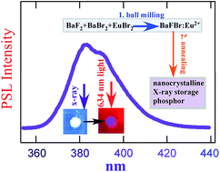
A straightforward and direct preparation route for nanocrystalline BaFBr:Eu2+ by ball-milling is reported. We demonstrate that the as-prepared nanocrystalline powder becomes an efficient photostimulable X-ray storage phosphor upon annealing at temperatures in the range of 400 to 900 °C. The dependence of crystallite size, photoluminescence and storage phosphor properties on annealing temperature and annealing time was investigated and the optimal conditions were determined. After annealing at 900 °C for 1 h, the average crystallite size was found to be ∼200 nm. In comparison with the typical ∼5 μm particles for conventional phosphors, the shape and size distribution of the crystallites may allow higher packing densities, resulting in higher sensitivities, and resolution in computed radiography. The photostimulable storage properties compare favorably with results obtained for a commercial BaFBr(I):Eu2+ imaging plate.

1 Introduction

BaFBr1−xIx (x = 0 to 0.05) doped with divalent europium has been the most widely used and successful X-ray storage phosphor in computed radiography to date and was investigated by the Fuji Film Company as early as the 1980s.1–3 The readout mechanism of this storage phosphor employs photostimulated luminescence, which has been extensively reviewed in the literature.4–7 The storage mechanism is based on the generation of electron–hole pairs upon x-irradiation with subsequent trapping at two centres. In BaFBr:Eu2+, F+-centres (halide vacancies) act as electron traps upon x-irradiation and the electrons can be released by subsequent photostimulation from these traps. There are two types of F-centres: F(Br) and F(F), but there appears to be some disagreement whether both types are involved.4,5 There has also been some controversy about the hole trap centres and their role in the photostimulation process. Takahashi et al.2 considered Eu2+ as the hole trap and reported a mechanism that results in the ionization of Eu2+ to Eu3+. Upon subsequent photostimulation Eu3+ centres are then thought to trap the electrons released from the F-centres, yielding excited Eu2+ activators that relax to the ground state and emit the broad band 4f65d–4f7 luminescence around 390 nm. However, other researchers reported that no Eu3+ can be observed in their experiments.8–10 For example, von Seggern et al.4 and Meijerink et al.9 could not observe any reduction or increase of Eu2+ or Eu3+ luminescence, respectively, as a function of x-irradiation. Based on EPR measurements Hangleiter et al.8 proposed a mechanism that involves a spatial correlation of isolated F centres and hole centres. In particular, it was suggested that aggregates of Eu2+ with F centres, aggregates of Eu2+ with hole centres, and a low concentration of aggregates of Eu2+ with both F and hole centres were formed upon X-ray irradiation. It was also found that O2− ions, an ubiquitous impurity in BaFX (X = Cl, Br) compounds, can act as hole trap centres.11,12 Notwithstanding the vast number of investigations into BaFBr:Eu2+ as an X-ray storage phosphor, some details of the mechanism for photostimulated luminescence are still not fully understood and warrant further investigations.

BaFBr(I):Eu2+ was initially, and typically still is, synthesized by high temperature sintering of stoichiometric mixtures of the solid reagents under an inert atmosphere.1–3 Various methods have been reported in the literature thereafter, such as non-stoichiometric sintering13–15 and co-precipitation of water based solutions.16 Recently, von Seggern et al.17 introduced a single step synthesis method by annealing BaCO3 and ammonium halides with trace amounts of EuF3 in a reducing atmosphere. They investigated the correlation between the intensity of photoluminescence and photostimulated luminescence with the annealing temperature and reported that the optimal particle size for microcrystalline BaFBr:Eu2+ is about 5 μm after annealing at 800 °C.

Recently, a one-step ball-milling method was reported for preparing nanocrystalline BaFCl:Sm3+,18 BaFCl:Sm2+,19 and Ba0.5Sr0.5FCl0.5Br0.5:Sm3+.20 In the present paper, a similar method was applied for the preparation of nanocrystalline BaFBr:Eu2+ and the as-prepared sample was annealed at temperatures up to 900 °C to investigate its properties as a potentially improved X-ray storage phosphor.

2 Experimental

Reagents obtained from Sigma-Aldrich Pty Ltd were used without further purification. First, 0.877 g (5 mmol) barium fluoride (BaF2, 99.99% trace metals basis) was ground and dried under vacuum at 400 °C for 3 h. Then 1.486 g (5 mmol) anhydrous barium bromide (BaBr2, beads, 99.999% trace metals basis, from ampoule), 0.048 mmol europium bromide (EuBr2, 99.99% trace metals basis; 1 weight% of BaBr2) and the vacuum dried BaF2 were mixed and ground in a nitrogen purged glove box to avoid oxidation of the Eu2+ ions. The mixture was subsequently placed into a 10 mL zirconia jar with two 12 mm diameter zirconia balls under nitrogen and the jar was sealed with parafilm. The solid reagents were then ball-milled on a Retsch Mixer Mill 200 at a frequency of 20 Hz for 120 min to yield nanocrystalline BaFBr:Eu2+ as a white powder. Subsequently, the as-prepared nanocrystalline BaFBr:Eu2+ was annealed in separate batches under vacuum for 1 h at temperatures between 150 °C and 900 °C; it is noted here that the latter is just ∼80 °C below the melting temperature of BaFBr.21 In a second experiment, separate batches of the sample were also annealed under vacuum at 900 °C for different time periods between 1 to 6 h.

From an analysis by employing an energy dispersive X-ray fluorescence spectrometer (Shimadzu EDX-800HS), we found that the samples prepared by the ball-milling method were only very slightly contaminated by zirconia (from the jar and balls) at an insignificant level of <0.03 weight%.

The powder X-ray diffraction (XRD) patterns of the samples were measured using a Rigaku MiniFlex 600 benchtop diffractometer operating at 40 kV and 15 mA with CuKα radiation (λ = 0.154 nm). All data were collected at room temperature in a step-scanning mode with a speed of 0.75° min−1 and a 2-theta range from 10° to 90°. The measured XRD patterns were compared with the standard reference data ICDD card 24-90.

Scanning electron microscopy (SEM) was conducted on a JEOL 7001 Field Emission Scanning Electron Microscope. Each samples was dispersed in acetone in an ultrasonic bath and then dropped onto a silicon wafer. The samples were all coated with 10 nm of Pt to make the samples conductive.

Photoluminescence (PL) spectra were measured on a Horiba Jobin-Yvon Spex Fluoromax-3 fluorometer. The excitation wavelength was set to 270 nm, and the spectra were collected in single scans with 0.5 nm steps and 0.5 s integration time.

The 270 nm wavelength is well within a main absorption feature of Eu2+ in the BaFBr host and also efficiently excites Eu3+ via an O2−–Eu3+ charge transfer transition. It is safe to assume that any Eu3+ is due to the unintentional contamination of the system with oxygen i.e. oxide defects.

The photostimulated luminescence (PSL) spectra were accumulated on a Spex 500M monochromator equipped with an Andor iDus Model DV401A-BV CCD camera and a Thorlabs FES0500 short pass interference filter was used at the entrance slit. In a typical experiment, the sample was firstly x-irradiated by a Sirona HELIODENT Plus dental system with an exposure time of 3.2 s corresponding to about ∼30 mGy entry surface dose. The x-irradiated sample was then immediately stimulated for 10 s by a red LED (634 nm; ∼5 mW cm−2) to measure its photostimulated luminescence spectrum. The photostimulated luminescence intensities of the as-prepared and annealed samples were compared with results obtained for a commercial dental imaging plate (Duerr Dental), which was x-irradiated using the same dental X-ray source with an exposure time of 0.32 s (∼3 mGy surface dose). A ten times higher dose was chosen for our samples in order to allow monitoring the weak photostimulated luminescence after annealing at temperatures <400 °C.

3 Results and discussion

The powder X-ray diffraction patterns of the as-prepared and annealed nanocrystalline BaFBr:Eu2+ phosphors are shown in Fig. 1. Prominent peaks in all the patterns could be indexed to the pure PbFCl tetragonal matlockite structure with space group P4/nmm.22 The XRD patterns confirm the purity of the BaFBr phase in the as-prepared and annealed samples. Prominent diffraction peaks become narrower and more intense with higher annealing temperature. From a Williamson–Hall analysis23 of the prominent peaks, and using a Gaussian line shape approximation, the average crystallite size of the as-prepared sample was estimated to be around 30 nm. This is good agreement with the average particle size as observed in the SEM image shown in Fig. 2a. As is seen in Fig. 1, the (002) peak shows a significantly increased intensity compared to the ICDD pattern. This is likely due to preferential orientation of the particles/crystallites with increasing size on the XRD holder.
image file: c5ra16107j-f1.tif
Fig. 1 Powder X-ray diffraction patterns of (a) the standard BaFBr data (ICDD card no. 24-90) and (b) the as-prepared nanocrystalline BaFBr:Eu2+, (c) after annealing for 1 h at 300 °C, (d) 600 °C and (e) at 900 °C for 1 h, (f) 3 h and (g) 6 h. Miller-indices are indicated for the prominent peaks.

image file: c5ra16107j-f2.tif
Fig. 2 SEM images of BaFBr:Eu2+ as-prepared (a), after subsequent annealing for 1 h (b) and 3 h (c) at 900 °C. Bars are indicating the resolution.

The crystallite size appears to grow quadratically (T2) with annealing temperature as is illustrated in the inset of Fig. 4, reaching a size of about 200 nm after annealing at 900 °C for 1 h. The SEM image displayed in Fig. 2b is commensurate with this result from XRD. Individual batches of the phosphor were also annealed separately at 900 °C for different periods between 1 to 6 h to investigate the effect of annealing time. With prolonged annealing time, the crystallites are sintered/fused into larger aggregates. Whilst after 3 h annealing at 900 °C the average particles are still submicron with crystallites on the 200–300 nm scale (see Fig. 2c), particles on the order of about 5 μm were observed after 6 h annealing.

However, these particles consist of fused crystallites that are still mostly on submicron scale.

The photoluminescence spectrum of the as-prepared nanocrystalline BaFBr:Eu2+ is shown in Fig. 3. The intense broad-band emission centered at 390 nm is due to the 4f65d → 4f7 Eu2+ transition. There are also some weak peaks observed in the longer wavelength range due to the 5DJ7FJ Eu3+ transitions,24 most probably a result of oxidation during the storage of chemicals and the ball-milling process. As indicated in the Experimental section, this oxidation is most likely due to the contamination with oxygen, yielding oxide ions that result in a strong O2−–Eu2+ charge-transfer excitation around 270 nm.


image file: c5ra16107j-f3.tif
Fig. 3 Photoluminescence spectrum at 298 K of the as-prepared nanocrystalline BaFBr:Eu2+ excited at 270 nm. The inset shows a magnification in the region of Eu3+ transitions.

Importantly, the spectrum clearly demonstrates that europium ions are mainly incorporated into the BaFBr lattice in their divalent state by our ball-milling method and that the Eu2+ photoluminescence is not affected by this minor impurity, for example, by excitation energy transfer from the Eu3+ to the Eu2+ ions. The photoluminescence spectra of the as-prepared nanocrystalline BaFBr:Eu2+ after individually annealing batches for 1 h from 150 °C up to 900 °C are displayed in Fig. 4. The Eu2+ transition becomes stronger with annealing temperature, and the integrated photoluminescence intensity of the emission follows a T4.3 power law. Thus the photoluminescence is about quadratically dependent on the crystallite size (the latter grows quadratically with annealing temperature). An increase in photoluminescence intensity of the Eu2+ fluorescence with increased annealing temperature was previously observed by von Seggern et al.17 for microcrystalline BaFBr:Eu2+ prepared by high-temperature sintering.


image file: c5ra16107j-f4.tif
Fig. 4 Photoluminescence spectra at 298 K of nanocrystalline BaFBr:Eu2+ excited at 270 nm after annealing for 1 h at (bottom to top) RT, 300, 600, 700, 800, 900 °C. The inset shows the dependence on annealing temperature of the average crystallite size as obtained from XRD (red trace, left axis) and the integrated photoluminescence intensity for the Eu2+ transition around 390 nm (black trace, right axis).

However, when the nanocrystalline BaFBr:Eu2+ phosphor was annealed at 900 °C for longer periods of time (2–6 h), no further increase of photoluminescence intensity was observed for the Eu2+ transition around 390 nm compared to the result after annealing for 1 h.

The photostimulated luminescence spectra upon x-irradiation for the various nanocrystalline BaFBr:Eu2+ samples are shown in Fig. 5 in comparison with the spectrum of a commercial imaging plate. The efficiency of an X-ray storage phosphor can be defined as the number of photostimulated luminescence photons generated per incident X-ray photon.17 The annealed samples were x-irradiated by the dental X-ray source for 3.2 s, while the commercial plate was x-irradiated for 0.32 s. The two exposures correspond to ∼30 and ∼3 mGy surface doses, respectively. As shown from Fig. 5, there is very little photostimulated luminescence observed for the as-prepared powder and for samples annealed at temperatures <400 °C. The photostimulated luminescence intensity starts to become significant if the powder is annealed at 400 °C with a dramatic increase with annealing temperature that follows a T5.6 power law. This is in qualitative accord with the observations by von Seggern et al.17 made for microcrystalline BaFBr:Eu2+ prepared by high temperature sintering. According to ref. 17, X-ray induced F-centres are only efficiently generated if samples have been annealed at high temperatures i.e. annealing at high temperatures must create F+-centres (halide vacancies); this appears to cause the dependence of photostimulated luminescence intensity on the annealing temperature. We stress here that the minor variation in the lineshape of the photostimulated spectrum of the commercial imaging plate in comparison with our samples is based on the variation in composition (commercial phosphor contains up to ∼5% iodide) and the thin blue filter film that covers the storage phosphor in the commercial plate.


image file: c5ra16107j-f5.tif
Fig. 5 Photostimulated luminescence spectra at 298 K of nanocrystalline BaFBr:Eu2+ as a function of annealing temperature (bottom to top, RT, 150, 300, 450, 600, 800, 900 °C) compared to the spectrum of a commercial imaging plate (black dotted line). The inset shows the dependence of integrated photostimulated luminescence signal on annealing temperature for the Eu2+ transition around 390 nm.

The photostimulated luminescence spectra of batches of nanocrystalline BaFBr:Eu2+ as a function of annealing time at 900 °C are shown in Fig. 6, again compared to the commercial imaging plate. The increase of photostimulated luminescence intensity with annealing time follows a simple exponential law of the form

 
f(t) = A[1 − exp(−kt)] (1)
with parameter values of A = 531 (in a.u.) and k = 0.89 h−1.


image file: c5ra16107j-f6.tif
Fig. 6 Photostimulated luminescence spectra at 298 K of nanocrystalline BaFBr:Eu2+ after annealing at 900 °C for 1, 2, 3, 4, 6 h (bottom to top) compared to the spectrum of the commercial imaging plate (black dotted line). The inset shows the dependence of the integrated photostimulated luminescence intensity for the Eu2+ transition around 390 nm as a function of annealing time.

In particular, the intensity of the sample after annealing for 6 h is ∼10 times higher than that of the commercial imaging plate. However, it was x-irradiated with a 10 times higher dose. In addition, the nanocrystalline BaFBr:Eu2+ samples were pressed into an aluminium sample holder with a depth of 500 μm, while the commercial film is ∼100 μm thick. The sample used in our experiment is thus about 5× thicker than a commercial film. Nevertheless, even when we take these factors into account, it follows that the sensitivity is on the same order of magnitude (approximately 5 times less) and the slightly higher sensitivity of the commercial phosphor plate is due to the co-doping with iodide ions. Also, the europium concentration in our samples is most likely too high leading to self-quenching of the photostimulated emission. The increase in storage efficiency as revealed by a continuing increase of PSL intensity (inset of Fig. 6) is likely due to a continuing creation of F+-centers (halide vacancies) at high temperatures. For the storage phosphor to work efficiently, such centres serve as the electron traps after the electron–hole pair creation upon x-irradiation.

4 Conclusions

Photostimulable nanocrystalline BaFBr:Eu2+ X-ray storage phosphor was directly prepared by ball milling a mixture of BaF2, BaBr2 and trace of EuBr2 under nitrogen. The as-prepared sample was annealed in vacuum under different conditions. It appears that the annealing temperature dependences of the crystallite size, and photoluminescence and photostimulated luminescence follow simple power laws. The intensity of the photostimulated luminescence also depends on the annealing time due to the continued formation of F+-centres whereas the photoluminescence remains approximately constant after 1 h. The maximum intensity of photostimulated luminescence is reached after annealing at 900 °C for 4–6 h.

The present mechanochemical method with subsequent annealing has the potential to result in photostimulable phosphors with much higher sensitivity and spatial resolution. For example, we expect that the addition of a few percent of BaI2 in the ball milling process will lead to higher sensitivity. The ball milling preparation method with subsequent annealing at 400–900 °C may also allow the tailoring of the particle shape and size distribution in order to maximize the packing density.25 This in turn may render higher sensitivities and also higher resolution in computed radiography, due to the smaller particle size, in comparison with the ∼5 μm particles that are typically used in the preparation of commercial imaging plates. The ball milling method has also the advantage of yielding homogenous distributions of the elements on the atomic level.26

In conclusion, the present preparation route appears to be much more straightforward than currently used methods and potentially leads to more efficient BaFX:Eu2+ X-ray storage phosphors plates.

Acknowledgements

The Australian Research Council is acknowledged for financial support via an ARC Linkage Project (LP110100451). We thank Simon Hager and Katie Levick from the Mark-Wainwright Analytical Centre of UNSW for providing the SEM images. Kate Badek is acknowledged for providing the XRF analysis.

Notes and references

  1. M. Sonoda, M. Takano, J. Miyahara and H. Kato, Radiology, 1983, 148, 833–838 CrossRef CAS PubMed .
  2. K. Takahashi, K. Kohda, J. Miyahara, Y. Kanemitsu, K. Amitani and S. Shionoya, J. Lumin., 1984, 31–32, 266–268 CrossRef CAS .
  3. K. Takahashi, J. Miyahara and Y. Shibahara, J. Electrochem. Soc., 1985, 132, 1492–1494 CrossRef CAS .
  4. H. von Seggern, Braz. J. Phys., 1999, 29, 254–268 CrossRef CAS .
  5. J. M. Spaeth, T. Hangleiter, F. K. Koschnick and T. Pawlik, Radiat. Eff. Defects Solids, 1995, 135, 1–10 CrossRef CAS .
  6. S. Schweizer, Phys. Status Solidi A, 2001, 187, 335–393 CrossRef CAS .
  7. J. M. Spaeth, Radiat. Meas., 2001, 33, 527–532 CrossRef CAS .
  8. T. Hangleiter, F. Koschnick and J. Spaeth, J. Phys.: Condens. Matter, 1990, 2, 6837–6846 CrossRef CAS .
  9. A. Meijerink and G. Blasse, J. Phys. D: Appl. Phys., 1991, 24, 626–632 CrossRef CAS .
  10. F. K. Koschnick, J. M. Spaeth, R. S. Eachus, W. G. McDugle and R. H. D. Nuttall, Phys. Rev. Lett., 1991, 67, 3571–3574 CrossRef CAS PubMed .
  11. R. S. Eachus, R. H. D. Nuttall, M. T. Olm, W. G. McDugle, F. K. Koschnick, T. Hangleiter and J. M. Spaeth, Phys. Rev. B: Condens. Matter Mater. Phys., 1995, 52, 3941–3950 CrossRef CAS .
  12. M. Salis, J. Lumin., 2003, 104, 17–25 CrossRef CAS .
  13. R. J. Klee, J. Phys. D: Appl. Phys., 1995, 28, 2529–2533 CrossRef CAS .
  14. A. Meijerink, Mater. Chem. Phys., 1996, 44, 170–177 CrossRef CAS .
  15. S. Schweizer, J. M. Spaeth and T. J. Bastow, J. Phys.: Condens. Matter., 1998, 10, 9111–9122 CrossRef CAS .
  16. Q. Liang, Z. Li, W. Ma, Y. Shi and X. Yang, Mater. Res. Bull., 2012, 47, 2357–2363 CrossRef CAS .
  17. H. von Seggern, S. Hesse, J. Zimmermann, G. A. Appleby, X. Meng, C. Fasel and R. Riedel, Radiat. Meas., 2010, 45, 478–484 CrossRef CAS .
  18. Z. Liu, M. A. Stevens-Kalceff, X. Wang and H. Riesen, Chem. Phys. Lett., 2013, 588, 193–197 CrossRef CAS .
  19. X. Wang, Z. Liu, M. A. Stevens-Kalceff and H. Riesen, Inorg. Chem., 2014, 53, 8839–8841 CrossRef CAS PubMed .
  20. X. Wang, H. Riesen, M. A. Stevens-Kalceff and R. P. Rajan, J. Phys. Chem. A, 2014, 118, 9445–9450 CrossRef CAS PubMed .
  21. R. Kolb, M. Schlapp, S. Hesse, R. Schmechel, H. von Seggern, C. Fasel, R. Riedel, H. Ehrenberg and H. Fuess, J. Phys. D: Appl. Phys., 2002, 35, 1914–1918 CrossRef CAS .
  22. H. Hagemann, F. Kubel and H. Bill, Mater. Res. Bull., 1993, 28, 353–362 CrossRef CAS .
  23. G. K. Williamson and W. H. Hall, Acta Metall., 1953, 1, 22–31 CrossRef CAS .
  24. Q. Ju, Y. Liu, R. Li, L. Liu, W. Luo and X. Chen, J. Phys. Chem. C, 2009, 113, 2309–2315 CrossRef CAS .
  25. H. Y. Sohn and C. Moreland, Can. J. Chem. Eng., 1968, 46, 162–167 CrossRef .
  26. L. J. Yin, W. W. Hu, X. Xu and L. Y. Hao, Ceram. Int., 2013, 39, 2601–2604 CrossRef CAS .

This journal is © The Royal Society of Chemistry 2015
Click here to see how this site uses Cookies. View our privacy policy here.