Insights into the enhancement mechanism coupled with adapted adsorption behavior from mineralogical aspects in bioleaching of copper-bearing sulfide ore by Acidithiobacillus sp.

Shoushuai Feng, Hailin Yang* and Wu Wang
The Key Laboratory of Industrial Biotechnology, Ministry of Education, School of Biotechnology, Jiangnan University, 1800 Lihu Road, Wuxi, 214122, People’s Republic of China. E-mail: fengss@jiangnan.edu.cn; Fax: +86 85918119; Tel: +86 85913671

Received 9th August 2015 , Accepted 2nd November 2015

First published on 4th November 2015


Abstract

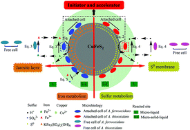
The enhancement mechanism of adapted adsorption behavior in the bioleaching of copper-bearing sulfide ore by Acidithiobacillus sp. was systematically investigated from a mineralogical viewpoint and compared to adsorption-deficient (DF) and adsorption-unadapted (UA) systems. With the assistance of the adapted adsorption behavior, both iron and sulfur metabolism was enhanced, which was proven by a series analysis of key chemical parameters, including scanning electron microscopy (SEM) and X-ray diffraction (XRD). SEM analysis revealed smaller jarosite and S0 granules as well as more potential adsorption sites on the ore’s surface, thus indicating a stronger “contact” mechanism. XRD analysis showed that more chemical derivatives were generated owing to active iron/sulfur metabolism. Additionally, the attached and free biomasses of A. ferrooxidans and A. thiooxidans were increased by 33.3–58.9% and 25.0–33.9%, respectively. Moreover, the final concentration of the extracted copper ions was improved by 22.8% (A. ferrooxidans) and 28.9% (A. thiooxidans). All results proved that the adsorption behavior coupled to the attached cells was greatly stimulated and accelerated by the adapted evolution and further contributed to a higher bioleaching efficiency. The adapted method and its mechanism will be useful to further guide similar bioleaching processes in the near future.


1. Introduction

A vast majority of low-grade ores cannot be economically utilized by the traditional smelting method and are deposited at mines.1–3 Substantial amounts of discarded ores are accumulating, resulting in resource waste and potential environmental problems. Copper-bearing sulfide ores such as chalcopyrite (CuFeS2), (>70% of world copper reserves), in general are common low-grade and refractory ores that also face the aforementioned problems.4,5 In the last few decades, bioleaching was recognized as a green and economical technology for recovering these waste ores.6–8 However, owing to its complicated and refractory nature, commercial application of bioleaching of copper-bearing sulfide ore is still not satisfactory.9,10 Recently, bioleaching of copper-bearing sulfide ore has attracted increasing attention, in particular because of growing copper consumption and environmental stress worldwide.

To improve the bioleaching process, it is essential to have a deep understanding of the bioleaching mechanism in detail. Various mechanisms have been proposed for illustrating the bioleaching process of sulfide ore (pyrite, sphalerite, chalcopyrite etc.), such as surface attaching, oxidation reactions, elemental transformation, interfacial evolution, bio-molecular changes, and surface erosions.11 Two indirect mechanisms via thiosulfate or via polysulfides were found in pyrite bioleaching with A. thiooxidans.12 The sphalerite bioleaching process was divided into two steps.13 The rapid surface attaching of microorganisms was the key to enhancing leaching efficacy, resulting in the oxidation of the pyrite and concomitant bio-generation of ferric ions and protons. Then, the continued regeneration of ferric ions by planktonic bacteria and the oxidation of the elemental sulfur reaction product further contributed to the higher leaching efficacy. Currently, “indirect contact” and “direct contact” mechanisms have been proposed to better understand the bioleaching process.14,15 The two mechanisms derive from bio-oxidation reactions in different spaces. In the former mechanism, bacteria oxidize soluble ferrous ions to ferric ions and sulfur to sulfate ions in the micro-liquid environment. Ferric ions oxidize the sulfide ore in an acidic environment. In the latter mechanism, bacterial attachment is important physiologically, but ferric ions oxidize the sulfide minerals in the solid–liquid interface environment. The specifics of bacterial (electro) chemical interactions with mineral surfaces and/or their direct contact (enzymatic) contribution to sulfide dissolution are unknown. In these mechanisms, the adsorption behavior of the attached cells was a prerequisite for the subsequent iron/sulfur metabolism.16 Previous research has also intensively studied the performance of the adsorption behavior, such as the effect of a single factor such as extracellular polymeric substance (EPS), mineral or bacterial attachment selection in various bioleaching processes.16,17 It was reported that a multilayered biofilm with the EPS of A. ferrooxidans, A. thiooxdians and Leptospirillum ferrooxidans was pivotal in “contact” mechanisms.18,19 The attached behaviors of different strains even with a closely related genetic relationship were diverse, while the mineral-selection in the attached process of the same strain was also different.14,20 However, most researchers focused on the adsorption kinetics, association factor and adsorption characteristics between different species. To date, to the best of our knowledge, the detailed mechanism of the adsorption behavior in the bioleaching of copper-bearing sulfide ore, especially the efficient strategy for enhancing the performance of adsorption behavior, remains poorly understood.

In our previous study, an acidophilic strain A. thiooxidans ZJJN-3 was isolated from industrial bio-heap leachate.21 A. thiooxidans ZJJN-3 and A. ferrooxidans CUMT-1 were applied in chalcopyrite bioleaching to form an efficient catalytic system.22 An integrated fermentation strategy was also proposed for enhancing chalcopyrite bioleaching in a 7 L bioreactor.23 In this study, two typical bioleaching strains (A. ferrooxidans and A. thiooxidans) were employed for exploring the enhancement mechanism of the adapted adsorption behavior in the bioleaching of copper-bearing sulfide ore. First, the effects of the adapted adsorption behavior on sulfur and iron metabolism were analyzed and compared to the adsorption-deficient (DF) and adsorption-unadapted (UA) systems. Second, the effects of the adaptive adsorption behavior on ore, such as morphological, componential and functional group differences were also investigated by scanning electron microscopy (SEM) and X-ray diffraction (XRD). Finally, the efficiency of the adaptive adsorption behavior on improving cell growth and bioleaching efficiency was further verified.

2. Materials and methods

2.1. Strain and growth conditions

A. ferrooxidans CUMT-1 was kindly donated by Professor Leng from the China University of Mining and Technology, Xuzhou, Jiangsu, China. A. thiooxidans ZJJN-3 was isolated from the leachate of an industrial bio-heap (low-grade secondary sulfide, 20 million m3) in Zijinshan Copper Mine, Longyan, Fujian, China. The strain was previously identified using an analysis of physiological and molecular characteristics.21 It was deposited in the China Center for Type Culture Collection with the number M2012104. The detailed strain characteristics are listed in Table 1. A. ferrooxidans was cultured in 9K media and A. thiooxidans was cultured in Starkey media. The basal salts of 9K media were listed as follows (g L−1): (NH4)2SO4 3.0, K2HPO4 0.5, MgSO4·7H2O 0.5, KCl 0.1, Ca(NO3)2 0.01. Energy substrate: 44.7 g L−1 FeSO4·7H2O. The basal salts of Starkey media were listed as follows (g L−1): (NH4)2SO4 3.0, KH2PO4 3.5, MgSO4 0.5, CaCl2·2H2O 0.25. Energy substrate: 10 g L−1 S0. Trace elements were listed as follows (mg L−1): Na2SO4 50.0, FeCl3·6H2O 11.0, H3BO3 2.0, MnSO4·H2O 2.0, ZnSO4·7H2O 0.9, Na2MoO4·2H2O 0.8, CoCl2·6H2O 0.6, CuSO4 0.5, Na2SeO4 0.1. For both bacterial systems, the initial pH of the media was adjusted to 2.2 and the strains were adapted by adding 3.0% (w/v) of copper-bearing sulfide ore sample at 30 °C and 170 rpm. Strains were incubated into fresh media once a month.
Table 1 The main characteristics of the strains used in the study
Species Strain Energy type Optimal T/pH Description and source
A. ferrooxidans CUMT-1 Ferrous and sulfur oxidizer 30–35 °C, pH 1.8–2.5 Waste acid mine drainage from coal ore, Jiangsu, China
A. thiooxidans ZJJN-3 Sulfur oxidizer 28–30 °C, pH 0–2.0 Leachate from Zijinshan copper mine, Fujian, China


2.2. Ore sample composition and pretreatment

The copper-bearing sulfide ore sample was collected from Dongguashan copper mine, Tongling, Anhui, China. The main mineralogical compositions of the primary ore were chalcopyrite, pyrite, pyrrhotite, and magnetite. The detailed elements and contents of the ore sample were assayed by atomic absorption spectrometry (Spectr AA-220, Varian, USA), as shown in Table 2. The ore sample was sieved through a 300-mesh grid, keeping the particle diameter at <48 μm. The ore sample was sequentially washed with 2 M HCl, distilled water, and pure ethanol. Then, the ore sample was dried at room temperature and preserved in a vacuum desiccator.
Table 2 The main characteristics of the ore sample used in the studya
Parameter and unit Value and description
a The ore sample was collected from the Dongguashan copper mine, Tongling, Anhui, China; the values of Ag, Au, Co, Cd and Hg were all below the detection limit (<0.0002).b The ore sample was ground and sieved through a 300-mesh grid, which kept the particle diameter at <48 μm.
Cu (%) 1.03 ± 0.05
S (%) 12.8 ± 0.21
Fe (%) 32.3 ± 0.53
Ca (%) 3.78 ± 0.32
Mg (%) 3.53 ± 0.25
Al (%) 1.66 ± 0.39
Zn (%) 0.051 ± 0.01
Mn (%) 0.044 ± 0.01
Ni (%) 0.029 ± 0.005
Pb (%) 0.028 ± 0.005
As (%) 0.0042 ± 0.001
Particle diameter (μm) <48b


2.3. Experimental procedure

2.3.1. Procedure for DF system. The DF system was designed as described here. The leaching solution was allowed to rest without agitation for 1 h to allow the ore particles to settle. Then the supernatant was transferred into another flask. The leached ore sample was collected by centrifugation (3K15, Sigma, Germany) at 380 × g for 2 min. The ore sample was suspended in 30 mL of fresh basal salts of 9K or Starkey media. Then 1.0 g of 0.2 mm glass beads was added and shaken with a vortex (lab-dancer, IKA, Germany) for 5 min. The ore sample was centrifuged and shaken again. The supernatant was then transferred into another tube. Almost no dissociative DNA was tested in the supernatant by this method according to the report of Gehrke et al. (1998), indicating that all attached cells were removed without breaking.24 Moreover, no additional cells were separated with an additional shaken-operation process, proving that all attached cells were separated from the ore surface with the above operation. The ore sample was added back into its original bioleaching supernatant. The above procedure was completed every five days.
2.3.2. Procedure of adsorption-adapted (AD) evolution. The AD evolution was processed as described here. A. ferrooxidans and A. thiooxidans were independently cultured with 3.0% (w/v) copper-bearing sulfide ore for 15 days. The leaching solution was stilled for 1 h and the supernatant was transferred into another flask. The ore sample was collected by centrifugation (3K15, Sigma, Germany) at 380 × g for 2 min, and suspended in 30 mL of fresh basal salts of 9K or Starkey media. Then, 1.0 g of 0.2 mm glass beads was added and shaken with a vortex (lab-dancer, IKA, Germany) for 5 min. Most of the attached cells with a poor adsorption performance were washed off while stronger ones were preserved. The ore sample was centrifuged, as before, and added into fresh media. This above process was repeated once every two weeks. After being repeated for 6 months, the attached cells were shed and collected, as in 2.3.1. The attached cells were used as the adapted strain for the bioleaching experiment.
2.3.3. Bioleaching experiment. Six bioleaching experiments with different performances of adsorption behavior were designed as follows: A. ferrooxidans CUMT-1 (DF); A. ferrooxidans CUMT-1 (unadapted; UA); A. ferrooxidans CUMT-1 (AD); A. thiooxidans ZJJN-3 (DF); A. thiooxidans ZJJN-3 (UA); A. thiooxidans ZJJN-3 (AD). The bioleaching experiments were carried out in 500 mL shaker flasks; 100 mL of media was added into the A. ferrooxidans (9K basal salts media) and A. thiooxidans (Starkey basal salts media) systems. Then, 3.0 g of copper-bearing sulfide ore sample was added into each flask. The cell density in each system was controlled at 5.0 × 10⁷ cells per mL after inoculation. The bioleaching experiments were carried out at 30 °C and 170 rpm. To balance the system from evaporation loss, 2.0 mL of sterile water was supplemented into each system once a day. The whole bioleaching cycle lasted 40 days.

2.4. Analytical methods

2.4.1. pH and Eh measurement. The pH value was measured by a pH meter (PHB-3TC, Sartorius, Germany). The Eh value was monitored by a Pt electrode (E-431Q, ASI, USA) with a calomel electrode (Hg/Hg2Cl2) as a reference.
2.4.2. Sulfate ion assay. The concentration of sulfate ions was detected according to the chromic acid-barium colorimetric assay using a spectrophotometer (IV-1100D, Meipuda, China). Concentration of sulfate ions (mg L−1) = 201.6 × OD420 nm − 26.029 (r2 = 0.998).
2.4.3. Ferrous and ferric ion assay. The concentrations of ferrous and ferric ions were measured according to the o-phenanthroline spectrophotometry assay using a spectrophotometer. Concentration of ferrous ions (mg L−1) = 5.077 × OD508 nm − 0.0765 (r2 = 0.999). Concentration of ferric ions (mg L−1) = 5.102 × OD508 nm − 0.143 (r2 = 0.998).
2.4.4. SEM analysis. The ore sample was previously dried at room temperature and preserved in a vacuum desiccator. The morphology and surface of the ore were observed with an SEM (Quanta-200, FEI, Netherlands). The suspension solution of the ore sample was added to the specimen holder. After natural volatilization, the sample was firmly immobilized and a 30 nm thick conductive coating of gold was applied to the surface. The scanning condition was set at 25 kV.
2.4.5. XRD analysis. The ore sample was previously washed with deionized water and dried at room temperature under vacuum conditions. Then the center depression of the detection plate was covered with the ore. The ore sample was scanned according to 2-theta range 3–90 (°) by an X-ray diffractometer (D8, AXS, Germany). The detailed data were analyzed by the MDI Jade 5.0 (Materials Data Ltd., USA) integrating PDF card library.
2.4.6. Detection of free, attached and total cell density. One milliliter of bioleaching sample was centrifuged at 380 × g for 2 min to separate the supernatant from the ore precipitation. The free cell density was counted by a single span microscope. Meanwhile, the bottom ore sample was re-suspended in 5.0 mL of fresh basal media. Then, 0.2 g of 0.2 mm glass beads was added and shaken with a vortex for 5 min. This sample was centrifuged and the mixing process was repeated once. The attached cell density was counted in the supernatant as before. The total cell density was the sum of the free and attached cell densities.
2.4.7. Copper ion assay. The concentration of copper ions was monitored by flame atomic absorption spectrometry (Spectr AA-220, Varian, USA). Concentration of copper ions (mg L−1) = 6.852 × λ325 nm − 0.0301 (r2 = 0.999).

2.5. Statistical analysis

All experiments were performed in triplicate. The statistical analysis of the experimental data was performed by one-way analysis of variance and expressed as mean values ± SD. The software SPSS 17.0 (SPSS Inc., Chicago, USA) was used for the statistical analysis.

3. Results and discussion

3.1. Effects of adapted adsorption behavior on iron and sulfur metabolisms

3.1.1. Iron metabolism. The changes in ferrous and ferric ions, the main parameters of iron metabolism, in different systems are shown in Fig. 1. The main biochemical reactions in copper-bearing sulfide ore samples (mainly chalcopyrite as an example) are summarized as eqn (1)–(7). In the A. ferrooxidans systems, the highest concentrations of ferrous ions in each system were (in mg L−1) 200.5 (DF), 327.4 (UA) and 356.3 (AD). In the A. thiooxidans systems, the values were (in mg L−1) 65.4 (DF), 75.3 (UA) and 94.5 (AD). It was reported that the adsorption behavior of the attached cells at the early stage is beneficial to further concentrate ferric ions and attack chalcopyrite, as shown by eqn (1).9,21 The greatest increase in ferrous ions was 63.3%, which was tested in the A. ferrooxidans-DM system. This is because the ferrous ion metabolism was more closely related to A. ferrooxidans than the pure sulfur oxidizer A. thiooxidans. Furthermore, compared to reductive sulfur, ferrous ion was more easily utilized by A. ferrooxidans in a multiple-energies system.25,26 A similar trend to ferrous ions was observed in ferric ions. In the A. ferrooxidans systems, the highest concentrations of ferric ions in each system were (in mg L−1) 435.2 (DF), 637.0 (UA) and 673.2 (AD). In the A. thiooxidans systems, the values were (in mg L−1) 82.4 (DF), 123.2 (UA) and 137.8 (AD). These results indicated that the adapted evolution of adsorption behavior was favorable for enhancing iron metabolism.
 
CuFeS2 + 4Fe3+ = Cu2+ + 2S0 + 5Fe2+ (1)
 
CuFeS2 + 4H+ + O2 = Cu2+ + 2S0 + Fe2+ + 2H2O (2)
 
4Fe2+ + 4H+ + O2 = 4Fe3+ + 2H2O (3)

image file: c5ra15934b-f1.tif
Fig. 1 Changes in key chemical parameters in different bioleaching systems. (A): A. ferrooxidans-DF; (B): A. ferrooxidans-UA; (C): A. ferrooxidans-AD; (D) A. thiooxidans-DF; (E) A. thiooxidans-UA; (F) A. thiooxidans-AD. (■) pH; (▲) ferrous ion; (□) ferric ion; (△) sulfate ion.
3.1.2. Sulfur metabolism. Similarly, the changes in pH and sulfate ion concentrations in different systems were also investigated (Fig. 1). After the adaptive phase, the pH gradually decreases along with the dissolution of the ore. In both the A. ferrooxidans and A. thiooxidans systems, the pH of the DM system was always the lowest, while the highest value was tested in the DF systems. In the A. ferrooxidans systems, the final pHs were 1.92 (DF), 1.82 (UA), and 1.74 (AD). In the A. thiooxidans systems, the final pHs were 1.82 (DF), 1.63 (UA), and 1.51 (AD). In the DF system, the granular sulfur on the ore surface could not be used by attached cells and thus formed the S0 passivation layer. The subsequent oxidization process was greatly inhibited. Conversely, in the AD system, the adsorption behavior of the attached cells was enhanced by the directionally domesticated evolution. Most of the sulfur could be more efficiently utilized, as shown in eqn (4) and (5), and produced sulfuric acid. Moreover, the pH decline range of A. thiooxidans (1.82 to 1.51) was higher than that of A. ferrooxidans (1.92 to 1.74), with the assistance of enhanced adsorption behavior.

Comparatively, the concentration of the sulfate ions steadily increased and achieved stability on day 20. The trend of the sulfate ions in both the A. ferrooxidans and A. thiooxidans systems was similar to the pH data. In the A. ferrooxidans systems, the highest concentrations of sulfate ions in each system were (in g L−1) 2.93 (DF), 3.56 (UA) and 3.88 (AD). In the A. thiooxidans systems, the highest concentrations of sulfate ions in each system were (in g L−1) 1.24 (DF), 2.83 (UA) and 3.86 g L−1 (AD). Compared to A. ferrooxidans-AD, the increased range of sulfate ion concentration in the A. thiooxidans-AD system, via enhancing adsorption behavior, was more significant. The dependence on the adsorption behavior with a pure-sulfur oxidizer such as A. thiooxidans was generally stronger, especially in eliminating the S0 membrane compared to a multiple-energies oxidizer.21 It was also verified that the attached biomass of the A. thiooxidans system on the sulfur surface was almost twice that of the A. ferrooxidans system.22 The above result was also closely consistent with the data on the changes in pH. These results all indicated the higher efficacy of the adaptive adsorption behavior on sulfur metabolism, especially for A. thiooxidans. A detailed comparison of key chemical parameters in different systems is listed in Table 3.

 
2S0 + 3O2 + 2H2O = 2SO42− + 4H+ (4)
 
SxOyn + O2 + H2O → SO42− + H+ (SxOyn represents reduced sulfur, such as SO32−) (5)
 
3Fe3+ + 2SO42− + 6H2O + K+ = KFe3(SO4)2(OH)6 + 6H+ (6)
 
2FeS2 + 2Fe3+ + 3H2O = 4SO32− + 4Fe2+ + 6H+ (7)

Table 3 Comparison of key chemical and biological parameters between pre-leaching and after-leaching in DF, UA and AD systems
  Parameter and unit Pre-leaching After-leaching
A. ferrooxidans A. thiooxidans
DF UA AD DF UA AD
a Represents soluble ions (sulfate, ferrous, ferric, and total iron ions) in the bioleaching system.
Chem-indexes pH 2.20 1.92 1.82 1.74 1.82 1.63 1.51
ORP/mV 270 278 298 312 275 286 303
Sulfate ion (g L−1) 2.4/0.6 2.9 3.3 3.7 1.2 2.8 3.8
Conversion ratio of sulfate iona (%) None 4.3 7.8 11.3 5.2 19.1 27.8
Ferrous ion (mg L−1) None 162.5 312.5 356.3 65.4 75.3 94.5
Conversion ratio of ferrous iona (%) None 1.7 3.2 3.7 0.7 0.8 1.0
Ferric ion (mg L−1) None 432.2 564.5 675.3 82.0 123.2 137.8
Conversion ratio of ferric iona (%) None 4.5 5.8 7.0 0.8 1.3 1.4
Total iron (mg L−1) None 594.7 877.0 1031.6 147.4 198.5 232.3
Conversion ratio of total irona (%) None 6.2 9.0 10.7 1.5 2.1 2.4
Final copper ion (mg L−1) None 25.06 39.50 48.51 24.11 44.25 54.34
Mineral color Black Tawny Tawny Tawny Gray Gray Gray
Mineral weight (g) 3.00 3.01 3.04 2.92 3.03 2.78 2.65
Bio-indexes Free biomass (107 cells per mL) 5.0 17.9 21.6 27.0 8.3 11.8 15.8
Attached biomass (107 cells per mL) None 0.62 1.29 1.72 0.81 1.65 2.62
Attached ratio (%) None 3.34 5.97 6.37 8.90 12.27 14.22
Total biomass (107 cells per mL) 5.0 18.51 22.89 28.72 9.11 13.45 18.42
Daily productivity (107 cells per mL) None 0.46 0.57 0.72 0.23 0.34 0.46


3.2. Effects of adaptive adsorption behavior on mineralogy

3.2.1. Ore morphology. To better understand the mineralogical effects of adapted adsorption behavior, the morphologies of ore samples in different bioleaching systems were observed by SEM (Fig. 2). The morphological differences between the DF, UA, and AD systems were significant, from either A. ferrooxidans or A. thiooxidans.In the A. ferrooxidans-DF system, the ore surface was smooth and the jarosite precipitation was tiny. Owing to the absence of attached cells, the “contact” mechanism was greatly inhibited and there was not enough energy substrate (ferrous ions) released from the ore for cell growth. The concentration of ferric ions sequentially decreased and further reduced the jarosite production. In the A. ferrooxidans-UA system, more compact jarosite appeared on the ore’s surface. With the assistance of adsorptive behavior, the iron metabolism was accelerated and produced more jarosite. In the A. ferrooxidans-AD system, the jarosite precipitation became significantly smaller and more potential adsorption sites were observed on the ore’s surface; jarosite formation was moderately inhibited by a lower pH, as shown in eqn (6), although with a more active metabolism. The potential adsorption site indicated a stronger adsorption behavior via the adapted evolution.
image file: c5ra15934b-f2.tif
Fig. 2 Morphological surface differences of the ore samples between different bioleaching systems. (A): A. ferrooxidans-DF; (B): A. ferrooxidans-UA; (C): A. ferrooxidans-AD; (D) A. thiooxidans-DF; (E) A. thiooxidans-UA; (F) A. thiooxidans-AD.

The same phenomenon was more obvious in the A. thiooxidans system, to some extent. In the A. thiooxidans-DF system, the ore surface was smoother and the sulfur granules were extremely exiguous. In the A. thiooxidans-UA system, some rills and micro-pores appeared on the ore surface. In the A. thiooxidans-AD system, there were more sulfur granules and the ore surface was significantly rougher. Meanwhile, more potential adsorption sites indicated a stronger “contact” mechanism with the adaptive attached cells. It was reported that the dependence on adsorption behavior with A. thiooxidans was stronger because most of the main energy source (S0) was generated on the ore surface.26 Additionally, due to the active chemical ion status, more unknown derivatives were also generated and coupled with the ore.

3.2.2. Ore components. The XRD analysis was performed to investigate the composition of the ore samples in different bioleaching systems (Fig. 3). The main components were CuFeS2, KFe3(SO4)2(OH)6, S, Fe7S8, Fe3O4, FeS2 and CaSO4·2H2O. Compared to the A. ferrooxidans-DF system, there were larger amounts of precipitate peaks such as jarosite and granular sulfur in the UA system and particularly in the AD system. The higher activity of the iron/sulfur metabolism was achieved by a stronger adsorption behavior and further produced more crystal forms. The result was also closely consistent with the morphological differences (Fig. 2). Compared to the A. thiooxidans-DF system, there were more sulfur peaks in the UA system, and also particularly in the AD system, indicating that more crystal forms of elemental sulfur were generated with more active sulfur metabolism. It was reported that, to some extent, sulfur was generally coupled with amorphous iron or other oxy-hydroxides.18 Meanwhile, the CaSO4·2H2O peak was significant in the UA and AD systems, indicating a higher concentration of sulfate ions. Additionally, the minor accumulation of other unknown peaks in these precipitates was due to previous washes with deionized water prior to XRD detection. It was also reported that the abundant inorganic ions and microbial organic compounds in the bioleaching systems contribute to more complicated derivatives.20
image file: c5ra15934b-f3.tif
Fig. 3 XRD analysis of the ore samples in different bioleaching systems. (A): A. ferrooxidans-DF; (B): A. ferrooxidans-UA; (C): A. ferrooxidans-AD; (D) A. thiooxidans-DF; (E) A. thiooxidans-UA; (F) A. thiooxidans-AD. (■) Fe3O4; (▲) FeS2; (●) CuFeS2; (□) CaSO4·2H2O; (△) KFe3(SO4)2(OH)6; (○) Fe7S8 (★) S.

3.3. Efficacy of adapted adsorption behavior for enhancing biomass and copper recovery

3.3.1. Biomass. With the assistance of adapted evolution, the amount of attached biomass was significantly improved (Fig. 4A). In the A. ferrooxidans systems, the highest attached biomass of each system was 0.62 (DF), 1.29 (UA) and 1.72 × 10⁷ cells per mL (AD). The amount of attached biomass was improved by 33.3% via the adapted evolution. Free biomass was also increased sequentially from 21.6 to 27.0 × 10⁷ cells per mL (25.0%); more energy and nutrients were released owing to a stronger “direct contact” mechanism. The phenomenon was more obvious in the A. thiooxidans systems. The highest attached biomass of each system was 0.81 (DF), 1.65 (UA) and 2.62 × 10⁷ cells per mL (AD). The attached biomass amount was improved by 58.9% via adapted evolution. Moreover, the free biomass increased from 11.8 to 15.8 × 10⁷ cells per mL (33.9%) with a stronger adsorption behavior because A. thiooxidans is a pure-energy oxidizer (sulfur), which is different from the multiple energy oxidizer A. ferrooxidans. A. thiooxidans relied more on its attached cells because the main energy substrate (S0) largely existed on the ore’s surface. Sulfur granules were primarily oxidized into intermediate states such as S4O62− or S4O52−, with the assistance of adsorption behavior. Then, the reduced and soluble sulfur was thoroughly utilized via a “non-contact” mechanism. These data are consistent with the TEM images of flagella and capsule, which also indicated a stronger requirement of attached cells by A. thiooxidans ZJJN-3.21
image file: c5ra15934b-f4.tif
Fig. 4 The highest biomass and final recovery efficiencies of copper ions in different systems. (A) The highest free and attached biomass; (B) the final recovery efficiency of copper ions. a–c and A–C represent statistically significant differences (c > b > a; C > B > A). The recovery efficiency of the A. ferrooxidans-DF or A. thiooxidans-DF system was selected as the standard and defined as 100%. The relative recovery efficiency of the A. ferrooxidans-DF/AD system and A. thiooxidans-DF/AD system was calculated by dividing the recovery efficiency of the A. ferrooxidans-DF or A. thiooxidans-DF system.
3.3.2. Copper recovery. The copper recovery efficiency was significantly enhanced by the adaptive evolution of the attached cells (Fig. 4B and Table 3). In the A. ferrooxidans system-AD, the efficiency was improved by 93.5% and 22.8%, compared to the DF and UA systems, respectively. The improvement in the A. thiooxidans-AD system was more significant. The efficiency was improved by 154.2% and 28.9%, compared to the DF and UA systems, respectively. In other words, more than 48.3–60.7% of the bioleaching efficiency was contributed by directly domesticating the attached cells. Compared to A. ferrooxidans, the efficacy of adaptive evolution was more prominent with A. thiooxidans. The result was also closely consistent with the chemical, mineralogical, and biological parameters, thereby proving the efficacy of adapted evolution.

3.4. Overall assessment effects of adsorption behavior in bioleaching copper-bearing sulfide ore

The microenvironments of bioleaching of copper-bearing sulfide ore were divided into solid–liquid and liquid microenvironments based on the biochemical reaction site (Fig. 5, chalcopyrite as an example). Apparently, attached cells adsorbed onto the ore’s surface in the solid–liquid microenvironment. The biochemical reaction in the liquid microenvironment was subsequently influenced by the surface adsorption process. The “direct contact” and “indirect contact” bioleaching mechanisms were derived from these two different microenvironments.25,27 The role of the attached biomass in bioleaching of copper-bearing sulfide ore was characterized by the aspects of iron and sulfur metabolisms.
image file: c5ra15934b-f5.tif
Fig. 5 Overall effects of the adsorption behavior in the bioleaching of copper-bearing sulfide ore (chalcopyrite as an example).

In the “direct contact” mechanism’s iron metabolism (A. ferrooxidans CUMT-1), attached cells adsorbed onto the ore’s surface and oxidized ferrous ions into ferric ions, as shown in eqn (3). The ore’s surface was sequentially attacked by the generated ferric ions, as shown in eqn (1), and dissolved copper ions. The resulting ferrous ions entered into the ion cycle again. The dissolution process of the ore took place at the interface between the cells and the ore’s surface. Extracellular polymeric substance (EPS), consisting of some polysaccharides, proteins, and nucleic acids, generally served as the reaction space.28 The increasing concentrations of the ferrous and ferric ions in the liquid microenvironment gradually initiated and enhanced the “indirect contact” mechanism.29,30 Also, the accumulated ferric ions partly participated during the formation of jarosite, as shown in eqn (6).

In the “direct contact” mechanism’s sulfur metabolism (A. thiooxidans ZJJN-3/A. ferrooxidans CUMT-1), sulfur colloids were subsequently oxidized into intermediate compounds such as S4O62− or S4O52−. Also, redundant sulfur gathered as micro-particles (S8) and formed a passivation layer. This reduced sulfur dissolved into the liquid microenvironment and was oxidized, as shown in eqn (5). Then, the hydrogen ions entered into the solid–liquid microenvironment and attacked the ore’s surface, as shown in eqn (2). The copper ions were finally released. Reduced sulfur and hydrogen ions in the liquid microenvironment gradually initiated and enhanced the “indirect contact” mechanism. With the assistance of adsorption behavior, more hydrogen ions, ferrous ions, ferric ions, sulfur compounds, and free biomass were created in the bioleaching system. The whole bioleaching system was directly or indirectly affected by these oxidizing and reductive agents. Therefore, in both the iron and sulfur metabolisms, adsorption behavior acted as an initiator and accelerator. Our research was the first to reveal the enhancement mechanism coupling with the adapted adsorption behavior from the mineralogical aspects of the bioleaching of copper-bearing sulfide ore.

4. Conclusions

Bioleaching of copper-bearing sulfide ore was improved by directly adapting the adsorption behavior, and its mineralogical enhancement mechanism was also successfully investigated and compared to the DF and UA systems. With the assistance of adapted evolution, both iron and sulfur metabolism was greatly enhanced. The amount of jarosite (A. ferrooxidans) and S0 (A. thiooxidans) became significantly smaller, along with more potential adsorption sites on the ore’s surface. More compound derivatives were generated because of active biochemical reactions. Attached biomass was increased and further contributed to higher free biomass. Moreover, the efficiency of copper recovery was improved by 22.8% (A. ferrooxidans) and 28.9% (A. thiooxidans). Taken together, these results indicate that this mechanism can be applicable to directly domesticate the adsorption behavior for improving the bioleaching of copper-bearing sulfide ore, especially with A. thiooxidans.

Acknowledgements

This work was supported by grants from the Natural Science Foundation of Jiangsu Province (No. BK20150133), the Scientific Program of Jiangnan University (No. JUSRP11538), the National Natural Science Foundation of China (Grant No. 31301540 and 21306064), the Priority Academic Program Development of Jiangsu Higher Education Institutions, and the 111 Project (No. 111-2-06).

References

  1. C. L. Zhu, L. Z. Liu, M. M. Fan, L. Liu, B. B. Dai, J. Z. Yang and D. P. Sun, RSC Adv., 2014, 4, 55044–55048 RSC.
  2. S. K. Behera, P. P. Panda, S. Singh, N. Pradhan, L. B. Sukla and B. K. Mishra, Int. Biodeterior. Biodegrad., 2011, 65, 1035–1042 CrossRef CAS.
  3. D. B. Johnson and C. A. du Plessis, Miner. Eng., 2015, 75, 2–5 CrossRef CAS.
  4. H. R. Watling, D. M. Collinson, J. Li, L. A. Mutch, F. A. Perrot, S. M. Rea, F. Reith and E. L. J. Watkin, Miner. Eng., 2014, 56, 35–44 CrossRef CAS.
  5. M. Vera, A. Schippers and W. Sand, Appl. Microbiol. Biotechnol., 2013, 97, 7529–7541 CrossRef CAS PubMed.
  6. S. Ghassa, Z. Boruomand, H. Abdollahi, M. Moradian and A. Akcil, Sep. Purif. Technol., 2014, 136, 241–249 CrossRef CAS.
  7. M. J. Chen, J. Q. Wang, J. X. Huang and H. Y. Chen, RSC Adv., 2015, 5, 34921–34926 RSC.
  8. S. O. Rastegar, S. M. Mousavi and S. A. Shojaosadati, RSC Adv., 2015, 5, 41088–41097 RSC.
  9. W. M. Zeng, G. Z. Qiu, H. Z. Zhou, J. H. Peng, M. Chen, S. N. Tan, W. L. Chao, X. D. Liu and Y. S. Zhang, Bioresour. Technol., 2010, 101, 7068–7075 CrossRef CAS PubMed.
  10. J. Y. Zhu, Q. Li, W. F. Jiao, H. Jiang, W. Sand, J. L. Xia, X. D. Liu, W. Q. Qin, G. Z. Qiu, Y. H. Hua and L. Y. Chai, Colloids Surf., B, 2012, 94, 95–100 CrossRef CAS PubMed.
  11. H. R. Watling, Hydrometallurgy, 2006, 84, 81–108 CrossRef CAS.
  12. A. Schippers and W. Sand, Appl. Environ. Microbiol., 1999, 65, 319–321 CAS.
  13. F. K. Crundwell, Hydrometallurgy, 2003, 71, 75–81 CrossRef CAS.
  14. C. L. Brierley and J. A. Brierley, Appl. Microbiol. Biotechnol., 2013, 97, 7543–7552 CrossRef CAS PubMed.
  15. N. Pradhan, K. C. Nathsarma, K. Srinivasa Rao, L. B. Sukla and B. K. Mishra, Miner. Eng., 2008, 21, 355–365 CrossRef CAS.
  16. S. N. Tan and M. Chen, Hydrometallurgy, 2012, 119–120, 87–94 CrossRef CAS.
  17. C. J. Africa, R. P. van Hille and S. T. L. Harrison, Appl. Microbiol. Biotechnol., 2013, 97, 1317–1324 CrossRef CAS PubMed.
  18. S. Mangold, K. Harneit, T. Rohwerder, G. Claus and W. Sand, Appl. Environ. Microbiol., 2008, 74, 410–415 CrossRef CAS PubMed.
  19. K. Harneit, A. Göksel, D. Kock, J. H. Klock, T. Gehrke and W. Sand, Hydrometallurgy, 2006, 83, 245–254 CrossRef CAS.
  20. M. A. Ghauri, N. Okibe and D. B. Johnson, Hydrometallurgy, 2007, 85, 72–80 CrossRef CAS.
  21. S. S. Feng, H. L. Yang, Y. Xin, L. Zhang, W. L. Kang and W. Wang, J. Ind. Microbiol. Biotechnol., 2012, 25–1635 Search PubMed.
  22. S. S. Feng, H. L. Yang, Y. Xin, L. K. Gao, J. W. Yang, T. Liu, L. Zhang and W. Wang, Bioresour. Technol., 2013, 129, 456–462 CrossRef CAS PubMed.
  23. S. S. Feng, H. L. Yang, X. Zhan and W. Wang, Bioresour. Technol., 2014, 161, 371–378 CrossRef CAS PubMed.
  24. T. Gehrke, J. Telegdi, D. Thierry and W. Sand, Appl. Environ. Microbiol., 1998, 64, 2743–2747 CAS.
  25. P. Devasia and K. A. Natarajan, Int. J. Miner. Process., 2010, 94, 135–139 CrossRef CAS.
  26. P. Martínez, S. Gálvez, N. Ohtsuka, M. Budinich, M. P. Cortés, C. Serpell, K. Nakahigashi, A. Hirayama, M. Tomita, T. Soga, S. Martínez, A. Maass and P. Parada, Metabolomics, 2013, 9, 247–257 CrossRef PubMed.
  27. R. H. Lara, J. V. García-Meza, R. Cruz, D. Valdez-Pérez and I. González, Appl. Microbiol. Biotechnol., 2012, 95, 799–809 CrossRef CAS PubMed.
  28. H. C. Flemming and J. Wingender, Nat. Rev. Microbiol., 2010, 8, 623–633 CAS.
  29. A. B. Vakylabad, M. Schaffie, M. Ranjbar, Z. Manafi and E. Darezereshki, J. Hazard. Mater., 2012, 241–242, 197–206 CrossRef CAS PubMed.
  30. L. Reyes-Bozo, M. Escudey, E. Vyhmeister, P. Higueras, A. Godoy-Faúndez, J. L. Salazar, H. Valdés-González, G. Wolf-Sepúlveda and R. Herrera-Urbina, Miner. Eng., 2015, 78, 128–135 CrossRef CAS.

Footnote

Electronic supplementary information (ESI) available. See DOI: 10.1039/c5ra15934b

This journal is © The Royal Society of Chemistry 2015
Click here to see how this site uses Cookies. View our privacy policy here.