Biodegradable containers composed of anionic liposomes and cationic polypeptide vesicles

A. A. Yaroslavov*a, O. V. Zaborovaa, A. V. Sybachina, I. V. Kalashnikovaa, E. Kesselmanb, J. Schmidtb, Y. Talmonb, A. R. Rodriguezc and T. J. Demingcd
aDepartment of Chemistry, M. V. Lomonosov Moscow State University, Leninskie Gory 1-3, 119991 Moscow, Russian Federation. E-mail: yaroslav@genebee.msu.su
bDepartment of Chemical Engineering, Technion-Israel Institute of Technology, 32000 Haifa, Israel
cDepartment of Bioengineering, University of California, Los Angeles, CA 90095, USA
dDepartment of Chemistry and Biochemistry, University of California, Los Angeles, CA 90095, USA

Received 7th August 2015 , Accepted 8th November 2015

First published on 11th November 2015


Abstract

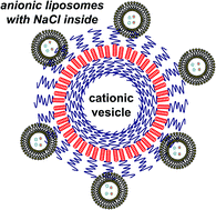
An electrostatic complexation of liposomes, composed of anionic palmitoyloleoyl phosphatidylserine (POPS1−) and zwitterionic dioleoylphophatidylcholine (DOPC), with bilayer vesicles composed of cationic poly(L-lysine)-b-poly(L-leucine) block copolypeptides has been investigated. The complexation was characterized by several physicochemical methods with the following main conclusions: (a) all added liposomes are totally adsorbed on the polypeptide vesicles up to a certain saturation concentration. (b) The calculated number of liposomes per a single polypeptide vesicle is about 60. (c) The liposomes remain intact (i.e., do not leak) after being complexed with the vesicles. (d) Complexes are stable in physiological ionic strength solution with [NaCl] = 0.15 M. (e) The vesicles are effectively digested by proteolytic enzyme trypsin even when covered by liposomes. These findings as well as the high potential for loading of anionic liposomes and cationic vesicles with biologically active compounds make these multi-liposomal complexes promising in the drug delivery field.


Introduction

A bilayer lipid vesicle (liposome) represents a widely-used container for drug delivery.1,2 The unique structure of liposomes allows encapsulation of both hydrophilic and hydrophobic compounds,3,4 and liposomes with targeting moieties on their surfaces show preferable binding to target cells.1,3,5 Vesicles have been described that release their contents in response to external stimulus such as heat or light6 or dissolve in water gases7 as well as pH-sensitive liposomes that release entrapped drugs in more acidic areas, e.g. in cancer tumors.8

Concentration of liposomes within a rather small volume could lead to multi-liposomal containers capable of simultaneously carrying diverse drugs and therefore, lead to an increase of their therapeutic effect. Recently we have described electrostatic immobilization of anionic liposomes on the surface of polystyrene particles covered by grafted polycationic chains (“spherical polycationic brushes”, SPB).9 With this approach, several dozens of intact (undisrupted) liposomes with various contents could be concentrated within an approx. 350 nm sphere. However, the non-(bio)degradable polystyrene core strongly restricts the use of liposome/SPB conjugates for biomedical applications.

In the present communication, we describe for the first time a multi-liposomal container, prepared by complexation of multiple anionic liposomes with individual cationic polypeptide vesicle (CPV). These containers remained stable in a 0.15 M NaCl solution, but could eventually be destroyed in the presence of the proteolytic enzyme trypsin, which is well known to digest poly(L-lysine). The biodegradability of these complexes makes them promising as a multi-functional drug carriers.

Experimental

A poly(L-lysine)60-b-poly(L-leucine)20 block copolypeptide, K60L20, (see ESI, Fig. S1A) was synthesized using transition metal-mediated amino acid N-carboxyanhydride polymerization, which allows control over copolypeptide chain length and composition.10 The Mw/Mn ratio determined using gel permeation chromatography/light scattering was found to be 1.13.11

In order to prepare the copolymer vesicles, we followed the procedure described elsewhere.11 Briefly, the lyophilized copolymer was first swelled in THF and dispersed using a sonication bath, followed by addition of an equal volume of deionized water that yielded a slightly turbid suspension. The latter was exhaustively dialyzed against deionized water to remove the THF and then against a 10−3 M Tris buffer with pH = 7 that finally gave a suspension of cationic polypeptide vesicles (CPVs) (see their structure in ESI, Fig. S1B). The dynamic light scattering (DLS) study of the vesicle suspension with a Brookhaven Zeta Plus showed two populations with the main contribution of particles with diameter of 200 nm (see ESI, Fig. S2).

Results & discussion

The number (concentration) of cationic groups in CPVs, exposed into the surrounding water solution and available for electrostatic coupling with anionic liposomes, was estimated via electrophoretic titration of a CPV suspension in a pH = 7 Tris buffer by a solution of sodium poly(styrene sulfonate) (NaPSS) in a pH = 7 Tris buffer with use of a Brookhaven Zeta Plus. As shown earlier,12 a degree of protonation of poly(L-lysine) amino groups at pH = 7 is close to maximum (100%). The interaction of two oppositely charged polyions, positive protonated poly(L-lysine) ions and negative poly(styrene sulfonate) anions, was accompanied by mutual neutralization of their charges and corresponding change in the electrophoretic mobility (EPM) of the resulting complex particles. Complete neutralization of CPV cationic charges by NaPSS anionic charges (i.e. the EPM = 0 point) was achieved at a concentration of sulfonate subunits [Sulfo] = 7 × 10⁻³ M that is equal to the effective concentration of cationic L-lysine on the outer CPV side, [Lys+]outer. According to the assumed unilamellar structure of the copolypeptide vesicles11 (see also ESI, Fig. S1B), poly-L-lysine chains are nearly uniformly (fifty-fifty) exposed into the surrounding solution and inside water cavity of vesicles. From there, a total lysine concentration in the CPV sample, titrated by NaPSS, turns to be [Lys+] = 2 × [Lys+]outer = 1.4 × 10⁻² M.

In a typical experiment, a pH = 7 buffer solution of CPV with [Lys+]outer = 10−4 M was mixed with a varying amount of aqueous suspension of unilamellar liposomes (ca. 40–50 nm in diameter) prepared by sonication (see synthetic procedure in ESI, S1) and composed of a zwitterionic dioleoylphophatidylcholine (DOPC) and an anionic palmitoyloleoyl phosphatidylserine (POPS1−) (see chemical structures of lipids in ESI, Fig. S3A and B). The molar fraction of anionic lipid ν = [POPS1−]/([POPS1−] + [DOPC]) in the liposomes was equal to 0.1. Interaction of DOPC/POPS1− liposomes with the CPVs altered the surface charge of vesicles, which was monitored using a Brookhaven Zeta Plus (Fig. 1). Adsorbed liposomes caused the EPM of liposome/CPV complex particles to decrease down to zero at 0.89 mg mL−1 liposome concentration or [POPS1−]EPM=0 = 1.13 × 10⁻⁴ M. At higher liposome concentrations, the charge of particles became negative due to adsorption of an excess of liposomes over the amount needed for complete neutralization of the vesicle charge.


image file: c5ra15863j-f1.tif
Fig. 1 EPM of copolymer vesicles vs. ν = 0.1 DOPC/POPS1− liposome concentration. Copolymer concentration 0.05 mg mL−1; [Lys+]outer = 1 × 10⁻⁴ M; 10−3 M Tris buffer, pH = 7.

A key question concerns the integrity of adsorbed liposomes that relates to the quality of immobilized liposomal containers. In order to study complexed liposome integrity, a suspension of DOPC/POPS1− liposomes filled with a 1 M NaCl solution was prepared. Formation of defects in the liposomal membrane should lead to leakage of salt from the liposomes into the surrounding solution, thus increasing suspension conductivity. Initially, a control experiment was carried out in which a NaCl-loaded liposome suspension was treated with a Triton X-100 solution that resulted in disruption of liposomes as expected, and resulted in NaCl leakage and a subsequent rise in the conductivity detected by a CDM 83 Radiometer conductometer. A NaCl-loaded liposome suspension was next mixed with a suspension of the CPVs that resulted in only a negligible increase in the conductivity. We concluded from there that the anionic DOPC/POPS1− liposomes maintain their integrity after adsorption on the surface of cationic polypeptide vesicles.

Liposome adsorption on CPVs was quantified using the earlier described procedure.9 A series of mixtures was prepared with a constant CPV concentration (0.05 mg mL−1 or [Lys+]outer = 1 × 10⁻⁴ M) and varying concentrations of DOPC/POPS1− liposomes with 0.05 wt% of 1,2-dioleoyl-sn-glycero-3-phosphoethanolamine-N-(1-pyrenesulfonyl) (pyrene-PE), (see chemical structure in ESI, Fig. S3C), a fluorescent lipid incorporated into the bilayer during the liposome preparation. After 30 minutes the mixtures were centrifuged at 18[thin space (1/6-em)]000 rpm for 40 min using a J2-21 Beckman micro centrifuge, the fluorescent intensities in the clear supernatants were measured. No fluorescence, i.e. no liposome, was detected in the supernatants up to 0.98 mg mL−1 lipid concentration or [POPS1−]max = 1.24 × 10⁻⁴ M, which reflected complete liposome binding to the CPVs within the indicated lipid interval (see ESI, Fig. S4).

The above electrophoretic, conductivity and fluorescence data allows a deeper insight into the structure and composition of the liposome/CPV complex. As shown in Fig. 1, the liposome/CPV complex became electroneutral (with EPM = 0) at a concentration of adsorbed liposomes of 0.89 mg mL−1 or a total concentration of POPS1− from both liposome leaflet [POPS1−] = 1.13 × 10⁻⁴ M. The latter was equal to a concentration of L-lysine subunits on the outer side of vesicles [Lys+]outer = 1 × 10⁻⁴ M. Taking into account a uniform (fifty-fifty) distribution of POPS1− molecules between both leaflets in the membrane of the initial DOPC/POPS1− liposomes13 and preservation of the liposome integrity after adsorption, the above mentioned equality of the two concentrations, [POPS1−] and [Lys+]outer, at EPM = 0 can only be interpret in terms of the CPV-induced migration of POPS1− molecules from the inner to outer membrane leaflet (flip-flop). Simultaneously, an equivalent amount of zwitterionic (neutral) DOPC molecules move in the opposite direction: from the inner to outer leaflet, which allows the adsorbed liposomes to retain their integrity. Earlier, we have described complexation of anionic liposomes with polystyrene microspheres grafted by polycationic chains, “spherical polycationic brushes”.14 We measured binding of intact (undisrupted) liposomes and the anionic lipid flip-flop in the bound liposomes. We see now that complexation of anionic liposomes with the CPVs occurs by a similar process. In both cases, a hydrophilic layer of grafted macromolecules prevents a direct contact of immobilized liposomes with the solid polystyrene and poly-L-leucine core and thereby ensures the integrity of immobilized liposomes.

Additional information about the morphology of the liposome/CPV complex was obtained by cryogenic transmission electron microscopy (cryo-TEM). Fig. 2A shows a cryo-TEM micrograph of the polypeptide vesicle. A dark ring in the figure reflects the hydrophobic polyleucine layer of the vesicular membrane. Polylysine chains projecting from the polyleucine layer are not visible in the figure since no contrast-enhancing material was used. A cryo-TEM of the DOPC/POPS1− liposomes by themselves (Fig. 2B) gives sizes in agreement with the DLS measurements. Fig. 2C is a cryo-TEM micrograph of liposome/CPV complexes in which CPVs (bigger rings) are covered by multiple liposomes (smaller rings). Clearly, both CPVs and liposomes retain their normal spherical shape (do not disrupt) upon complexation.


image file: c5ra15863j-f2.tif
Fig. 2 Cryo-TEM images of (A) CPV suspension, (B) a 1 mg mL−1 DOPC/POPS1− liposome suspension, (C) and liposomes/CPV complexes. CVP concentration 0.025 mg mL−1; [Lys+]outer = 5 × 10⁻⁵ M; liposomes concentration 1 mg ml−1; 10−3 M Tris buffer, pH = 7; all bars correspond 100 nm.

The capacity of CPV towards anionic liposomes, i.e. the maximum number of liposomes capable of complexing with a single CPV (N), can be estimated comparing the surface area of a single cationic vesicle SCPV = π(200)2 nm2 and the surface area occupied by an adsorbed liposome Sad_lip = π(25)2 nm2. Calculation gives N = SμG/Sad_lip ≈ 60 liposomes with mean diameter 50 nm per a cationic vesicle.

Another question of great importance concerns the stability of liposome/CPV complexes in aqueous salt media. When possible biomedical applications of the liposome/CPV complexes are considered, a special interest is paid to their stability in physiological ionic strength solution with [NaCl] = 0.15 M. We have investigated this question using a fluorescence technique. Cationic polymers are known to be effective fluorescence quenchers due to collisional quenching.15 We detected formation and dissociation of liposome/CPV complexes by measuring the fluorescence intensity of a rhodamine-labeled lipid, 1,2-dimyristoyl-sn-glycero-3-phosphoethanolamine-N-(lissamine rhodamine B sulfonyl) (Rh-PE) (see chemical structure in ESI, Fig. S3D), incorporated into the liposomal membrane. Addition of an aqueous CPV suspension to a suspension of rhodamine-labeled liposomes led to a decrease in the fluorescence intensity (Fig. 3A). Further addition of a NaCl solution to the complex suspension had no effect on the fluorescence intensity up to [NaCl] = 0.3 M (Fig. 3B). Preservation of the liposome/CPV complexes in physiological solution was thus confirmed.


image file: c5ra15863j-f3.tif
Fig. 3 (A) Fluorescence intensity of pyrene-labeled ν = 0.1 DOPC/POPS1− liposomes vs. CPV concentration. [POPS1−] = 1.4 × 10⁻⁴. (B) Fluorescence intensity of rhodamine-labeled liposomes complexed with CPV vs. NaCl concentration. Copolymer concentration 0.052 mg mL−1; [Lys+]outer = 1.4 × 10⁻⁴ M; [POPS1−] = 1.4 × 10⁻⁴. 10−3 M Tris buffer, pH = 7.

Biodegradability is a key requirement for carriers of biologically active substances. Liposomes are known to decompose when attacked by lipase, a common enzyme hydrolyzing ester bonds in lipid molecules and involving in diverse biological processes.16,17 Biodegradability of the polypeptide core was shown in model experiments where decomposition of the positive complex particles (prepared in an excess of CPVs) was initiated by addition of the proteolytic enzyme trypsin, capable of cleaving amide bonds in the polypeptide copolymer. The proteolysis was monitored by DLS measurement of particle size in the suspension (Fig. 4A). Initially, only a slight change in the particle size was found in the absence of trypsin (curve 1). Injection of trypsin (curve 2) had a negligible effect on the particle size within first 1 hour. Then the particle size began to increase. Approx. 1 hour later, large aggregates were detected in the system, which finally disintegrated down to 50 nm particles. In parallel, the EPM of complex particles was monitored (Fig. 4B) with a negligible change of particle EPM in the absence of trypsin (curve 1′) and a significant change of their EPM after addition of trypsin (curve 2′). In the latter case, the EPM eventually decreased down to zero and then became negative 2 hours after trypsin addition.


image file: c5ra15863j-f4.tif
Fig. 4 Time-dependent changes in size (A) and EPM (B) of liposome/CPV complex in the presence (curves 1 and 1′) and in the absence of trypsin (curves 2 and 2′). ν = 0.1 DOPC/POPS1− liposome concentration 1.76 mg mL−1; copolymer concentration 0.3 mg mL−1 ([Lys+]outer = 6 × 10⁻⁴ M); trypsin concentration 0.032 mg mL−1; 10−2 M Tris buffer, pH = 7.9.

Based on electrophoretic and light scattering data, the following metamorphosis of the liposome/CPV complex can be proposed (Fig. 5). In the absence of trypsin, only slight changes in the particle size and their EPM are detected showing the stability of polypeptide blocks and the vesicular structure of complex particles (I). Addition of enzyme induces the cleavage of amide bonds in poly(L-lysine) chains thus decreasing a relative positive charge of the complex particles (II). In the beginning of the enzymatic reaction, the total charge of complex particles is high enough in order to suppress particle aggregation (II). The latter becomes appreciable when EPM of complex particles approaches zero; partially destroyed CPVs can be also involved in the aggregates (III). Finally, the unilamellar structure of CPV is completely destroyed and only free anionic liposomes are found in the suspension (IV) and the remaining hydrophobic poly leucine fragments precipitated. In biological environment, liposomes will be hydrolyzed by diverse lipases down to smaller species thus ensuring biodegradability of the entire liposome/CPV complex particle.


image file: c5ra15863j-f5.tif
Fig. 5 Trypsin-induced metamorphosis of liposome/CPV complex (schematical presentation). Complex before addition of trypsin (I), initial stage of complex reorganization (II), aggregates of electroneutral complex particles (III), particles after disruption of polypeptide vesicles (IV).

Conclusions

We have shown for the first time electrostatic complexation of anionic liposomes with cationic unilamellar vesicles whose membranes consist of a poly(L-lysine)-b-poly(L-leucine) block copolypeptides. The liposomes were found to retain their integrity after complexation. A single 200 nm copolypeptide vesicle binds several dozens of 50 nm liposomes; the resulting multi-liposomal complex remains stable (does not dissociate) to same time, the complex disintegrates when digested by the proteolytic enzyme trypsin. Facile preparation, stability in physiological solution, biodegradability and the high potential for loading of anionic liposomes and cationic vesicles with biologically active compounds make these multi-liposomal complexes promising in the drug delivery field.

Acknowledgements

This work was in part supported by the Russian Science Foundation (Project 14-13-00255)

References

  1. V. P. Torchilin, Nat. Rev. Drug Discovery, 2005, 4, 145 CrossRef CAS PubMed.
  2. T. M. Allen and P. R. Cullis, Adv. Drug Delivery Rev., 2014, 65, 36 CrossRef CAS PubMed; G. Gregoriadis and A. T. Florence, Drugs, 1993, 45(1), 15 CrossRef PubMed.
  3. S. Clerc and Y. Barenholz, Biochim. Biophys. Acta, 1995, 1240(2), 257 CrossRef CAS; Y. Yao, M. Xue, Z. Zhang, M. Zhang, Y. Wang and F. Huang, Chem. Sci., 2013, 4, 3667 RSC.
  4. V. Weissig, Liposomes: Methods and Protocols, Vol. 1: Pharmaceutical Nanocarriers, Humana Press, Springer, New York, 2010, p. 559 CrossRef CAS PubMed; V. Joguparthi, T.-X. Xiang and B. D. Anderson, J. Pharm. Sci., 2008, 97(1), 400 CrossRef CAS PubMed.
  5. F. Perche and V. P. Torchilin, J. Drug Delivery, 2013, 2013, 705265 Search PubMed.
  6. B. M. Dicheva, T. L. M. ten Hagen, L. Li, D. Schipper, A. L. B. Seynhaeve, G. C. van Rhoon, A. M. M. Eggermont, L. H. Lindner and G. A. Koning, Nano Lett., 2013, 13(6), 2324 CrossRef CAS PubMed; A. Pashkovskaya, E. Kotova, Y. Zorlu, F. Dumoulin, V. Ahsen, I. Agapov and Y. Antonenko, Langmuir, 2010, 26(8), 5726 CrossRef PubMed; A. Puri, Pharmaceutics, 2014, 6, 1 CrossRef PubMed.
  7. K. Jie, Y. Zhou, Y. Yao, B. Shi and F. Huang, J. Am. Chem. Soc., 2015, 137, 10472 CAS.
  8. O. Warburg, K. Posener and E. Negelein, Biochem. Z., 1924, 152, 319 CAS; G. Helmlinger, A. Sckell, M. Dellian, N. S. Forbes and R. K. Jain, Clin. Cancer Res., 2002, 8(4), 1284 Search PubMed.
  9. A. V. Sybachin, O. V. Zaborova, M. Ballauff, E. Kesselman, J. Schmidt, Y. Talmon, F. M. Menger and A. A. Yaroslavov, Langmuir, 2012, 28(46), 16108 CrossRef CAS PubMed.
  10. T. J. Deming, Nature, 1997, 390, 386 CrossRef CAS PubMed; J. Deming, J. Polym. Sci., Polym. Chem. Ed., 2000, 38, 3011 CrossRef.
  11. E. P. Holowka, D. J. Pochan and T. J. Deming, J. Am. Chem. Soc., 2005, 127, 12423 CrossRef CAS PubMed.
  12. A. A. Yaroslavov, O. Y. Kuchenkova, I. B. Okuneva, N. S. Melik-Nubarov, N. O. Kozlova, V. I. Lobyshev, F. M. Menger and V. A. Kabanov, Biochim. Biophys. Acta, Biomembr., 2003, 1611(1–2), 44 CrossRef CAS.
  13. A. A. Yaroslavov, A. V. Sybachin, O. V. Zaborova, V. N. Orlov, M. Ballauff, Y. Talmon and F. M. Menger, Chem.–Eur. J., 2013, 1(41), 13674 CrossRef PubMed.
  14. A. V. Sybachin, O. V. Zaborova, V. N. Orlov, P. I. Semenyuk, M. Ballauff, E. Kesselman, J. Schmidt, Y. Talmon, F. M. Menger and A. A. Yaroslavov, Langmuir, 2014, 30(9), 2441 CrossRef CAS PubMed.
  15. J. R. Lakowicz, Principles of Fluorescence Spectroscopy, Springer, New York, 3rd edn, 2006, p. 278 Search PubMed.
  16. M. Kokkona, P. Kallinteri, D. Fatouros and S. G. Antimisiaris, Eur. J. Pharm. Sci., 2000, 9(3), 245 CrossRef CAS PubMed.
  17. G. Bozzuto and A. Molinari, Int. J. Nanomed., 2015, 10, 975 CrossRef CAS PubMed.

Footnote

Electronic supplementary information (ESI) available. See DOI: 10.1039/c5ra15863j

This journal is © The Royal Society of Chemistry 2015
Click here to see how this site uses Cookies. View our privacy policy here.