DOI:
10.1039/C5RA15629G
(Paper)
RSC Adv., 2015,
5, 79715-79723
A {110} facet predominated Bi6O6(OH)3(NO3)3·1.5H2O photocatalyst: selective hydrothermal synthesis and its superior photocatalytic activity for degradation of phenol†
Received
5th August 2015
, Accepted 10th September 2015
First published on 11th September 2015
Abstract
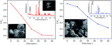
A basic bismuth(III) nitrate photocatalyst with the composition of Bi6O6(OH)3(NO3)3·1.5H2O (BBN) was facilely synthesized using a hydrothermal strategy via incomplete hydrolysis of bismuth nitrate. Characterization of the composition, morphology, microstructure, optical absorption, BET surface area, and photocatalytic behavior was systematically explored. The results indicated that BBN architectures built up of multilayered meshing-teeth structures with predominant {110} side facets can be selectively obtained by fine-tuning the reaction parameters. The sample exhibits an obviously superior photocatalytic activity for the degradation of phenol compared with BBN sheets with dominant top {001} planes and commercial P25, with the rate constant k improved by 3.6 and 2.8 fold, respectively. The excellent photocatalytic behavior combined with the rather low BET surface area of 0.0453 m2 g−1 indicate that the highly reactive {110} facets in BBN are responsible for the photocatalysis. The active oxidation species and main intermediates in the phenol/BBN system are ascertained using scavenger experiments and high performance liquid chromatography (HPLC) techniques. Combining the band edge of BBN and the redox potentials of the active species, a possible migration mechanism of photogenerated e−/h+ pairs on the surface of BBN is proposed. This work provides some new insights for the rational design and synthesis of active-facet exposed basic salt photocatalysts with excellent efficiency.
1. Introduction
Phenol and its derivatives are categorized as some of the most serious environmental contaminants discharged by industrial plants due to their toxicity, carcinogenicity and difficulty to degrade.1,2 In recent years, semiconductor photocatalysis has gradually become one of the most effective technologies for the degradation of phenolic wastewater since it can completely treat the pollutants to provide harmless end products under mild conditions.3–6 The key issues in the photocatalytic degradation of phenol contaminants in wastewater depend on the performance of the photocatalytic materials used. Developing a new efficient photocatalyst to meet future environmental requirements is still one of the focuses of current studies.
Among the numerous semiconductors, bismuth-containing materials have received great attention in environmental remediation over the past few years. Bismuth (Bi) is a p-block metal with a d10 configuration, and the Bi 6s orbitals can interact with O 2p orbitals to form a preferable hybridized valence band (VB), which favors the mobility of photogenerated holes in the VB and benefits the enhancement of the photocatalytic performance of Bi3+-based oxides.7 So far, bismuth compounds with satisfactory photocatalytic properties for the degradation of organic pollutants have been gradually researched in the photocatalytic field such as Bi2O3,8,9 Bi2WO6,10,11 Bi2MoO6,12,13 BiVO4,14,15 BiOX (X = Cl, Br, I),16–18 etc. Bi-based basic salts are also members of the bismuth compound family and have shown very rich crystal chemistry with almost 15 different structures discovered.19 They were preliminarily studied for medical applications and as a precursor for bismuth oxides,20 seldom attention has been given in the domain of photocatalysis.
Just recently, basic bismuth nitrate nanosheets with a complex composition of Bi6O6(OH)3(NO3)3·1.5H2O were found to be a new type of photocatalyst for UV-light degradation of dyes such as malachite green and methyl orange.21,22 But for nanosized photocatalysts, it is difficult to thoroughly recollect them from the degradation solution and thus this brings about secondary pollution. Also, organic dyes can often be extensively degraded under UV illumination even in the absence of a photocatalyst. Our recent research has shown that BBN is also an efficient co-catalyst to Bi2WO6 for degradation of Rhodamine B due to band-gap coupling effects.23 Although a double-layered BBN architecture has been synthesized by Liu et al. for removal of phenol, it only exhibited comparable UV photocatalytic activity with the classic P25.24 In the very limited reports, no systematical exploration has been carried out to clarify the influence of the preparation parameters on the structure, which is a prerequisite for better understanding of the relationship between the specific structure and photocatalytic performance. Usually, the photocatalytic reaction occurs at the interface of the reactant and catalyst, and photocatalytic activity is strongly dependent on the surface morphology and exposed crystal facets.25–27 Pioneering works on the synthesis of anatase28,29 and BiOCl30,31 with highly reactive facets have paved a new way for the enhancement of photocatalytic performance. Undoubtedly, facet engineering is not only an exciting direction to pursue for highly active new-generation photocatalysts but also offers opportunities to investigate the relationship between the surface properties and the photocatalytic properties.
In this paper, we first report on the controllable synthesis of BBN architectures built up of multilayered tooth-meshing structures with predominantly {110} side surfaces and the facet-dependent photoreactivity for the degradation of phenol under UV light. The {110} facet predominated BBN photocatalyst exhibits an obviously superior photocatalytic activity over less layered counterparts and individual BBN sheets with dominant {001} crystal planes. It also presents a much higher photocatalytic behavior than P25 with the rate constant improved by 2.8 fold. The migration direction of photoinduced carriers at the reaction interface is furthermore provided based on the results of the active species and intermediates.
2. Experimental section
Preparation
All the reagents were of analytical grade and used without further purification. BBN with predominant {110} facets was prepared as follows: 1 mmol of Bi(NO3)3·5H2O was dissolved in 17.0 mL of distilled water. After stirring for 30 min, the resulting suspension was transferred into a 25 mL Teflon-lined stainless steel autoclave and heated at 120 °C for 8 h. After the autoclave cooled to room temperature naturally, the solid products were collected by centrifugation, washed repeatedly with deionized water and absolute ethanol several times, and then dried at 60 °C in air. For comparison, a series of parallel experiments was designed. The temperature series samples were treated at a temperature between 80 and 200 °C for 8 h and the time series samples were prepared at 120 °C for a different number of hours, keeping the other conditions unchanged (Table S1†).
Characterization
The crystal phase and composition of the samples were characterized using X-ray diffraction (XRD) on a Bruker D8-Advance diffractometer with Cu Kα (λ = 0.15418 nm) radiation. The morphologies and microstructures of the prepared products were examined with a field-emission scanning electron microscope (FEI, NOVA Nano SEM 230) and a high-resolution transmission electron microscope (FEI, Tecnai G2 F20). The BET surface area measurements were obtained through N2 adsorption–desorption isotherms collected at liquid nitrogen temperature using a Micromeritics ASAP2020 surface area and porosity analyzer. The UV-vis diffuse reflectance spectra were recorded on a JASCO V-550/V-570 UV-vis spectrophotometer fitted with an integrating sphere accessory.
Photocatalytic tests
The photocatalytic activities of the samples were evaluated for the degradation of a phenol aqueous solution under UV irradiation (300 W Hg lamp) in a photoreactor equipped with a water circulation facility (Fig. S1†). In each test, 10.0 mg of photocatalyst was added into a phenol solution with an initial concentration of 10−4 M in a quartz tube. Before illumination, the suspension was magnetically stirred in the dark for 30 min to establish an adsorption–desorption equilibrium. Then after given time intervals of UV irradiation, one quartz tube was taken out and the photocatalyst was immediately separated by centrifugation to analyze the supernate using a Shimadzu 2550 UV-vis spectrophotometer.
The degradation intermediates from phenol were monitored using an HPLC instrument of the Agilent 1100 series (Palo Alto, CA, USA) equipped with an Agilent Zorbax Eclipse XDB-C18 column (150 mm × 4.6 mm, 5 μm). During sample analysis, the column was maintained at 30 °C. The intermediates were detected using an isocratic elution program. Methanol and water with a volume ratio of 30
:
70 were used as the mobile phase at a flow-rate of 1 mL min−1 and the injection volume was 20 μL.
3. Results and discussion
Phase, morphology and microstructure analysis
Fig. 1 shows a typical XRD pattern of the hydrothermal sample selectively prepared at 120 °C for 8 h. All diffraction peaks match well with those of tetragonal basic bismuth(III) nitrate BBN (a = b = 3.818 Å, c = 17.149 Å, JCPDS: 53-1038). No signs of impurity phases such as Bi, BiO(NO3), Bi2O3, and Bi(NO3)3 were detected, showing the high purity of the product. The sharp and narrow diffraction peaks indicate the high crystallinity of BBN. But it was interestingly noted that the intensity ratios of the diffraction peaks show an obvious difference to that of the standard values. The (002) peak is greatly inhibited but the (110) and (102) peaks are dramatically strengthened. In particular, the diffraction intensity ratio of the (110)/(002) planes in the as-obtained BBN sample is 2.5, increased 25 fold compared with the standard value of 0.1. The intensity ratio of (102)/(002) has also been enhanced by 12.2 times. The XRD results demonstrate the preferential growth of a specific crystal plane in BBN.
 |
| Fig. 1 XRD pattern of the as-synthesized BBN sample and at the bottom is the JCPDS standard. | |
The morphology and microstructure of BBN were identified using SEM and TEM. The panoramic SEM image in Fig. 2a indicates that the sample consists of monodispersed globular-like structures mostly ranging from 20 to 37 μm in diameter with a coarse surface. The close-up view of a few BBN architectures in Fig. 2b shows that the spheres are built from the multilayered tooth-meshing of 10–15 layers. The coarse surface comes from the ragged arrangement of triangle-shaped teeth with a side length of 1–3 μm. Further investigations were carried out using TEM to give detailed insights into the microstructures of BBN. The cog-wheel outline of an isolated BBN particle shown in the TEM image (Fig. 2c) agrees well with the tooth-meshing structure indicated in SEM. Fig. 2d shows the HRTEM image of the edge part obtained from the top surface of a BBN meshing-tooth. A clear lattice fringe with a spacing of 0.270 nm corresponds to the {110} crystal planes and implies high crystallinity of the meshing-tooth. The selected-area electron diffraction (SAED) in Fig. 2e with a well-ordered dot pattern further confirms the single crystal nature of the building unit and can be well indexed to {110} equivalent planes. The ED pattern was projected from the [001] zone axis of a BBN meshing-tooth. Therefore, the exposed side surface of the tooth structure contains dominant {110} facets, while the top surface of the tooth unit belongs to {001} facets. The TEM data coupled with the XRD results strongly confirm that the BBN sample with a multi-layered meshing-teeth structure exhibits predominantly {110} crystal planes.
 |
| Fig. 2 (a) Panoramic FE-SEM image of the product, (b) magnified FE-SEM image, (c) TEM image of an isolated structure, (d) HRTEM image of the edge part in (c), and (e) the corresponding SAED pattern. | |
Influence of the preparation parameters
To gain a better understanding of the formation process of the BBN superstructures, products obtained at different growth stages were collected for SEM and XRD measurements. As shown in Fig. 3a, the precursor presents as orderly rods with lengths of 10–25 μm and widths of 2–5 μm, respectively. As the reaction proceeds for 30 min (Fig. 3b), the rods become disordered and tend to construct a few clusters. After 50 min of reaction, some multilayered tooth-meshing architectures have appeared with the coexistence of precursor rods and transitional clusters. The fast transformation from clusters to tooth-meshing superstructures may disclose the metastable properties of the intermediate morphology. The XRD patterns show that the products before 50 min are complex and difficult to index due to the complicated hydrolysis of bismuth nitrate.32 After 2 h of treatment, the rods and clusters disappear completely and all the particles present a tooth-meshing structure but with much less layers and non-uniform diameters. At this stage, the XRD pattern can be ascribed to tetragonal BBN and the (110) and (102) peaks show preferential growth compared with the standard file. Finally, BBN architectures of well-defined multilayered tooth-meshing are formed via the Ostwald ripening process33 at 8 h. The prevailing (110) diffraction was in sharp contrast to great inhibition of the (002) peak. It seems that the more exposed side faces in the multilayered meshing-teeth structures account for the obvious change in facet orientation. It has been ascertained using HRTEM that the side surface belongs to {110} planes.
 |
| Fig. 3 SEM images and XRD patterns of the time series samples: (a) precursor, (b) 30 min, (c) 50 min, (d) 2 h, (e) 8 h, and (f) corresponding XRD patterns. | |
The influence of the hydrothermal temperature on the morphology and structure of BBN were also investigated. As shown in Fig. 4a, the sample obtained at 80 °C still consisted of randomly constructed rods with a complicated XRD pattern. For elevated temperatures of 100–120 °C, the samples were distinguished from the former with morphologies of multilayered tooth-meshing (Fig. 4b and c). But the architectures formed at 120 °C are more uniform and obviously have more layers of meshing-teeth. Although the BBN products both exhibit preferential (110) planes, a vivid comparison gives a 1.3-fold higher intensity ratio of (110)/(002) planes at 120 °C. Further increase of the hydrothermal temperature leads to great changes in both morphology and structure. As shown in Fig. 4d, the compact BBN architectures become cracked with 3D meshing-teeth gradually replaced by flattened sheets at 160 °C. The corresponding XRD pattern gives an obviously strengthened (002) diffraction peak. At a high temperature of 200 °C, the BBN architectures have been extensively destroyed, accompanied by the growth of isolated and irregular sheets with a lateral size of 20–40 μm. The XRD pattern displays a predominant (002) peak with the ratio of (002)/(110) approaching the standard value. The results further indicate that the exposed side surface of the meshing-tooth structure should be mainly attributed to {110} facets. So an appropriate hydrothermal temperature and time are both key factors to achieve BBN structures with preferentially oriented {110} facets.
 |
| Fig. 4 SEM images and XRD patterns of the temperature series samples: (a) 80 °C, (b) 100 °C, (c) 120 °C, (d) 160 °C, (e) 200 °C, and (f) the corresponding XRD patterns. | |
Optical properties
To confirm the optical absorption properties of the BBN architectures, a UV-vis DRS spectrum was obtained and the results are shown in Fig. 5. According to the spectrum, the BBN presents photoabsorption of UV light with a threshold of 375 nm. The steep shape of the spectrum indicates that the light absorption is not due to a transition from the impurity level but comes from the intrinsic band-gap transition.34 Thus the BBN with the composition of Bi6O6(OH)3(NO3)3·1.5H2O behaves like a bulk semiconductor. For a crystalline semiconductor, the optical absorption near the band edge follows the equation αhν = A(hν − Eg)n/2, where α, hν, A, and Eg are the absorption coefficient, photonic energy, proportionality constant, and band gap, respectively.35 The value of n indicates the characteristics of the transition in a semiconductor.36 According to previous literature24 and the clear region of linearity in (αhν)1/2 vs. hν, the value of n for BBN is ascertained to be 4, as for an indirect transition. As can be expected based on the absorption spectra, the sample is white in color. The intercept of the tangent to the plot gives a good approximation of the band gap energy of 3.3 eV. The DRS spectrum indicates that the BBN can be used as a photocatalyst under UV irradiation.
 |
| Fig. 5 UV-vis diffuse reflectance spectrum of a BBN sample with Eg analysis in the inset. | |
Photocatalytic properties
Photocatalytic degradation of phenol over the temperature and time series samples was evaluated under the irradiation of a 300 W Hg lamp with the results shown in Fig. 6. A contrast test showed that only ca. 6.5% of the phenol was degraded in the dark in the presence of BBN, indicating that the adsorption of phenol can be negligible. The blank test in the absence of BBN conversely presents an increased absorption at 270 nm in the initial stage (Fig. S2†). This phenomenon has been reported in the study of UV photolysis of phenol and it is ascribed to excessively accumulated intermediates with a similar structure to phenol.37 The slightly decreased absorbance after 60 min irradiation in the absence of BBN shows the ineffectiveness of direct photolysis. The BBN sample prepared at 120 °C for 8 h gives the best photocatalytic behavior with 98.1% removal of phenol in 45 min, which is much better than commercial P25 under the same conditions. As shown in Fig. 6b, the pseudo first-order kinetics constant k of the phenol degradation over BBN was calculated to be 0.08945 min−1, improved by a factor of 2.8 in comparison with that of P25 (0.03192 min−1). It should be noted that the BET surface area of BBN was detected to be merely 0.045 m2 g−1, which is considerably smaller than the 43.26 m2 g−1 of the most widely used P25. The superior photocatalytic activity and rather low surface area may suggest a preferential orientation of the reactive facets in BBN. The as-obtained sample also exhibits a higher degradation efficiency than the reported double-layered BBN architecture.24 For example, 38% of the phenol has been degraded after 15 min irradiation over the multilayered BBN in comparison with the value of only 6.0% over double-layered BBN. Also, there is still 18% of the phenol left after 45 min of photocatalysis for the double-layered BBN in contrast to the small residue of 1.9% in our work. The results may imply an important role of the side face of the BBN meshing-teeth in photocatalysis, which is further confirmed by the following photocatalytic results over the temperature and time series BBN samples.
 |
| Fig. 6 Photocatalytic degradation of phenol and the corresponding pseudo first-order plots over: (a and b) the temperature series samples, and (c and d) the time series samples. | |
The hydrothermal temperature has a significant influence on the photocatalytic activity of BBN. As shown in Fig. 6a and b, the degradation of phenol increases at first and then decreases with the increase of the temperature, and the best activity is achieved at 120 °C. The rate constant k for the 120 °C sample is as much as 2.4 and 3.6 times those for the BBN samples obtained at 100 and 200 °C, respectively. From the above SEM and XRD analysis, it is known that the BBN-120 °C sample presents the most layers of meshing-teeth with predominantly {110} side surfaces. However, BBN-200 °C exhibits prevailing microsheets with extremely diminished side faces but dominant {100} top surfaces. The BET surface area of the sheet-shaped BBN is 0.245 m2 g−1, which is 5.4-fold higher than that of BBN-120 °C. The much improved photocatalytic activity combined with the lowered surface area confirms the high reactivity of the predominant {110} facets in BBN-120 °C. Fig. 6c and d show that the reaction time is another important factor for the photocatalytic activity of BBN. The 50 min sample gives a rather low photocatalytic activity due to the partial formation of tooth-meshing architectures as shown in Fig. 4. The degradation of phenol is accelerated over BBN for prolonged hydrothermal times with more layers of meshing-teeth. But a conversely lowered activity was detected for BBN-12 h with the rate constant k only 34% of that of BBN-8 h. The structure and morphology analysis (Fig. S3†) implied that BBN-12 h shows decreased layers of meshing-teeth and much inhibited (110) planes. The results further confirm the highly reactive {110} tooth surface of BBN.
Effects of the process parameters
The photocatalytic performance of a catalyst is affected by various process parameters. The influence of BBN dosage and the initial concentration of the phenol solution was investigated. As shown in Fig. 7a, the degradation efficiency for phenol is firstly increased and then decreased with the enhancement of the BBN amount and the best BBN dosage was determined to be 10 mg. The corresponding rate constant k (inset) over 10 mg of BBN was improved 3.5 and 3.0 fold compared with those of 5 and 20 mg of BBN, respectively. Obviously, a lower catalytic activity is observed when a smaller amount of catalyst is used because less catalytically active sites are available. However, excessive BBN powder would cause an increase in the opacity of the degradation solution and light scattering, and even a decrease in the number of catalytic surface active sites caused by aggregation of the photocatalyst particles.38 So there is often an optimum amount of photocatalyst to achieve the maximum photocatalytic efficiency. These phenomena have also been reported in previous literature.39,40
 |
| Fig. 7 The degradation of phenol over BBN under different operating conditions: (a) BBN dosage, (b) the initial concentration of phenol. | |
Fig. 7b presents the influence of the initial phenol concentration (C0) on the photocatalytic activity. It can be seen that a lower phenol concentration gives a more efficient degradation. A higher phenol concentration of 1.5 × 10⁻⁴ M gives a decreased degradation ratio by ca. 20%. It is believed that as the C0 of phenol increases, the surface active sites of BBN would be over loaded with additional phenol molecules. As a result, the photo-generation of reactive oxygen species would be reduced. In addition, more incident photons would be intercepted by the increase in phenol molecules before they reach and react with the photocatalyst.41 So 10 mg of BBN per 10 mL of phenol solution (10−4 M) was selected as the optimum photocatalytic parameter.
Photocatalytic mechanism
To evaluate the role of the primary reactive species, scavenger experiments were performed by adding individual scavengers to the phenol/BBN system and the results are shown in Fig. 8. The addition of tert-butanol (TBA), a well-known scavenger of ˙OH radicals,42 into the photoreaction system causes the degradation efficiency to be decreased by 16.6% after 45 min irradiation. This weak inhibition indicates that ˙OH may be not the major species in the oxidation process. However, introduction of the holes scavenger ethylene diamine tetra-acetic acid (EDTA)42 gives a greatly depressed degradation efficiency of 50.1% at 45 min. The presence of benzoquinone (BQ), a superoxide radical (O2−˙) scavenger,43 leads to the strongest inhibition for phenol degradation. In addition, the photocatalysis was carried out with continuous N2-sparging due to the non-volatility of phenol. Under the anoxic conditions, the degradation of phenol is also significantly suppressed. The presence of oxygen is important and its role is to primarily act as an efficient e− trap, leading to the generation of O2−˙ and preventing the recombination of e− and h+.44,45 The similar results from added BQ and the N2-saturated suspension together confirm the most important effects of O2−˙. Accordingly, the reactive species are believed to play their oxidation roles in a sequence of importance of O2−˙, h+, and then ˙OH in the phenol/BBN system.
 |
| Fig. 8 Photocatalytic degradation of phenol over BBN with the addition of scavengers of different reactive species and in N2-saturated solution. | |
The photocatalytic oxidation of phenol often involves complicated processes. HPLC was utilized to monitor the evolution of intermediates during phenol degradation over the BBN with dominant {110} facets. Fig. 9a–c show typical HPLC chromatograms of the phenol degradation solutions before and after irradiation for different times. The original solution exhibits only one strong peak at the retention time of 9.5 min corresponding to phenol (Fig. 9a). The intensity of the phenol peak decreases gradually with prolonged irradiation and disappears after 45 min (Fig. 9b and c), indicating full degradation of phenol and in good consistency with the results shown in Fig. 6a. In addition to the undegraded phenol, three major aromatic products were detected in the initial stage with a retention time of 2.2, 3.1 and 4.6 min, respectively. The intermediates were identified to be hydroquinone, resorcinol and catechol by comparison with the chromatograms of standard materials.
 |
| Fig. 9 (a–c) HPLC chromatograms of phenol photodegradation by BBN for different times, (d) concentrations of the intermediates formed from phenol during the photocatalysis. | |
Corresponding concentration–time curves calculated using internal standard calibration with good linear correlation (Table S2†) are shown in Fig. 9d. Accompanied with a continuously decreasing concentration of phenol (Fig. S4†), the concentrations of the intermediates are enhanced at an earlier stage and then reduce quickly. Specifically, catechol and hydroquinone achieve a maximum concentration of 0.00113 and 0.00045 mg mL−1 at 8 min, respectively, and resorcinol reaches a peak concentration of only 0.00029 mg mL−1 at 3 min. According to substitution rules, ˙OH radicals attack the phenol molecules with higher probability at the 2 and 4 positions versus the OH group of the aromatic ring. So it is rational that the m-substituted resorcinol presents a lower concentration. In previous reports on the traditional TiO2, benzoquinone is also a common organic byproduct generated during the degradation of phenol.46 It is different that no obvious benzoquinone peak was detected in the phenol/BBN system. O2−˙ and h+ have been ascertained to be the dominant active species, which are nonselective for organic degradation with strong oxidation abilities. It may be inferred that the life span of benzoquinone formed at different stages of the reaction is short due to the fast oxidation by O2−˙ and h+. The transient presence of benzoquinone with a low concentration would make it difficult to be detected.24 The aromatic intermediates would undergo further photocatalytic oxidation quickly to form nontoxic aliphatic acids by ring cleavage47 and subsequently be completely transformed to CO2 and H2O.
It is well known that the effective separation and transfer of photoinduced electrons and holes are important factors for a desired photocatalyst. The key to causing the migration of carriers is the difference between the redox potential of active species and the positions of the valence band (VB) and conduction band (CB) of the semiconductor. Combining the band edges of BBN with the ascertained oxidative species, a proposed migration mechanism of the photogenerated carriers on the surface of BBN is illustrated in Fig. 10. The BBN is excited by UV irradiation to generate electrons in the CB and holes in the VB, which then migrate to the surface of the photocatalyst. It has been reported that BBN exhibits n-type characteristics with a CB potential at −0.90 V,22 which is more negative than the redox potential of O2/O2−˙ (−0.28 V vs. NHE).48 Then forced by the potential difference, the CB electrons can be quickly captured by adsorbed molecular oxygen from the water to form O2−˙, which has been confirmed to be the most important active species. The VB holes are believed to play vital roles, through two different routes, at the phenol/BBN interface. The major one is the direct degradation of phenol due to the strong oxidative ability of BBN holes with a calculated VB position of 2.40 V based on the estimated Eg of 3.3 eV from the DRS spectra. The direct oxidation has been affirmed by the greatly inhibited degradation efficiency with added EDTA. In addition, the holes can also oxidize the OH− or H2O in the phenol solution to form ˙OH owing to the slightly anodic VB position compared with the redox potential of ˙OH/OH− (2.38 V vs. NHE).49 The ˙OH has also been confirmed to be one of the active species in the phenol/BBN system although not the most important. Then the ˙OH reacts with phenol molecules in the initial stage of photocatalysis, leading to the formation of benzenediol intermediates as monitored by HPLC. Then with the synergism of the active species of O2−˙, h+, and ˙OH, the phenol and intermediates would subsequently be decomposed into nontoxic aliphatic acids47,50 and finally transformed to H2O and CO2.
 |
| Fig. 10 Schematic illustration of the charge transfer on the BBN surface and the degradation process for phenol. | |
4. Conclusions
In summary, the BBN photocatalyst Bi6O6(OH)3(NO3)3·1.5H2O with predominately exposed {110} facets was selectively prepared. The sample exhibits much higher photocatalytic activities for phenol than that of BBN with dominant {001} planes and commercial P25, with the rate constant k improved by 3.6 and 2.8 fold, respectively. This suggests that the {110} facets should be reactive high-energy crystal planes in BBN. This work provides a better understanding of the correlations between the preparation parameters, specific structure and photocatalytic performance of the novel photocatalyst. The scavenger experiments and HPLC techniques disclose the dominant active species and main intermediates in the initial stage of photocatalysis. A possible migration mechanism of the photogenerated carriers on the surface of BBN was proposed.
Acknowledgements
The project was supported by the National Natural Science Foundation of China (No. 21303122), the Program for Innovative Research Team in University of Tianjin (TD12-5038), and the Open Project Program of Tianjin Key Laboratory of Structure and Performance for Functional Molecules in 2014.
References
- L. H. Keith and W. A. Telliand, Environ. Sci. Technol., 1979, 13, 416–423 CrossRef.
- F. J. Diaz, A. T. Chow, A. T. O’Geen, R. A. Dahlgren and P. K. Wong, Water Res., 2009, 43, 2750–2760 CrossRef CAS PubMed.
- M. R. Hoffmann, S. T. Martin, W. Choi and D. W. Bahnemann, Chem. Rev., 1995, 95, 69–96 CrossRef CAS.
- S. W. Liu, J. G. Yu and M. Jaroniec, J. Am. Chem. Soc., 2010, 132, 11914–11916 CrossRef CAS PubMed.
- M. Mecklenburg, A. Schuchardt, Y. K. Mishra, S. Kaps, R. Adelung, A. Lotnyk, L. Kienle and K. Schulte, Adv. Mater., 2012, 24, 3486–3490 CrossRef CAS PubMed.
- G. W. Cui, W. L. Wang, M. Y. Ma, M. Zhang, X. Y. Xia, F. Y. Han, X. F. Shi, Y. Q. Zhao, Y. B. Dong and B. Tang, Chem. Commun., 2013, 49, 6415–6417 RSC.
- J. W. Tang, Z. G. Zou and J. H. Ye, Angew. Chem., Int. Ed., 2004, 43, 4463–4466 CrossRef CAS PubMed.
- L. Zhang, W. Wang, J. Yang, Z. Chen, W. Zhang, L. Zhou and S. Liu, Appl. Catal., A, 2006, 308, 105–110 CrossRef CAS PubMed.
- X. Xiao, R. P. Hu, S. H. Tu, C. X. Zheng, H. Zhong, X. X. Zuo and J. M. Nan, RSC Adv., 2015, 5, 38373–38381 RSC.
- F. Amano, A. Yamakata, K. Nogami, M. Osawa and B. Ohtani, J. Am. Chem. Soc., 2008, 130, 17650–17651 CrossRef CAS PubMed.
- L. S. Zhang, H. L. Wang, Z. G. Chen, P. K. Wong and J. S. Liu, Appl. Catal., B, 2012, 47, 1919–1924 Search PubMed.
- G. Tian, Y. Chen, W. Zhou, K. Pan, Y. Dong, C. Tian and H. Fu, J. Mater. Chem., 2011, 21, 887–892 RSC.
- M. Zhang, C. Shao, P. Zhang, C. Su, X. Zhang, P. Liang, Y. Sun and Y. Liu, J. Hazard. Mater., 2012, 225–226, 155–163 CrossRef CAS PubMed.
- D. Tang, H. C. Zhang, H. Huang, R. H. Liu, Y. Z. Han, Y. Liu, C. Y. Tong and Z. H. Kang, Dalton Trans., 2013, 42, 6285–6289 RSC.
- D. Ke, T. Peng, L. Ma, P. Cai and K. Dai, Inorg. Chem., 2009, 48, 4685–4691 CrossRef CAS PubMed.
- Y. Xu, S. C. Xu, S. Wang, Y. X. Zhang and G. H. Li, Dalton Trans., 2014, 43, 479–485 RSC.
- J. Xiong, Q. Dong, T. Wang, Z. Jiao, G. Lu and Y. Bi, RSC Adv., 2014, 4, 583–586 RSC.
- R. Hao, X. Xiao, X. Zuo, J. M. Nan and W. D. Zhang, J. Hazard. Mater., 2012, 209–210, 137–145 CrossRef CAS PubMed.
- N. Henry, M. Evain, P. Deniard, S. Jobic, O. Mentré and F. Abraham, J. Solid State Chem., 2003, 176, 127–136 CrossRef CAS.
- N. Henry, O. Mentre, J. C. Boivin and F. Abraham, Chem. Mater., 2001, 13, 543–551 CrossRef CAS.
- L. Y. Xie, J. X. Wang, Y. H. Hu, Z. Y. Zheng, S. X. Weng and P. Liu, Mater. Chem. Phys., 2012, 136, 309–312 CrossRef CAS PubMed.
- L. Y. Xie, J. X. Wang, Y. H. Hu, S. Y. Zhu, Z. Y. Zheng, S. X. Weng and P. Liu, RSC Adv., 2012, 2, 9881–9886 RSC.
- Y. Cui, L. M. Yang, G. Y. Zhang and Y. Feng, Catal. Commun., 2015, 59, 83–87 CrossRef CAS PubMed.
- Y. X. Yang, H. Y. Liang, N. Zhu, Y. P. Zhao, C. S. Guo and L. Liu, Chemosphere, 2013, 93, 701–707 CrossRef CAS PubMed.
- S. He, S. Zhang, J. Lu, Y. Zhao, J. Ma, M. Wei, D. G. Evans and X. Duan, Chem. Commun., 2011, 47, 10797–10799 RSC.
- Z. Y. Jiang, Q. Kuang, Z. X. Xie and L. S. Zheng, Adv. Funct. Mater., 2010, 20, 3634–3645 CrossRef CAS PubMed.
- Z. Y. Zhou, N. Tian, J. T. Li, I. Broadwell and S. G. Sun, Chem. Soc. Rev., 2011, 40, 4167–4185 RSC.
- H. G. Yang, C. H. Sun, S. Z. Qiao, J. Zou, G. Liu, S. C. Smith, H. M. Cheng and G. Q. Lu, Nature, 2008, 453, 638–641 CrossRef CAS PubMed.
- G. Liu, H. G. Yang, X. Wang, L. Cheng, J. Pan, G. Q. Lu and H. M. Cheng, J. Am. Chem. Soc., 2009, 131, 12868–12869 CrossRef CAS PubMed.
- J. Jiang, K. Zhao, X. Y. Xiao and L. Z. Zhang, J. Am. Chem. Soc., 2012, 134, 4473–4476 CrossRef CAS PubMed.
- Y. Xu, S. C. Xu, S. Wang, Y. X. Zhang and G. H. Li, Dalton Trans., 2014, 43, 479–485 RSC.
- A. N. Christensen, M. A. Chevallier, J. Skibsted and B. B. Iversen, J. Chem. Soc., Dalton Trans., 2000, 265–270 RSC.
- Z. Chen, L. W. Qian, J. Zhu, Y. P. Yuan and X. F. Qian, CrystEngComm, 2010, 12, 2100–2106 RSC.
- A. Kudo, I. Tsuja and H. Kato, Chem. Commun., 2002, 1958–1959 RSC.
- Y. Ohko, K. Hashimoto and A. Fujishima, J. Phys. Chem. A, 1997, 101, 8057–8062 CrossRef CAS.
- J. Zhang, F. J. Shi, D. F. Chen, J. M. Gao, Z. X. Huang, X. X. Ding and C. C. Tang, Chem. Mater., 2008, 20, 2937–2941 CrossRef CAS.
- C. X. Li, Y. T. Li and Z. H. Wang, Tech. Equip. Environ. Pollut. Control, 2002, 8, 48–51 Search PubMed.
- M. A. Gondal, X. F. Chang and Z. H. Yamani, Chem. Eng. J., 2010, 165, 250–257 CrossRef CAS PubMed.
- X. Chang, G. Ji, Q. Sui and J. Huang, J. Hazard. Mater., 2009, 166, 728–733 CrossRef CAS PubMed.
- G. Y. Zhang, Y. Feng, Y. Y. Xu, D. Z. Gao and Y. Q. Sun, Mater. Res. Bull., 2012, 47, 625–630 CrossRef CAS PubMed.
- I. K. Konstantinou and T. A. Albanis, Appl. Catal., B, 2004, 49, 1–14 CrossRef CAS PubMed.
- H. J. Lu, L. L. Xu, B. Wei, M. Y. Zhang, H. Gao and W. J. Sun, Appl. Surf. Sci., 2014, 303, 360–366 CrossRef CAS PubMed.
- M. Ge, N. Zhu, Y. P. Zhao, J. Li and L. Liu, Ind. Eng. Chem. Res., 2012, 51, 5167–5173 CrossRef CAS.
- Z. J. Zhang, W. Z. Wang, E. P. Gao, M. Shang and J. H. Xu, J. Hazard. Mater., 2011, 196, 255–262 CrossRef CAS PubMed.
- H. B. Fu, C. S. Pan, W. Q. Yao and Y. F. Zhu, J. Phys. Chem. B, 2005, 109, 22432–22439 CrossRef CAS PubMed.
- L. Liu, H. J. Liu, Y. P. Zhao, Y. Q. Wang, Y. Q. Duan, G. D. Gao, M. Ge and W. Chen, Environ. Sci. Technol., 2008, 42, 2342–2348 CrossRef CAS.
- Z. Deng, C. Zhang and L. Liu, Intermetallics, 2014, 52, 5–14 CrossRef PubMed.
- Y. L. Zhang, D. Y. Li, Y. G. Zhang, X. F. Zhou, S. J. Guo and L. B. Yang, J. Mater. Chem. A, 2014, 2, 8273–8280 CAS.
- Y. Y. Liu, Z. Y. Wang, B. B. Huang, X. Y. Zhang, X. Y. Qin and Y. Dai, J. Colloid Interface Sci., 2010, 348, 211–215 CrossRef CAS PubMed.
- X. Y. Li, Y. H. Cui, Y. J. Feng, Z. M. Xie and D. J. Gu, Water Res., 2005, 39, 1972–1981 CrossRef CAS PubMed.
Footnote |
† Electronic supplementary information (ESI) available. See DOI: 10.1039/c5ra15629g |
|
This journal is © The Royal Society of Chemistry 2015 |
Click here to see how this site uses Cookies. View our privacy policy here.