Silybin, a flavonolignan from milk thistle seeds, restrains the early and advanced glycation end product modification of albumin

Saurabh Awasthi and N. T. Saraswathi*
Molecular Biophysics Lab. School of Chemical and Biotechnology, SASTRA University, Thanjavur-613401, Tamilnadu, India. E-mail: saras@scbt.sastra.edu

Received 4th August 2015 , Accepted 9th October 2015

First published on 9th October 2015


Abstract

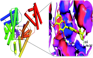
We report the inhibitory activity of silybin, a flavonolignan against the formation of early and advanced glycation end products based on fluorescence, circular dichroism and molecular interaction studies. Silybin was found to be a potent inhibitor for both the early and advanced glycation end products. The inhibition of glycation mediated fibrillation of BSA was determined based on the binding of amyloid specific dyes i.e. thioflavin T, congo red and confocal imaging. Also, we determined the protective effect of silybin towards the oxidative damage induced hemolysis of erythrocytes. The molecular interaction pattern of silybin revealed its better affinity with BSA when compared to the other natural inhibitors of glycation such as quercetin, epigallocatechin gallate, and curcumin.


Introduction

The Maillard reaction and formation of advanced glycation end products (AGEs) are the fundamental causes in the development of oxidative stress and many pathological conditions such as retinopathy, neuropathy, nephropathy and micro-vascular complications.1,2 The Maillard reaction is a multi step non-enzymatic process which is initiated due to the reaction of glucose with free amino groups in proteins leading to the formation of highly reactive AGEs. AGEs are characterized by their specific properties such as fluorescence, and cross linking.3,4 Albumin is the most abundant protein in the plasma and highly prone for non-enzymatic glycation. Glycation alters the structure and function of proteins leading to the cross linking and aggregation. Glycation accelerate fibrillation and formation of amyloid like aggregation in proteins and studies have shown that glycation exacerbates the neuronal toxicity of amyloid peptide.5 In the recent time research has been focused on small molecule inhibitors for prevention of non-enzymatic glycation. In spite of the strong antiglycation properties, the potent antiglycation compounds such as aminoguanidine (AG) and metformin (MET) have been found to be associated with severe side effects, which also led to the withdrawal of AG from the 3rd phase of clinical trial.6 Due to the side effects of synthetic compounds the natural products are promising towards development of a potent inhibitor for the formation of AGEs.

Silybin (SIB) is a flavonolignan and one of the major constituents of silymarin. SIB is a mixture of two diastereoisomers known as silybin A and silybin B in a ratio of 1[thin space (1/6-em)]:[thin space (1/6-em)]1.7 The anti-diabetic, antioxidant, anticancer and hepatoprotective activities of silybin have been reported from both in vitro and in vivo examinations. Also in vivo studies using mice have proven the clinical efficacy of silybin.8–10 In our earlier work, we evaluated the antiglycation activity of Sesbania grandiflora leaves and aspartic acid. Aspartic acid showed antiglycation effects by carbonyl trapping and chemical chaperon like activity.11,12

The present study was carried out to demonstrate the antiglycation potential of silybin using in vitro glycation model of BSA. Assays were carried out to determine the inhibition of early and advanced glycation end products based on measurements of fructosamine, and fluorescent AGEs such as argpyrimidine, pentosidine, crossline and vesperlysine. The protein bound carbonyl content was also estimated in order to get insights about the glycation modification of BSA. Circular dichroism (CD) analysis revealed that SIB imparted protective effects towards the native helical structure of BSA. The inhibition of glycation induced fibrillation was studied based on thioflavin T and congo red binding. Furthermore the protective effect of SIB towards the functional properties of BSA was determined based on the oxidative stress induced hemolysis of erythrocytes. Based on the observations it was evident that silybin restrains formation of advanced glycation end products and fibrillation of albumin.

Experimental

Chemicals

Bovine serum albumin (BSA), silybin (SIB), methylglyoxal (MG), aminoguanidine (AG), 2,4,6-trinitrobenzenesulfonic acid (TNBSA), thioflavin T (Th T), and congo red (CR) was purchased from Sigma, USA. 2,4-Di-nitro phenyl hydrazine (DNPH), guanidine hydrochloride and nitro blue tetrazolium (NBT) was obtained from Sisco Research Laboratory (SRL), India. All other chemicals of AR grade for the preparation of buffers were purchased from Merck, India.

In vitro glycation of BSA

Glycation of BSA was carried out using physiological concentrations of glucose and methylglyoxal under high sterile conditions. Briefly, 10 mg ml−1 of BSA prepared in sodium phosphate buffer (containing 0.02% sodium azide) was incubated in the dark with glucose (25 mM) and methylglyoxal (10 mM) for the period of 30 days. Incubation was carried out in closed capped glass vials. The control samples include BSA alone, BSA + glucose and BSA + methylglyoxal.

Determination of fructosamine adduct

Fructosamine assay was used to determine the level of early glycation product formed as described earlier13 with slight modifications. Briefly, the glycated protein sample was incubated with NBT (150 μM) reagent prepared in the sodium carbonate buffer for 30 min. The absorbance was recorded at 530 nm using Thermo Scientific Evolution-201 spectrophotometer. AG was used as a positive control.

Inhibition of fluorescent AGEs

The estimations of AGEs fluorescence were carried out for glycated protein (1 mg ml−1) sample using a Jasco-FP8200 spectrofluorimeter. The excitation (λex) and emission (λem) wavelengths for different AGEs have been given in Table 1. The results were expressed as percent inhibition of AGEs.
Table 1 The excitation and emission wavelengths of different AGEs
Advance glycation end products (AGEs) Excitation wavelength (nm) Emission wavelength (nm) Reference
Total AGEs 350 440 14
Argpyrimidine 320 380 14
Vesperlysine 350 405 14
Pentosidine 335 385 14 and 15
Crossline 380 440 16


Determination of protein bound carbonyl content

Protein bound carbonyl groups in glycated BSA sample was determined following the method as described by earlier using DNPH.17 The amount of carbonyl content was expressed as nmol carbonyl mg−1 protein based on the extinction coefficient for DNPH (ε = 22[thin space (1/6-em)]000 M−1 cm−1).

Determination of lysine modification

Quantification of lysine modification was carried out by using TNBSA assay.18 Briefly, the 500 μL of glycated BSA (1 mg ml−1) sample was incubated with 250 μL of 0.01% TNBSA for 2 hours at room temperature. After incubation 250 μL of 10% SDS and 1 N HCl was added and absorbance was read at 335 nm using Thermo Scientific Evolution-201 spectrophotometer. TNBSA was prepared in 100 mM sodium bicarbonate buffer (pH 8.5), and native BSA was used as a control. Results were presented as the percent lysine modification.

CD spectroscopy

CD spectra of BSA in the absence and presence of SIB was measured using Jasco J-715 Circular Dichroism Spectropolarimeter Jasco, Japan at room temperature. A quartz cell with 1 mm path length was used for far-UV (195–260 nm) measurements with 1 nm bandwidth at a scan speed of 100 nm min−1. All spectra were the average of three consecutive scans for each sample.

Determination of fibrillar state

The inhibition of glycation mediated fibrillation of BSA was determined based on the binding of amyloid specific dyes i.e. CR and ThT.
Congo red (CR) assay. CR binding assay was carried out by measuring the absorbance for AGE–BSA and BSA (control) separately, as well as for congo red background as described earlier.19 Briefly, 800 μL of protein solution (100 μM) was incubated with 200 μL of CR (100 μM congo red in the phosphate buffer saline–ethanol 10% (v/v)). Absorbance was recorded at 530 nm and results were expressed as the percent inhibition of amyloid formation.
Thioflavin T (ThT) fluorescence assay. To determine of inhibitory activity of SIB for β aggregation, ThT, another marker for the amyloid cross β structure was used. Glycated samples and positive control (100 μL) were incubated with 32 μM ThT for 1 h at room temperature.20 Fluorescence was measured at λex and λem wavelengths of 435 and 485 nm (slit, 10 nm) respectively with correction for background signals without ThT. The results were expressed as % inhibition, calculated by the formula – inhibition% = [(F0FSIB)/F0] × 100, where F0 is the fluorescence of the positive control and FSIB is the florescence of the glycated albumin samples co-incubated with silybin.
Confocal microscopy. Fluorescent imaging of glycation mediated amyloid like aggregation of BSA was carried out by using Carl Zeiss Confocal microscope (LSM-710). Imaging was carried out using ThT (excitation = 445 nm and emission = 480 nm) with protein samples (100 μM) glycated in the presence and absence of silybin.

Computational method

Simulations for molecular docking studies were carried out to determine the molecular interactions of silybin with BSA. AutoDock Vina 4.0 (ref. 21) was used to carry out docking studies. The structure of protein and ligands and were retrieved from the Protein Data Bank (PDB), and PubChem chemical compounds database. Both protein and ligands were pre-processed using AutoDcok tools, which involved the addition of Kollman charges, polar hydrogen and energy minimization. Docking score of SIB was compared with well known compounds with antiglycation activity such as epigallocatechin gallate (EGCG), curcumin (CUR) and quercetin (QUR). The complex structures were visualized using Pymol in order to make the images for the interacting residues and the binding pattern of SIB with BSA.

Protection to erythrocytes

Blood sample (1 ml) was withdrawn in citrate tube with a prior written informed consent and all experiments were performed in compliance with the relevant laws following ethical guidelines. The inhibitory potential of silybin for the oxidative stress induced erythrocyte hemolysis in the presence of glycated albumin was evaluated following method as described earlier.22 Blood sample was centrifuged at 3000 rpm for 10 min, followed by three times washing of erythrocyte with 0.15 M sodium chloride solution. Cells obtained were suspended in PBS (pH 7.4) and used for assays. Erythrocytes were treated with 100 μM hydrogen peroxide (H2O2) to obtain complete hemolysis. To the 100 μL of erythrocyte suspension 100 μL glycated BSA was mixed and 100 μL of H2O2 (100 μM) was added followed by incubation was carried out at 37 °C for 3 h. Three different concentrations of SIB were used to determine its protective effects. The absorbance was measured at 540 nm.

Results and discussion

Non-enzymatic glycation of albumin was carried out to determine the anti-glycation activity of silybin using an in vitro glycation model of BSA with glucose and methylglyoxal. The free amino groups in albumin react with sugars to give rise to the formation of early glycation intermediate called Amadori product (early glycation product). These Amadori products upon oxidation give rise to the formation of highly reactive and toxic AGEs. The assays were carried out to estimate the inhibition of both early and advanced glycation end products. BSA was incubated with glucose and methylglyoxal considering their physiological concentrations (25 mM and 5 mM respectively). In vitro, glycation of BSA was carried out for the period of 30 days under highly sterile conditions. Due to the high anti-glycation activity, amino-guanidine (AG) has been regularly used as a reference compound for in vitro assays.23

Effect of silybin on early glycation

Fructosamine is the early glycation product of lysine, which is formed due to the reaction of the free amino group with glucose. Fructosamine is also used as a marker in the management of glycemic levels in diabetes patients. Fructosamine assay was carried out using nitro blue tetrazolium (NBT) to determine the inhibition of early glycation products. NBT is used in the diagnosis of diabetes.24,25 NBT reacts with fructosamine and leads to the formation of blue color formazan, which can be read at 530 nm. Increasing concentrations (50, 100, & 500 μM) of SIB were used to study the dose-dependent inhibitory activity for early glycation and results of fructosamine assay are presented in the Fig. 1.
image file: c5ra15550a-f1.tif
Fig. 1 Effect of silybin on fructosamine formation. Results are means ± standard deviations of three different assays.

More than 70% inhibition was achieved in the presence of SIB at the concentration of 500 μM which suggests the significantly high inhibitory activity of silybin. Inhibition of non-enzymatic glycation at early stage could be helpful to prevent the formation of more reactive advanced glycation end products.

Effect of silybin on advanced glycation end products (AGEs)

AGEs are highly heterogeneous compounds, which are classified based on their characteristic properties such as fluorescence and cross-linking. AGEs specific fluorescence has been frequently used to quantify the extent of AGEs modification. SIB was effectual in reducing the formation of AGEs as observed at different concentrations (Fig. 2). With increasing studies on the glycation of proteins the fluorescence characteristics of many different AGEs have been well defined (Table 1). In addition to the total AGEs inhibition, we analyzed the effect of SIB on specific AGEs. It was seen that the presence of SIB showed significantly reduced fluorescence for all the four different AGEs, i.e. argpyrimidine, pentosidine, vesperlysine, and crossline. The reason for specifically analyzing these four AGEs was their role in the development of complications in body and well characterized fluorescence properties. The level of inhibitory potential of SIB for different fluorescent AGEs studied was found to be vesperlysine > argpyrimidine > pentosidine > crossline. The SIB showed comparatively better inhibitory activity for AGEs when compared to the aminoguanidine.
image file: c5ra15550a-f2.tif
Fig. 2 Effect of silybin on AGEs formation. Results are means ± standard deviations of three different assays.

Effect of silybin on protein bound carbonyl content

The di-carbonyls compounds such as glyoxylic acid and methylglyoxal are the glycation by-products formed due to the oxidation reaction have been regarded as the precursor in the formation of highly reactive AGEs.26 The protein bound carbonyl content was determined using the di-nitro phenyl hydrazine (2,4-DNPH) assay and results are presented in the Fig. 3. As shown in the figure, the BSA glycated without any inhibitor shows very high bound carbonyl content. On the other hand, SIB exhibited a dose-dependent reduction in the carbonyl content. The carbonyl trapping or scavenging potential of SIB was evident in the results which showed comparatively very low level of protein bound carbonyl when incubated along with BSA and glucose.
image file: c5ra15550a-f3.tif
Fig. 3 Effect of silybin on protein bound carbonyl content. Results are means ± standard deviations of three different assays.

Effect of silybin on lysine modification

Lysine residues are highly susceptible for non-enzymatic glycation and based on the mass spectroscopic studies, 34 of the 59 lysine residues in BSA have been identified to serve as major sites for glycation.27 Masking of lysine and arginine residues has been proposed to be as one of the key mechanism towards the antiglycation activity of phytochemicals.28 The quantification of modified lysine residues for glycated BSA incubated in the presence of SIB can provide insights about its antiglycation activity. The lysine quantification was carried out for the non-enzymatically glycated BSA in the presence and absence of SIB by TNBSA assay and results are presented as the extent of lysine modification in Fig. 4.
image file: c5ra15550a-f4.tif
Fig. 4 Effect of silybin on lysine modification in BSA. Results are means ± standard deviations of three different assays.

Glycated BSA showed up to 80% lysine modification which was lowered in the presence of SIB and AG. The extent of lysine modification was found to be as low as 20% at 500 μM of SIB. Reduction in the lysine modification can be a key mechanism in the inhibition of glycation and formation of AGEs as many AGEs compounds identified until now, are the derivatives of lysine modification.

Effect of silybin on glycation induced fibrillation of albumin

Glycation is known to induce amyloid like fibrillation,29 which is fundamental in the development of severe pathological condition such as Alzheimer's disease,30 Parkinson disease,31 and familial amyloidotic polyneuropathy.32 To determine the inhibitory effects of SIB on the development of fibrillar state of albumin studies were carried out using amyloid specific dyes i.e. CR and ThT. CR binds perpendicularly to the amyloid beta fibrils can be estimated based on the absorbance at 530 nm. The results of the CR binding assay are presented in the Fig. 5a. SIB showed reduced fibrillation of BSA.
image file: c5ra15550a-f5.tif
Fig. 5 Effect of silybin on the fibrillation state of BSA. (a) Binding of CR (b) binding of ThT. Results are means ± standard deviations of three different assays.

Further studies were carried out using thioflavin T, which has been extensively used in order to study the amyloid fibrillation of protein both in vitro and in vivo.33 Thioflavin T exhibits specific fluorescence on binding to the fibrillar state of protein, which can be read at the λex = 236 nm and λem = 282 nm. Glycated BSA showed very high ThT binding, which was reduced in the presence of SIB and AG (Fig. 5b). SIB exhibited better inhibitory activity when compared to aminoguanidine. More detailed studies will be needed in this regard in order to more efficiently, explore the anti-fibrillation activity of SIB.

Confocal microscopic imaging of amyloid fibrils by using ThT fluorescence has been extensively used in order to study the fibrillation of BSA.34 Imaging of BSA glycated in the presence and absence silybin was carried out using by fluorescent imaging in order to get more insights about the anti-fibrillation of effects of SIB and results are shown in Fig. 6.


image file: c5ra15550a-f6.tif
Fig. 6 Effect of silybin on glycation induced amyloid like aggregation and fibrillation of BSA.

BSA glycated without any inhibitor showed highest aggregation, whereas the presence of SIB exhibited inhibition of such aggregation and fibrillation. BSA glycated in the presence of SIB showed native like aggregation which suggests protective effects of SIB. These results were in total agreement with the CR and ThT binding assay.

Effect of silybin on glycation mediated conformational changes

Glycation induces structural modifications in BSA leading to the transition from the native helical state to the beta sheeted conformation. Circular dichroism (CD) study provides the best tool to study the glycation mediated secondary structural changes in BSA and protective effect of silybin. Fig. 7 presents the CD spectra for the native BSA, BSA + Glu and BSA + Glu + SIB500. Glycated BSA showed loss of alpha helicity and increased beta sheet formation. The presence of SIB showed close to native like spectra with very slight structural change. Outcome from CD analysis further affirmed the protective effect of silybin against the formation of advanced glycation end products and fibrillation of BSA.
image file: c5ra15550a-f7.tif
Fig. 7 Effects on silybin on the conformation of BSA. Far-UV CD spectra of native BSA, glycated BSA and in the presence of silybin (50, 100 and 500 μM).

Molecular interaction pattern of silybin with BSA

In order to get the insights about the interaction pattern of SIB and BSA molecular docking studies were carried out using AutoDock. The natural compounds such as epigallocatechin gallate (EGCG) and curcumin (CUM) (Fig. 8) which have been studied in past due to their potent anti-glycation and anti-fibrillation activity were used to compare the interaction of SIB with BSA.
image file: c5ra15550a-f8.tif
Fig. 8 Structures of (a) silybin A (b) silybin B (c) curcumin (d) epigallocatechin gallate.

The values for the binding affinities (kcal mol−1) of ligands with BSA are shown in the Fig. 9. SIB was found to have highest affinity with BSA i.e. −8.6 kcal mol−1 which was comparatively higher than the three reference compounds, i.e. epigallocatechin gallate (EGCG, −8.2), curcumin (CUR, −6.2) and quercetin (QUR, −5.7).


image file: c5ra15550a-f9.tif
Fig. 9 The binding affinity of SIB with BSA as determined from the molecular docking approach. Results are means ± standard deviations of three different assays.

The interaction pattern of SIB with BSA and residues involved in the hydrogen bonding are shown in Fig. 10. SIB binds with BSA by the forming two hydrogen bonds with arginine-194 and histidine-247 along with the hydrophobic forces. The distance of two hydrogen bonds was found 2.6 Å and 1.9 Å respectively with Arg-194 and His-247. The high affinity of SIB for BSA further provides insights about its inhibitory activity in the glycation mediated aggregation and fibrillation of BSA.


image file: c5ra15550a-f10.tif
Fig. 10 The molecular interactions of silybin with BSA. The H-bonds are shown as green dashed line and distance is given in Å.

Protective effect of silybin on oxidative hemolysis of erythrocytes

Besides being the major carrier of drugs albumin also exhibits antioxidant function in body. Non-enzymatic glycation is known to impair the antioxidant activity of albumin.35,36 Investigations were carried to evaluate protective effects SIB on towards the antioxidant properties of glycated albumin by determining its effect on H2O2 induced lysis of erythrocytes. 100 mM H2O2 was used to achieve 100% complete lysis of cells. In the presence of glycated albumin hemolysis was observed to be more than 75% (Fig. 11).
image file: c5ra15550a-f11.tif
Fig. 11 Effect of silybin on oxidative stress induced hemolysis of erythrocytes.

The presence of AG exhibited protective effects on albumin as there was only 25–29% hemolysis was observed. Silybin exerted similar protective effects at different concentrations (45–22%). From results it was evident that silybin exhibits protective effects on albumin retains its antioxidant property.

Conclusions

In summary, this study presents the first demonstration of antiglycation activity of silybin and its potential to restrain the glycation induced fibrillation of BSA. The protective effects of silybin on albumin structure and function were confirmed based on the observations from the fluorescence, CD and erythrocyte's hemolysis assay. Computational studies revealed the mode of interaction of SIB with BSA. SIB showed comparatively better antiglycation activity than AG and also exhibited comparatively higher affinity with BSA.

Acknowledgements

Authors thank SASTRA University for providing the infrastructure for research work and also to Dr Thajuddin for providing the Confocal Microscopy facility (DST-PURSE) at Bharathidasan University.

References

  1. N. Ahmed, D. Dobler, M. Dean and P. J. Thornalley, J. Biol. Chem., 2005, 280, 5724–5732 CrossRef CAS PubMed.
  2. S. Yamagishi, Exp. Gerontol., 2011, 46, 217–224 CrossRef CAS PubMed.
  3. R. Singh, A. Barden, T. Mori and L. Beilin, Diabetologia, 2001, 44, 129–146 CrossRef CAS PubMed.
  4. H. Vlassara and J. Uribarri, Curr. Diabetes Rep., 2014, 14, 453 CrossRef PubMed.
  5. X. H. Li, L. L. Du, X. S. Cheng, X. Jiang, Y. Zhang, B. L. Lv, J. Z. Wang and X. W. Zhou, Cell Death Dis., 2013, 13, 673 CrossRef PubMed.
  6. P. J. Thornalley, Arch. Biochem. Biophys., 2003, 419, 31–40 CrossRef CAS PubMed.
  7. A. Zarrelli, V. Romanucci, L. de Napoli, L. Previtera and G. Di Fabio, Helv. Chim. Acta, 2015, 98, 399–409 CrossRef CAS PubMed.
  8. Q. Wang, M. Liu, W. W. Liu, W. B. Hao, S. Tashiro, S. Onodera and T. Ikejima, J. Asian Nat. Prod. Res., 2012, 14, 413–423 CrossRef CAS PubMed.
  9. G. Marrazzo, P. Bosco, F. La Delia, G. Scapagnini, C. Di Giacomo, M. Malaguarnera, F. Galvano, A. Nicolosi and G. Li Volti, Neurosci. Lett., 2011, 504, 252–256 CrossRef CAS PubMed.
  10. R. Agarwal, C. Agarwal, H. Ichikawa, R. P. Singh and B. B. Aggarwal, Anticancer Res., 2006, 6, 4457–4498 Search PubMed.
  11. G. Prasanna and N. T. Saraswathi, Planta Med., 2013, 79, PN84 Search PubMed.
  12. G. Prasanna and N. T. Saraswathi, J. Biomol. Struct. Dyn., 2015, 1, 1–9,  DOI:10.1080/07391102.2015.1060160.
  13. J. R. Baker, D. V. Zyzak, S. R. Thorpe and J. W. Baynes, Clin. Chem., 1993, 39, 2460–2465 CAS.
  14. P. J. Thornalley and N. Rabbani, Biochim. Biophys. Acta, 2014, 1840, 818–829 CrossRef CAS PubMed.
  15. R. Meerwaldt, T. Links, R. Graaff, S. R. Thorpe, J. W. Baynes, J. Hartog, R. Gans and A. Smit, Ann. N. Y. Acad. Sci., 2005, 1043, 290–298 CrossRef CAS PubMed.
  16. M. Bohlooli, A. A. Moosavi-Movahedi, F. Taghavi, P. Maghami, A. A. Saboury, Z. Moosavi-Movahedi, M. Farhadi, J. Hong, N. Sheibani and M. Habibi-Rezaei, Int. J. Biol. Macromol., 2013, 62, 358–364 CrossRef CAS PubMed.
  17. R. L. Levine, D. Garland, C. N. Oliver, A. Amici, I. Climent, B. W. Lenz, S. Shaltiel and E. R. Stadman, Methods Enzymol., 1990, 186, 464–478 CAS.
  18. P. Cayot and G. Tainturier, Anal. Biochem., 1997, 249, 184–200 CrossRef CAS PubMed.
  19. W. E. Klunk, J. W. Pettegrew and D. J. Abraham, J. Histochem. Cytochem., 1989, 37, 1273–1281 CrossRef CAS PubMed.
  20. H. LeVine 3rd, Methods Enzymol., 1999, 309, 274–284 CAS.
  21. O. Trott and A. J. Olson, J. Comput. Chem., 2010, 31, 455–461 CAS.
  22. C. M. Ajila and U. J. Prasada Rao, Food Chem. Toxicol., 2008, 46, 303–309 CrossRef CAS PubMed.
  23. F. F. Hou, J. Boyce, G. M. Chertow, J. Kay and W. F. Owen Jr., J. Am. Soc. Nephrol., 1998, 9, 277–283 CAS.
  24. D. S. Kang, J. Park, J. K. Kim and J. Yu, Ann. Pediatr. Endocrinol. Metab., 2015, 20, 21–26 CrossRef PubMed.
  25. J. Windeler and J. Kobberling, J. Clin. Chem. Clin. Biochem., 1990, 28, 129–138 CAS.
  26. J. Liggins and A. J. Furth, Biochim. Biophys. Acta, 1997, 1361, 123–130 CrossRef CAS.
  27. J. Anguizola, R. Matsuda, O. S. Barnaby, K. S. Hoy, E. DeBolt, M. Koke and D. S. Hage, Clin. Chim. Acta, 2013, 425, 64–76 CrossRef CAS PubMed.
  28. M. M. Joglekar, S. N. Panaskar, A. D. Chougale, M. J. Kulkarni and A. U. Arvindekar, Mol. BioSyst., 2013, 9, 2463–2472 RSC.
  29. B. Bouma, L. M. Kroon-Baenberg, Y. P. Wu, B. Brunies, G. Posthuma, O. Kranenburg, P. G. de Groot, E. E. Voest and M. F. Gebbink, J. Biol. Chem., 2003, 278, 41810–41819 CrossRef CAS PubMed.
  30. D. Eisenberg and M. Jucker, Cell, 2012, 148, 1188–1203 CrossRef CAS PubMed.
  31. E. Guerrero, P. Vasudevaraju, M. L. Hegde, G. B. Britton and K. S. Rao, Mol. Neurobiol., 2013, 47, 525–536 CrossRef CAS PubMed.
  32. M. Biancalana and S. Koide, Biochim. Biophys. Acta, 2010, 1804, 1405–1412 CrossRef CAS PubMed.
  33. G. Da Costa, R. A. Gomes, A. Guerreiro, E. Mateus, E. Monteriro, E. Barroso, A. V. Coelho, A. P. Freire and C. Cordeiro, PLoS One, 2011, 6, 24850 Search PubMed.
  34. Z. Yaseen, S. U. Rehman, M. Tabish, A. H. Shallaa and Kabir-ud-Din, RSC Adv., 2015, 5, 58616–58624 RSC.
  35. E. Bourdon, N. Loreau and D. Blache, FASEB J., 1999, 13, 233–244 CAS.
  36. S. Awasthi, S. K. Gayathiri, R. Ramya, R. Duraichelvan, A. Dhason and N. T. Saraswathi, Appl. Biochem. Biotechnol., 2015 DOI:10.1007/s12010-015-1793-x.

This journal is © The Royal Society of Chemistry 2015