A turn-on fluorescent probe for hydrogen sulfide and its application in living cells

Miao Zhaoa, Hua Lib, Hui Lib, Qinglong Qiaoa, Cheng Caoa and Zhaochao Xu*a
aKey Laboratory of Separation Science for Analytical Chemistry of CAS, Dalian Institute of Chemical Physics, Chinese Academy of Sciences, Dalian 116023, China. E-mail: zcxu@dicp.ac.cn; Fax: +86-411-84379648; Tel: +86-411-84379648
bHebei Vocational & Technical College of Building Materials, Hebei, China

Received 2nd August 2015 , Accepted 4th October 2015

First published on 7th October 2015


Abstract

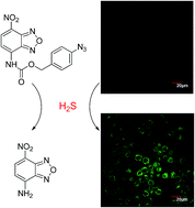
Hydrogen sulfide (H2S) is an important endogenous signalling molecule in cellular physiology and pathology. Accordingly, sensitive, selective and reliable methods for H2S detection are in high demand. Fluorescence-based methods thus have received great attentions in recent years. Herein we describe the design, synthesis and application of a probe for H2S detection based on the reduction of azide to amine. This probe is highly sensitive and selective toward H2S over biothiols and other biologically relevant species. Finally, the probe was successfully applied for H2S fluorescent imaging in living cells.


Introduction

Hydrogen sulfide (H2S) has been known as a toxic pollutant for many years. However, it now emerges as the third most important gasotransmitter following nitric oxide (NO) and carbon monoxide (CO).1,2 The endogenous H2S is biosynthesized from cysteine and homocysteine by three enzymes: cystathionine-γ-synthase (CBS), cystathionine-β-lyase (CSE), and 3-mercaptopyruvate sulfur transferase (3-MST).3,4 The significance of H2S has been validated in various physiological processes, such as vasodilation,5,6 antioxidation, anti-apoptosis and anti-inflammation.7,8 H2S has been revealed to relax vascular smooth muscle, induce vasodilation of isolated blood vessels, and reduce blood pressure.9–11 Additionally, H2S also performs as an antioxidant or a scavenger of reactive oxygen and nitrogen species in cells.12 And the abnormal H2S level is believed to be related with some diseases like Alzheimer's disease,13 Down's syndrome,14 diabetes15 and liver cirrhosis.16 Thus, visualization of H2S in living cells seems to be great important and helpful.

Fluorescence bioimaging technology is one of the most attractive molecular imaging methods for in vivo detection of biomolecules by its several advantages, such as high sensitivity, excellent selectivity, rapid response, non-invasiveness, and aptness for living cells, tissues, and living model animals.17–20 As we know, H2S is a reductant as well as a nucleophilic agent, which can specifically react with some probes. We classify these probes according to the reaction types with H2S. (a) H2S reductive reactions: reducing azides or nitro to amines. The pioneering reduction of azide to amine in fluorescent probes were reported by Chang21 and Wang22 in nearly the same time. Since then, several probes have been developed based on this concept by changing the fluorophores.23–29 (b) H2S nucleophilic reactions: Michael addition reaction, and tandem nucleophilic reaction. Xian constructed a H2S probe through a nucleophilic substitution reaction between H2S and the disulfidemoiety.30 He reported a probe which sensing H2S in cells through the tandem nucleophilic reaction of H2S with the aldehyde and α,β-unsaturated acrylate methyl ester.31 Lin constructed new NIR probes based on thiolysis of dinitrophenyl ether.32,33 (c) copper-sulfide precipitation reaction. Nagano reported a fluorescent probe by utilizing azamacrocyclic copper(II) complex for H2S detection.34 Shen constructed a red fluorescent probe based phenanthrene-fused dipyrromethene copper(II) complex.35

In this paper, we develop a new turn-on fluorescent probe for sensing H2S based on NBD fluorophore. The probe can be easily synthesized from 4-amino-7-nitro-2,1,3-benzoxadiazole (NBD-NH2) (Scheme 1). When introducing the electron withdrawing group carbamate, the electron density of NBD-NH2 was efficiently decreased, which resulted in a considerably turn-off effect of fluorescence intensity compared to NBD-NH2. While the azide group reduced to amino by H2S, the p-aminobenzyl moiety undergo a self-immolate through an intramolecular 1,6-elimination to release NBD-NH2, and the fluorescence would be recovered. The probe exhibits high fluorescence enhancement, relatively fast response and excellent selectivity toward H2S. Finally, the probe was successfully applied to image H2S in living HT-29 cells.


image file: c5ra15384k-s1.tif
Scheme 1 Proposed detection mechanism of 1.

Experimental section

Materials and methods

Unless otherwise noted, materials were obtained from Aldrich and were used without further purification. The synthesis of compound 4-amino-7-nitro-2,1,3-benzoxadiazole (NBD-NH2) (3) was according to the published procedure.36 Melting points were measured using a Büchi 530 melting point apparatus. 1H NMR and 13C NMR spectra were recorded using Bruker 400 MHz and 100 MHz, UV-Vis absorption spectra were obtained on Agilent Cary 60 UV-Vis Spectrophotometer. Fluorescence emission spectra were obtained using Cary Eclipse Fluorescence Spectrophotometer.

Synthesis and characterization of probe 1

To a mixture of Na2CO3 (636 mg, 6.0 mmol, 3.0 equiv.) and triphosgene (700 mg 2.4 mmol, 1.2 equiv.) in THF (10 mL), a solution of 3 (360 mg, 2.0 mmol) in THF (5 mL) was added dropwise in an ice bath for 1 h. Then the solution was stirred for additional 6 h at room temperature. The reaction solution was then filtered. Compound 2 (357 mg, 2.4 mmol, 1.2 equiv.) was added to the filtrate, and the solution was stirred at room temperature for 6 h. The solvent was evaporated and the crude product was purified by silica gel column chromatography (PE[thin space (1/6-em)]:[thin space (1/6-em)]DCM = 1[thin space (1/6-em)]:[thin space (1/6-em)]1) to afford the final compound 1 as a light yellow solid (220 mg, 31%); M.p. 164–166 °C. 1H NMR (400 MHz, CDCl3) δ 8.56 (d, J = 8.4 Hz, 1H), 8.15 (d, J = 8.4 Hz, 1H), 8.03 (s, 1H), 7.44 (d, J = 8.4 Hz, 2H), 7.07 (d, J = 8.4 Hz, 2H), 5.29 (s, 2H). 13C NMR (100 MHz, CDCl3) δ 151.8, 144.8, 143.0, 141.0, 134.0, 133.7, 131.2, 130.6, 130.4, 119.4, 111.1, 68.2. HRMS (ESI) calcd for C14H10N7O5 [MH+] 356.0743, found 356.0747.

Culture of HT-29 cells and fluorescent imaging

HT-29 cells were cultured in Dulbecco's modified Eagle's medium (DMEM, Invitrogen) supplemented with 10% FBS (fetal bovine serum) in an atmosphere of 5% CO2 and 95% air at 37 °C. The cells were seeded in 24-well flat-bottomed plates and then incubated for 48 h at 37 °C under 5% CO2. 1 (5 μM) was then added to the cells and incubated for 30 min. Then the cells were washed three times with PBS. After incubating with 20 μM H2S, fluorescence imaging was observed under a confocal microscopy (Olympus FV1000) with a 60× objective lens.

Results and discussion

Characterization of fluorescent probe 1 for H2S recognition

Absorbance and fluorescence studies in aqueous solution (CH3CN[thin space (1/6-em)]:[thin space (1/6-em)]PBS = 9[thin space (1/6-em)]:[thin space (1/6-em)]1, pH = 7.4) were performed firstly. The changes of the absorption spectra in the absence and presence of NaHS (a commonly used hydrogen sulfide source) (0–20 equiv.) were displayed in Fig. 1. The probe 1 showed an absorption band at 520 nm. When we added NaHS to the solution of probe 1, the absorbance at 520 nm decreased sharply to its limiting value, while an absorption band at 470 nm developed, which was visible to the naked eye with a clear colour change from pink to yellow. Additionally, if the concentration of NaHS reaches 20 equiv., the reaction can be completely finished in minutes.
image file: c5ra15384k-f1.tif
Fig. 1 UV-Vis absorption spectra of 10 μM probe 1 in the presence of 0–20 equiv. of H2S in aqueous solution (CH3CN[thin space (1/6-em)]:[thin space (1/6-em)]PBS = 9[thin space (1/6-em)]:[thin space (1/6-em)]1, 20 mM PBS, pH = 7.4). Inset: cuvette images of probe 1 before and after addition of H2S taken under ambient light.

The emission spectra and fluorescence titration experiments of probe 1 with H2S were then recorded in aqueous solution (CH3CN[thin space (1/6-em)]:[thin space (1/6-em)]PBS = 9[thin space (1/6-em)]:[thin space (1/6-em)]1, pH = 7.4) (Fig. 2). The free probe 1 displayed quite weak fluorescence. When H2S was added progressively from 0 to 20 equiv. to the solution of probe 1, the probe was converted efficiently to fluorescent NBD-NH2. And the fluorescence intensity of the emission band at 550 nm increased significantly. From Fig. 2b, it was found that if the concentration of H2S was over 20 equiv., the reaction can be finished in minutes. Therefore, the time-dependent fluorescence responses were next detected with 20 equiv. of H2S from 0 to 55 min, and the results showed that the reaction was completed within 50 min (Fig. 2c and d).


image file: c5ra15384k-f2.tif
Fig. 2 (a) Fluorescent emission spectra of 10 μM probe 1 in the presence of 0–20 equiv. of H2S in aqueous solution (CH3CN[thin space (1/6-em)]:[thin space (1/6-em)]PBS = 9[thin space (1/6-em)]:[thin space (1/6-em)]1, pH = 7.4) (NaHS was dissolved in water in the concentration of 1 mM). Excitation at 470 nm. (b) Fluorescence intensity of 10 μM probe 1 in the presence of 0–20 equiv. of H2S. (c) Time dependence of fluorescence profiles of probe 1 (10 μM) with 20 equiv. H2S. (d) Time dependence of fluorescence intensity of probe 1 (10 μM) at 550 nm with 20 equiv. H2S.

In addition, the formation of compound NBD-NH2 was confirmed by 1H-NMR. The cleavage reaction of probe 1 with 20 equiv. H2S revealed the characteristic NMR peaks almost the same as the NBD-NH2 (Fig. 3). The transformation of 1 to NBD-NH2 was also confirmed by TLC experiments.


image file: c5ra15384k-f3.tif
Fig. 3 Partial 1H NMR spectra of 1, NBD-NH2 and the reaction mixture of 1 with H2S in CD3CN.

The selectivity of the fluorescent response of 1 with various anions, sulfur-containing inorganic salts and biothiols including cysteine, homo-cysteine and glutathione were then conducted. The addition of 50 equiv. of F, Cl, Br, I, CO32−, HCO3, OAc, NO3, NO2, OH, citric acid, hydrogen citrate, dihydrogen citrate, N3, PO43−, HPO42−, H2PO4, P2O74−, SCN, SO32−, HSO3, SO42−, HSO4, S2O32−, S2O42−, S2O52− produced only a nominal change in the fluorescence spectra of 1. In addition, both biothiols (50 equiv. Cys, Hcys and GSH, the main competitive species in biological systems) and reducing conditions ascorbate acid (50 equiv.) showed no response to probe 1. By contrast, only H2S shows a dramatic fluorescent enhancement, suggesting very high selectivity of 1 towards H2S (Fig. 4).


image file: c5ra15384k-f4.tif
Fig. 4 Fluorescence responses of 10 μM 1 to various analytes in aqueous solution (CH3CN[thin space (1/6-em)]:[thin space (1/6-em)]PBS = 9[thin space (1/6-em)]:[thin space (1/6-em)]1, pH = 7.4). Excitation at 470 nm. Bars represent the final fluorescence intensity of 1 with 0.5 mM analytes over the original emission of free 1. (1) free probe 1; (2) F, (3) Cl, (4) Br, (5) I, (6) CO32−, (7) HCO3, (8) OAc, (9) NO3, (10) NO2, (11) OH, (12) citric acid, (13) hydrogen citrate, (14) dihydrogen citrate, (15) N3, (16) PO43−, (17) HPO42−, (18) H2PO4, (19) P2O74−, (20) SCN, (21) SO32−, (22) HSO3, (23) SO42−, (24) HSO4, (25) S2O32−, (26) S2O42−, (27) S2O52−, (28) Cys, (29) GSH, (30) Hcy, (31) ascorbic acid, (32) HS.

We also performed the competition experiments in the presence of biothiols, ascorbate acid and H2O2. In fact, when H2S and these analytes coexisted, almost the same fluorescence enhancement was observed as that only treated with H2S (Fig. 5).


image file: c5ra15384k-f5.tif
Fig. 5 Fluorescence response of 1 (10 μM) to NaHS (200 μM) and other reactive species, Cys (0.5 mM), GSH (0.5 mM), Hcy (0.5 mM), AC (0.5 mM), H2O2 (0.5 mM).

Encouraged by the above results, we next sought to apply probe 1 to image H2S in living cells. Analysis of human colon cancer biopsies and patient-matched normal margin mucosa revealed the selective up-regulation of the H2S-producing enzyme cystathionine-β-synthase (CBS) in colon cancer, resulting in an increased rate of H2S production. Then, HT-29 cells was selected to incubate with probe 1. The cytotoxicity of probe 1 was first examined toward HT-29 cells by a MTT assay (Fig. S6). The results showed that >90% HT-29 cells survived after 24 h (5.0 μM probe 1 incubation), demonstrating that 1 was of low toxicity toward cultured cell lines. When incubated with 5 μM 1 for 30 min at 37 °C, the cells were washed with PBS (pH = 7.4) to remove excess of 1. The HT-29 cells exhibited almost no fluorescence as shown in the confocal image Fig. 6a, indicating that biothiols and other biological species showed no interference. While after incubating with 20 μM NaHS for 20 min, the cells displayed enhanced green fluorescence (Fig. 6b). Another 20 min later, a higher turn-on fluorescence response was observed (Fig. 6c). All these experiments indicated that the probe 1 has the potential biological application for imaging H2S in living cells.


image file: c5ra15384k-f6.tif
Fig. 6 Images of H2S detection in HT-29 cells incubated with 1 (5 μM) at 37 °C. (a) HT-29 cells were incubated with 1 for 30 min; after which 20 μM H2S was added, further incubation for 20 min (b) and 40 min (c); (d) merged images of (c) and bright field.

Conclusion

In summary, we have reported a reaction-type fluorescent probe 1 for detection of H2S in aqueous solution based on a specific analyte-induced cleavage of carbamate. This new probe can be easily prepared and works effectively at physiological pH, which shows high selectivity for H2S over other biological thiols (such as Cys, Hcys and GSH). Concomitantly, the solution colour changes from pink to yellow. This probe can also detect H2S in living cells. The successful application of our probe to detect cellular H2S will help us to study the biological role of H2S and encourage the appearance of new H2S probes suitable for cell imaging.

Acknowledgements

We thank financial supports from the National Natural Science Foundation of China (21422606, 21276251, 21402191), Ministry of Human Resources and Social Security of PRC, the 100 talents program funded by Chinese Academy of Sciences.

Notes and references

  1. H. Kimura, Amino Acids, 2011, 41, 113–121 CrossRef CAS PubMed .
  2. T. Morita, M. A. Perrella, M.-E. Lee and S. Kourembanas, Proc. Natl. Acad. Sci. U. S. A., 1995, 92, 1475–1479 CrossRef CAS .
  3. M. J. Wang, W. J. Cai and Y. C. Zhu, Clin. Exp. Pharmacol. Physiol., 2010, 37, 764–771 CrossRef CAS PubMed .
  4. G. Yang, L. Wu, B. Jiang, W. Yang, J. Qi, K. Cao, Q. Meng, A. K. Mustafa, W. Mu and S. Zhang, Science, 2008, 322, 587–590 CrossRef CAS PubMed .
  5. W. Zhao, J. Zhang, Y. Lu and R. Wang, EMBO J., 2001, 20, 6008–6016 CrossRef CAS PubMed .
  6. D. J. Lefer, Proc. Natl. Acad. Sci. U. S. A., 2007, 104, 17907–17908 CrossRef CAS PubMed .
  7. J. W. Calvert, S. Jha, S. Gundewar, J. W. Elrod, A. Ramachandran, C. B. Pattillo, C. G. Kevil and D. J. Lefer, Circ. Res., 2009, 105, 365–374 CrossRef CAS PubMed .
  8. R. C. Zanardo, V. Brancaleone, E. Distrutti, S. Fiorucci, G. Cirino and J. L. Wallace, FASEB J., 2006, 20, 2118–2120 CrossRef CAS PubMed .
  9. A. Papapetropoulos, A. Pyriochou, Z. Altaany, G. Yang, A. Marazioti, Z. Zhou, M. G. Jeschke, L. K. Branski, D. N. Herndon and R. Wang, Proc. Natl. Acad. Sci. U. S. A., 2009, 106, 21972–21977 CrossRef CAS PubMed .
  10. G. Yang, L. Wu and R. Wang, FASEB J., 2006, 20, 553–555 CAS .
  11. L. Li, M. Bhatia, Y. Z. Zhu, Y. C. Zhu, R. D. Ramnath, Z. J. Wang, F. B. M. Anuar, M. Whiteman, M. Salto-Tellez and P. K. Moore, FASEB J., 2005, 19, 1196–1198 CAS .
  12. M. Whiteman, J. S. Armstrong, S. H. Chu, S. Jia-Ling, B. S. Wong, N. S. Cheung, B. Halliwell and P. K. Moore, J. Neurochem., 2004, 90, 765–768 CrossRef CAS PubMed .
  13. K. Eto, T. Asada, K. Arima, T. Makifuchi and H. Kimura, Biochem. Biophys. Res. Commun., 2002, 293, 1485–1488 CrossRef CAS .
  14. P. Kamoun, M. C. Belardinelli, A. Chabli, K. Lallouchi and B. Chadefaux-Vekemans, Am. J. Med. Genet., Part A, 2003, 116, 310–311 CrossRef PubMed .
  15. W. Yang, G. Yang, X. Jia, L. Wu and R. Wang, J. Physiol., 2005, 569, 519–531 CrossRef CAS PubMed .
  16. S. Fiorucci, E. Antonelli, A. Mencarelli, S. Orlandi, B. Renga, G. Rizzo, E. Distrutti, V. Shah and A. Morelli, Hepatology, 2005, 42, 539–548 CrossRef CAS PubMed .
  17. Z. Xu, J. Yoon and D. R. Spring, Chem. Soc. Rev., 2010, 39, 1996–2006 RSC .
  18. T. Ueno and T. Nagano, Nat. Methods, 2011, 8, 642–645 CrossRef CAS PubMed .
  19. J. Chan, S. C. Dodani and C. J. Chang, Nat. Chem., 2012, 4, 973–984 CrossRef CAS PubMed .
  20. Y. Tang, D. Lee, J. Wang, G. Li, J. Yu, W. Lin and J. Yoon, Chem. Soc. Rev., 2015, 44, 5003–5015 RSC .
  21. A. R. Lippert, E. J. New and C. J. Chang, J. Am. Chem. Soc., 2011, 133, 10078–10080 CrossRef CAS PubMed .
  22. H. Peng, Y. Cheng, C. Dai, A. L. King, B. L. Predmore, D. J. Lefer and B. Wang, Angew. Chem., Int. Ed., 2011, 50, 9672–9675 CrossRef CAS PubMed .
  23. Q. Qiao, M. Zhao, H. Lang, D. Mao, J. Cui and Z. Xu, RSC Adv., 2014, 4, 25790–25794 RSC .
  24. S. K. Das, C. S. Lim, S. Y. Yang, J. H. Han and B. R. Cho, Chem. Commun., 2012, 48, 8395–8397 RSC .
  25. L. A. Montoya and M. D. Pluth, Chem. Commun., 2012, 48, 4767–4769 RSC .
  26. Q. Wan, Y. Song, Z. Li, X. Gao and H. Ma, Chem. Commun., 2013, 49, 502–504 RSC .
  27. F. Yu, P. Li, P. Song, B. Wang, J. Zhao and K. Han, Chem. Commun., 2012, 48, 2852–2854 RSC .
  28. K. Zheng, W. Lin and L. Tan, Org. Biomol. Chem., 2012, 10, 9683–9688 CAS .
  29. L. He, W. Lin, Q. Xu and H. Wei, Chem. Commun., 2015, 51, 1510–1513 RSC .
  30. C. Liu, J. Pan, S. Li, Y. Zhao, L. Y. Wu, C. E. Berkman, A. R. Whorton and M. Xian, Angew. Chem., Int. Ed., 2011, 50, 10327–10329 CrossRef CAS PubMed .
  31. Y. Qian, J. Karpus, O. Kabil, S.-Y. Zhang, H.-L. Zhu, R. Banerjee, J. Zhao and C. He, Nat. Commun., 2011, 2, 495–501 CrossRef PubMed .
  32. X. Cao, W. Lin, K. Zheng and L. He, Chem. Commun., 2012, 48, 10529–10531 RSC .
  33. H. Chen, W. Lin, H. Cui and W. Jiang, Chem.–Eur. J., 2015, 21, 733–745 CrossRef CAS PubMed .
  34. K. Sasakura, K. Hanaoka, N. Shibuya, Y. Mikami, Y. Kimura, T. Komatsu, T. Ueno, T. Terai, H. Kimura and T. Nagano, J. Am. Chem. Soc., 2011, 133, 18003–18005 CrossRef CAS PubMed .
  35. X. Qu, C. Li, H. Chen, J. Mack, Z. Guo and Z. Shen, Chem. Commun., 2013, 49, 7510–7512 RSC .
  36. W. Jiang, Q. Fu, H. Fan, J. Ho and W. Wang, Angew. Chem., Int. Ed., 2007, 46, 8445–8448 CrossRef CAS PubMed .

Footnote

Electronic supplementary information (ESI) available. See DOI: 10.1039/c5ra15384k

This journal is © The Royal Society of Chemistry 2015
Click here to see how this site uses Cookies. View our privacy policy here.