DOI:
10.1039/C5RA15272K
(Paper)
RSC Adv., 2015,
5, 86551-86557
Self-assembled flower-like ZnCo2O4 hierarchical superstructures for high capacity supercapacitors†
Received
31st July 2015
, Accepted 28th September 2015
First published on 28th September 2015
Abstract
Self-assembled ZnCo2O4 nanosheets were synthesised by a one-step hydrothermal method and their electrochemical supercapacitor properties were investigated in 2 M aqueous KOH solution. Interesting capacitive properties were observed with a superb cyclic stability obtained even at a current density of 20 A g−1. A specific capacitance value of 1691 F g−1 was obtained at a current density of 1 A g−1 with an energy density of ∼57.4 W h kg−1. The experimental findings provide useful insights into the design of supercapacitors for potential high performance energy storage applications in the future.
1. Introduction
The hugely oppressive conditions of recent years due to the tremendous requirement of energy are further worsened by the rapid depletion of non-renewable energy sources, such as fossil fuels. In addition, complicated environmental issues are fast arising out of their continuous consumption, and have motivated the intense search for cleaner, renewable and eco-friendly energy storage and conversion options. Solar cells,1 fuel cells,2 Li-ion batteries3 and supercapacitors4 are some of the promising alternatives that have undergone critical research in recent years. Owing to their intriguing and tunable physical and chemical properties, two dimensional materials (in various forms) have drawn copious amount of attention from the scientific community; these two dimensional materials have thus found some direct applications in such areas.5,6 Among such two dimensional materials, graphene has been the most phenomenal since its discovery, owing to its fascinating physical and chemical properties,7–9 and has provided an ideal platform for the study of analogous two dimensional materials such as transition metal chalcogenides,10 metal oxides etc.11–13 Graphene along with its two dimensional analogues and other heterostructures (due to their hybridisation) have been studied intensively for their possible application as electrode materials for supercapacitors.14–17
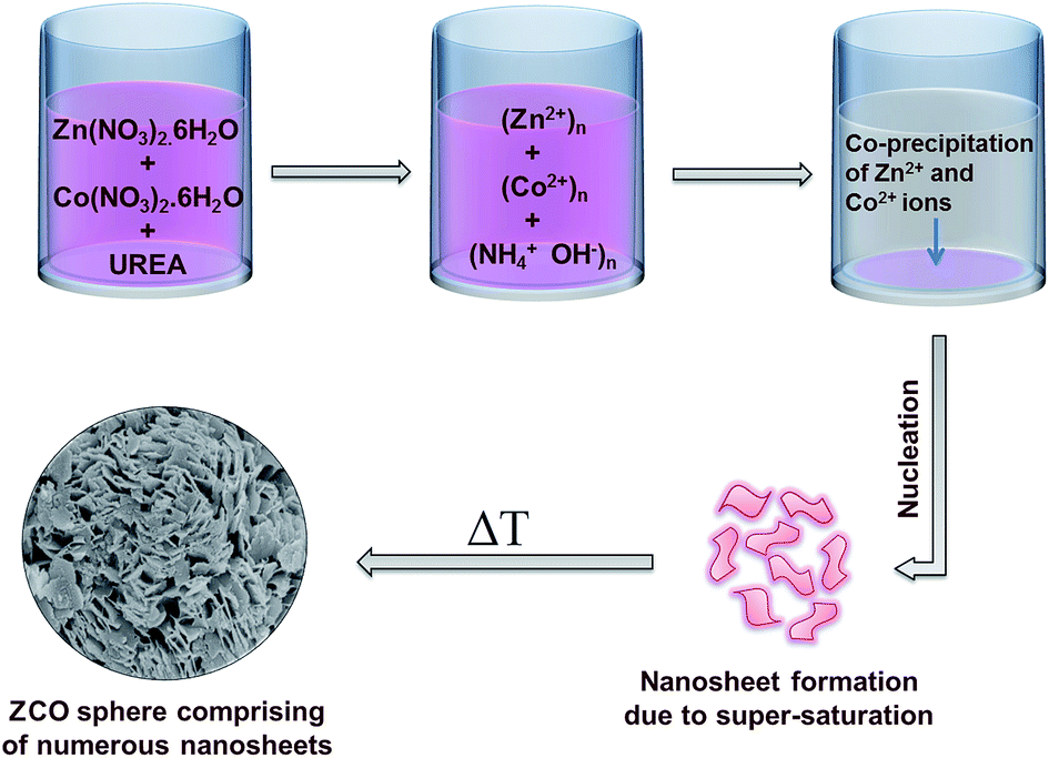
Many three dimensional materials are composed of numerous such two dimensional structures,18–20 which are chemically or physically aggregated in order to form a stable morphology. In this report we have demonstrated a facile route for the synthesis of a ternary metal oxide (spinel oxide) having a micro-spherical structure. The as obtained spherical spinel oxide is formed due to the growth and aggregation of a number of two dimensional nanosheets. Spinel oxides are a new class of materials, currently drawing huge attention due to their unique, complex structure and properties as well as broader applicability.21–24 The spinel oxide reported here is ZnCo2O4 (ZCO) which belongs to the group of normal spinels having a space symmetry group Fd![[3 with combining macron]](https://www.rsc.org/images/entities/char_0033_0304.gif) m ≡ O7h.25 Detailed electrochemical studies were performed in order to investigate the potential application of ZCO as supercapacitor electrodes.
m ≡ O7h.25 Detailed electrochemical studies were performed in order to investigate the potential application of ZCO as supercapacitor electrodes.
Supercapacitors store energy in terms of both electrostatic double layer capacitance (EDLC) and pseudocapacitance. The pseudocapacitance property of a material is characterised by the reversible faradic reaction caused by rapid redox activities inside an electrochemical cell. They have a much faster charging capability as compared to batteries and slower discharge rate as compared to a normal capacitor. In contrast to rechargeable batteries, they can deliver the enormous amounts of power required for electric vehicles and other high power consuming devices while having the additional advantage of long cyclic stability. Recently, various metal oxides such as Co3O4,26 MnO2,27 RuO2,28 along with many ternary metal oxides such as MnCo2O4,29 NiCo2O4,4,30 and CoMo2O4 (ref. 31) have shown interesting supercapacitive properties. The ZCO sample reported here is a ternary transition metal oxide synthesised by a facile hydrothermal method. The Zn and Co precursors along with urea were dissolved in de-ionised water. The dissolved urea decomposes to form CO2 and NH3. Further reaction of NH3 with water (H2O) forms NH4OH and subsequently increases the pH of the mixture. Under such hydrothermal conditions, super-saturation occurs among the co-precipitated particles, which favors the formation of nanosheet-like structures.32–34 Furthermore, self-assembly of these nanosheets forms thermally stable tiny spherical structures.
2. Experimental methods
All the chemicals were used as received without any alteration.
2.1. Synthesis of ZnCo2O4
3 mmol of Zn(NO3)2·6H2O and 6 mmol of Co(NO3)2·6H2O were first dissolved in 20 ml de-ionised (DI) water and kept under constant stirring conditions. An aqueous solution of urea (60 mmol of urea dissolved in 20 ml DI water) was then slowly added to the above solution and the mixture was further kept under constant stirring conditions for 2 h. The as prepared final solution (40 ml) was then transferred to a 50 ml Teflon lined stainless steel autoclave and kept at 200 °C for ∼12 h. The resultant colloidal mixture was then collected via filtration and washed with DI water and absolute ethanol several times to remove any spurious content and unreacted ions. The final precipitate was then dried by keeping it inside a vacuum oven at 70 °C overnight.
2.2. Characterisation
The as-prepared sample was characterised by the powder X-ray diffraction technique (Bruker D8 Advanced diffractometer having Cu-Kα radiation, λ = 0.154184 nm). The morphology and elemental composition of the sample were analysed by FESEM (Merlin compact with Gemini-I electron column, Zeiss Pvt. Ltd, Germany), EDAX and elemental mapping (INCA, Oxford Instruments, UK). The Raman spectrum was collected by a Renishaw inVia micro Raman spectrometer with an excitation source of 532 nm.
2.3. Electrochemical measurements
The electrochemical activities of the sample were studied by taking a glassy carbon electrode (GCE) modified with the sample as the working electrode, Ag/AgCl as the reference electrode and a Pt wire as the counter electrode. In a typical procedure, a glassy carbon electrode was polished thoroughly using micro-polishing powder (Al2O3, 0.05 μm) to get a mirror finish. Then it was ultrasonicated in dilute HNO3 to remove any kind of surface adsorbents. 1 mg of ZCO was added to a 100 μl mixture of ethanol (95 μl) and Nafion (5 μl) and was sonicated to get an ink like dispersion. 2 μl of the above dispersion was drop-casted onto the finely polished surface of the glassy carbon electrode and was vacuum dried (for about 1 h). The modified GCE was then taken as the working electrode. Cyclic voltammetry and charge–discharge measurements were carried out in 2 M aqueous KOH solution using a standard Potentiostat/Galvanostat (PG262A, Technoscience Instruments, Bangalore, India).
3. Results and discussion
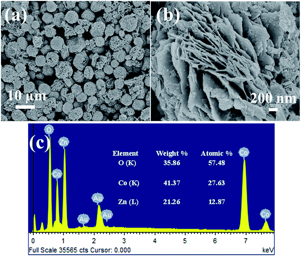
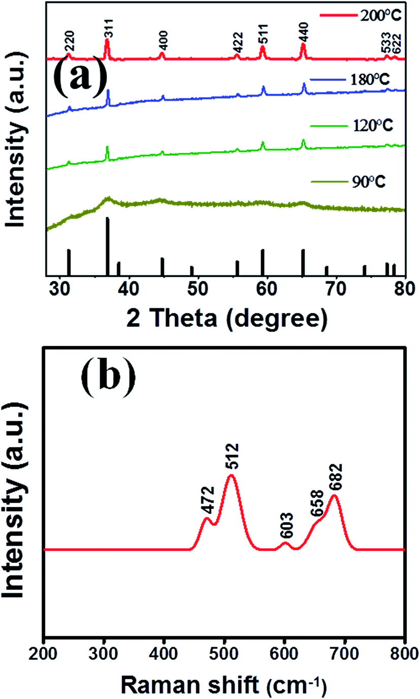
A schematic representation of the formation of ZCO spherical structures is shown in Scheme 1. A time varied synthesis of ZCO was also carried out to show the progressive stages of the formation of ZCO. Corresponding field emission scanning electron microscopy (FESEM) images have been provided in the ESI (Fig. S1†). Detailed morphology of the finally obtained ZCO sample was examined by FESEM along with energy dispersive X-ray spectroscopy (EDS). The FESEM images (Fig. 1a and b) elucidate the entanglement of the large and thin edged ZCO nanosheets. The EDS spectrum shows the composition of the sample and its atomic and weight percentages. The mapping data are shown in Fig. 2 which confirms the homogeneous distribution of the constituents of ZCO, throughout the mapping region. Fig. 3a shows the powder X-ray diffraction (XRD) patterns of ZCO nanosheets at different temperatures (i.e. 90 °C, 120 °C, 180 °C and 200 °C). It can be clearly observed that the sample synthesised at 200 °C has the best crystalline and clear phase growth, though samples at 120 °C and 180 °C are more or less the same. The XRD pattern obtained in the case of the sample synthesised at 200 °C confirmed the formation of the pure phase ZCO (JCPDS file no. 23-1390), which belongs to a cubic spinel group having space symmetry Fdm ≡ O7h. The Raman spectra for the sample (Fig. 3b) reveals five characteristic peaks, including a shoulder/satellite peak at 658 cm−1 which results from the A1g symmetry mode. A similar vibrational mode was also observed at 682 cm−1 which corresponds to the A1g mode. Peaks appearing at 472 cm−1 and 512 cm−1 are due to the strong vibration modes Eg and F(2)2g, respectively. The peak at 603 cm−1 is due to the stretching of the Co–O bond and belongs to the F(1)2g symmetry mode. All these Raman peaks can be assigned to the reported cubic spinel, ZCO.25
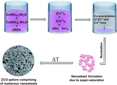 |
| | Scheme 1 Illustration of the mechanism behind the formation of ZCO spherical structures. | |
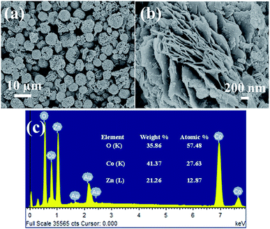 |
| | Fig. 1 FESEM images of the ZCO sample at (a) low magnification and (b) high magnification. (c) EDS with atomic and weight percentage compositions of the sample. | |
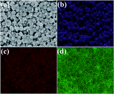 |
| | Fig. 2 Elemental mapping data for the ZCO sample. (a) The electron image over which the mapping has been done. The presence of (b) oxygen, (c) zinc and (d) cobalt is confirmed. | |
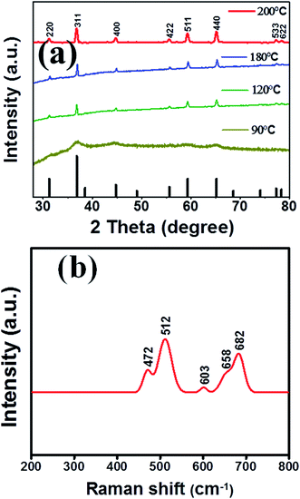 |
| | Fig. 3 (a) X-ray diffraction pattern for ZCO at different temperatures and (b) Raman spectroscopy data. | |
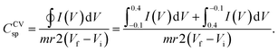
The electrochemical studies were performed by both cyclic voltammetry and charge–discharge measurements in 2 M aqueous KOH solution with the help of a three electrode configuration. From the cyclic voltammetry (CV) curves, the capacitance was calculated implementing the following formula (for a three electrode configuration):35
| |
 | (1) |
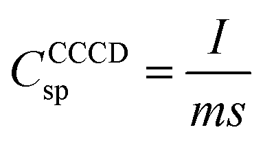
Similarly, from the constant current charge–discharge (CCCD) measurements, the specific capacitance of the material was evaluated using the following formula:
| |
 | (2) |
where
CCVsp and
CCCCDsp are the specific capacitances from the cyclic voltammetry and constant current charge–discharge measurements respectively,
I(
V)d
V is the area under the cyclic voltammetry curve,
m is the mass of the sample drop-cast on the GCE surface,
r is the scan rate,
s is the slope of the discharge curve and
Vf −
Vi is the working potential window. Similarly, the energy density (
Ed) and power density (
Pd) were calculated using the following equations (
eqn (3) and
(4)):
| |
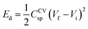 | (3) |
| |
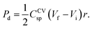 | (4) |
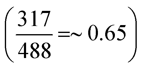
Throughout the measurement process, the potential window (Vf − Vi) was kept at a value of 0.5 V (from −0.1 V to 0.4 V). The values of the specific capacitance at different scan rates and current densities and the cyclic stability of the sample were calculated using the data plots obtained from the electrochemical measurements. A Ragone plot was plotted to observe the overall energy density and power density of the sample. Fig. 4 shows the cyclic voltammetry at different scan rates (Fig. 4a) and the charge–discharge at different current densities (Fig. 4b). The obtained cyclic voltammetry curves were not of rectangular/quasi-rectangular shape which reveals the pseudocapacitive property of the ZCO sample. However, the cyclic voltammetry curves do not show prominent/sharp redox peaks though mild, broad redox couples are slightly visible at lower scan rates (i.e. 1 mV s−1 and 2 mV s−1, see Fig. S2 in the ESI†). This property is not unique in the case of supercapacitor electrodes based on metal oxides. Unlike pure faradic materials such as metal hydrides/hydroxides, metal oxides are rather known as pseudocapacitive materials and possess altogether different electrochemical properties (approaching the EDLC domain with the additional faradic reactions, which might or might not be visually inspected).36 The pseudocapacitive property is also significantly affected by the size of the electrode material and the process of the supercapacitor device fabrication.37,38 In another report, Simon and Gogotsi have demonstrated that even if prominent redox peaks may not be visualised in the case of metal oxides having cations with a wide range of oxidation states, they still undergo a series of continuous oxidation and reduction reactions,39 eventually producing unprecedented smooth cyclic voltammetry curves. Incidentally, the subdued oxidation and reduction peaks in the case of the ZCO sample reported here, overlap, which shows that it has a good coulombic efficiency. Furthermore, it can clearly be observed that at higher scan rates, any such redox peaks are absent. The prime reason behind it can be attributed to the kinetically slow surface reactions at higher scan rates. And the major contribution is from the double layer effect arising due to the adsorption of OH− ions by ZCO.40 As the faradic reaction is on the slower side at higher sweep rates, therefore the capacitance decreases gradually. That is why the sample showed a specific capacitance of 1654 F g−1 at a sweep rate of 1 mV s−1, whereas at a scan rate of 200 mV s−1, the value obtained was 694 F g−1. The charge–discharge curves are nearly triangular and highly symmetric in shape, which suggests that the sample has a very high coulombic efficiency and the faradic reactions taking place are highly reversible in nature. Also the immediate discharge without any significant amount of IR drop suggests that the sample has a lower value of internal resistance Fig. 4c and d depict the variations of the specific capacitance with the scan rate and current density, respectively. The capacitance shown by the sample is by virtue of two different contributions; one is from the fast redox reaction and the other is from EDLC. These two contributions were calculated separately using the discharge curves by following a method detailed in a previous report.41 At a current density of 1 A g−1, the ratio of EDLC to pseudocapacitance was found to be  , while for a current density of 20 A g−1, the ratio obtained was
  , which clearly shows that at lower current densities, the contribution from the pseudocapacitance is much larger than that of EDLC, whereas for higher current densities, EDLC plays a major role in the electrochemical supercapacitor performance of the sample. A long cyclic stability test, in terms of 5000 charge–discharge cycles (at a current density of 20 A g−1) has been performed for the ZCO sample which is shown in Fig. 5. The cycle number versus capacity retention plot is illustrated in Fig. 5a. The first 5 cycles and last 5 cycles are provided in Fig. 5b and c, respectively, which show little distortion in the shape of the charge–discharge curves even after 5000 cycles. It was found that the retention power of the ZCO sample decreased to ∼78% of its initial value. The morphology of the sample was investigated after 5000 charge–discharge cycles with the help of field emission scanning electron microscopy (see Fig. S3 in the ESI†). The supercapacitor performance of the ZCO sample, in terms of the energy density and power density, has been elucidated in Fig. 5c, through a Ragone plot.
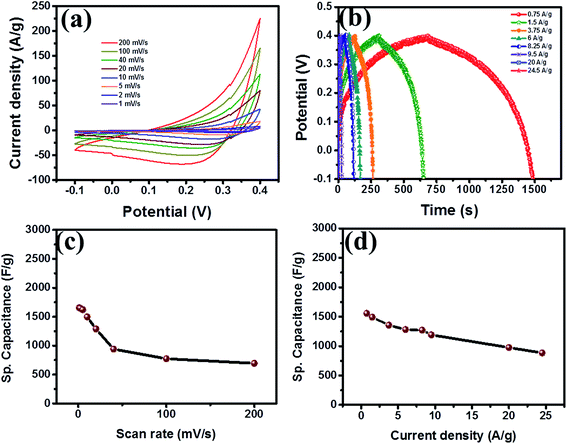 |
| | Fig. 4 Electrochemical measurement data for ZCO. (a) Cyclic voltammetry curves at different scan rates, (b) charge–discharge curves at different current densities and the variation of the specific capacitance with respect to the (c) scan rate and (d) current density. | |
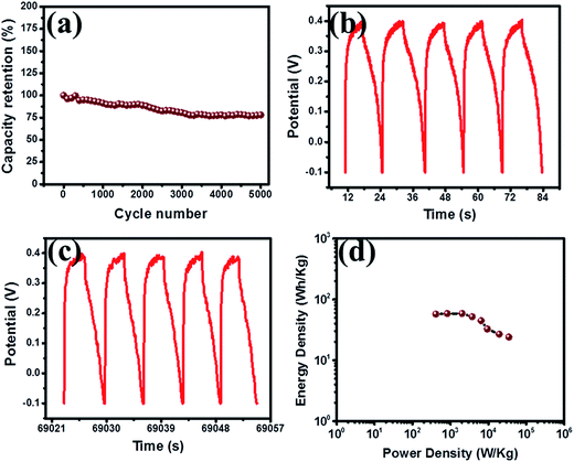 |
| | Fig. 5 (a) Capacity retention power of the ZCO sample in percent, calculated from 5000 charge–discharge cycles. (b) The initial and (c) last 5 charge–discharge curves taken from the 5000 cycle data. (c) The Ragone plot, establishing a relation between the energy density and power density. | |
The ZCO sample reported here possesses promising electrochemical supercapacitive performance comparable to the recently reported data on ZCO having different structures, and fabrication procedures. Nanorod structured ZCO grown on nickel foam has been reported to have a specific capacitance of ∼1400 F g−1 at a current density of 1 A g−1.42 A specific capacitance of 647 F g−1 at a current density of 1 A g−1 has also been reported for porous ZCO microspherical structures.43 The as reported ZCO sample has a micro-spherical shape consisting of numerous nanosheets which have a thickness in the nanometer range, and possess highly rough surfaces with imperfect growth at the edges, enabling the possibility of better ion-trapping capabilities. The three dimensional structure has also got numerous prominent pores which provide an active path for the diffusion of the electrolyte, facilitating a much improved ion/electron transportation. The morphological peculiarity is further corroborated by detailed electrochemical investigations in a three-electrode configuration, taking 2 M aqueous KOH solution as the electrolyte. The sample showed enhanced supercapacitor properties as compared to the previous reports and thus has enough potential for application as an active material for the fabrication of high performance supercapacitor devices. A detailed comparison of the supercapacitor performances of various reported metal oxides have been provided in Table 1.
Table 1 Comparison of supercapacitor performance of various metal oxides and their composite structures
| Electrode material |
Highest specific capacitance |
Capacity retention (<1000 cycles) |
Max. energy density (W h kg−1) |
Max. power density (kW kg−1) |
Reference |
| Self-assembled ZnCo2O4 nanosheets |
1691 F g−1 (at 1 A g−1) |
∼78% |
57.4 |
34.7 |
Present work |
| ZnCo2O4 porous nanotube |
770 F g−1 (at 10 A g−1) |
∼94.1% |
25 |
15.3 |
44 |
| ZnCo2O4 micro-spheres |
953.2 F g−1 (at 4 A g−1) |
97.8% |
33.1 |
8 |
45 |
| MnCo2O4 nanostructure |
346 F g−1 (1 A g−1) |
∼88% |
— |
— |
46 |
| 3D-nanonet Co3O4 |
739 F g−1 (at 1 A g−1) |
∼90.2% |
16.42 |
3 |
47 |
| NiMoO4 microsphere |
974.4 F g−1 (1 A g−1) |
∼74.5% |
32.2 |
2.1 |
48 |
| 3D NiCo2O4 microsphere |
1284 F g−1 (2 A g−1) |
∼97.5% |
— |
— |
30 |
| NiO nanocomposite |
429.7 F g−1 (0.2 A g−1) |
∼86.1% |
— |
— |
49 |
4. Conclusions
In summary, a facile synthesis method for ZCO nanosheets has been reported here and their supercapacitor performance was tested thoroughly in an alkaline medium (2 M aqueous KOH), showing an energy density of 57.4 W h kg−1 and high specific capacitance of 1691 F g−1. All the analyses show that these ZCO nanosheet structures have promising applicability as low-cost, environmentally friendly and high performance supercapacitor electrodes.
Acknowledgements
Dr C. S. Rout would like to thank DST (Government of India) for the Ramanujan fellowship (Grant No. SR/S2/RJN-21/2012). This work was supported by the DST-SERB Fast-track Young scientist (Grant No. SB/FTP/PS-065/2013), UGC-UKIERI thematic awards (Grant No. UGC-2013-14/005) and BRNS-DAE, (Grant No. 37(3)/14/48/2014-BRNS/1502). Also, part of this work is supported by the Indo-US Science and Technology Forum (IUSSTF) through a joint INDO-US centre grant and Ministry of Human Resources Development (MHRD), India through a center of excellence grant.
Notes and references
- H. Sun, J. Deng, L. Qiu, X. Fang and H. Peng, Energy Environ. Sci., 2015, 8, 1139–1159 CAS.
- H. Huang and X. Wang, J. Mater. Chem. A, 2014, 2, 6266–6291 CAS.
- S. Goriparti, E. Miele, F. de Angelis, E. Di Fabrizio, R. Proietti Zaccaria and C. Capiglia, J. Power Sources, 2014, 257, 421–443 CrossRef CAS PubMed.
- T.-Y. Wei, C.-H. Chen, H.-C. Chien, S.-Y. Lu and C.-C. Hu, Adv. Mater., 2010, 22, 347–351 CrossRef CAS PubMed.
- Y. Sun, Q. Wu and G. Shi, Energy Environ. Sci., 2011, 4, 1113–1132 CAS.
- F. Bonaccorso, L. Colombo, G. Yu, M. Stoller, V. Tozzini, A. C. Ferrari, R. S. Ruoff and V. Pellegrini, Science, 2015, 347, 1246501 CrossRef PubMed.
- A. H. Castro Neto, N. M. R. Peres, K. S. Novoselov and A. K. Geim, Rev. Mod. Phys., 2009, 81, 109–162 CrossRef CAS.
- A. K. Geim, Science, 2009, 324, 1530–1534 CrossRef CAS PubMed.
- A. K. Geim and K. S. Novoselov, Nat. Mater., 2007, 6, 183–191 CrossRef CAS PubMed.
- G. A. Muller, J. B. Cook, H.-S. Kim, S. H. Tolbert and B. Dunn, Nano Lett., 2015, 15, 1911–1917 CrossRef CAS PubMed.
- Z. Sun, T. Liao, Y. Dou, S. M. Hwang, M.-S. Park, L. Jiang, J. H. Kim and S. X. Dou, Nat. Commun., 2014, 5, 3813 CAS.
- E. Kan, M. Li, S. Hu, C. Xiao, H. Xiang and K. Deng, J. Phys. Chem. Lett., 2013, 4, 1120–1125 CrossRef CAS PubMed.
- M. Pandey, A. Vojvodic, K. S. Thygesen and K. W. Jacobsen, J. Phys. Chem. Lett., 2015, 6, 1577–1585 CrossRef CAS PubMed.
- H. Wang, H. Feng and J. Li, Small, 2014, 10, 2165–2181 CrossRef CAS PubMed.
- J. J. Yoo, K. Balakrishnan, J. Huang, V. Meunier, B. G. Sumpter, A. Srivastava, M. Conway, A. L. Mohana Reddy, J. Yu, R. Vajtai and P. M. Ajayan, Nano Lett., 2011, 11, 1423–1427 CrossRef CAS PubMed.
- S. Peng, L. Li, H. Tan, R. Cai, W. Shi, C. Li, S. G. Mhaisalkar, M. Srinivasan, S. Ramakrishna and Q. Yan, Adv. Funct. Mater., 2014, 24, 2155–2162 CrossRef CAS PubMed.
- S. Ratha and C. S. Rout, ACS Appl. Mater. Interfaces, 2013, 5, 11427–11433 CAS.
- B. Huang, M. Cao, B. Cheng, J. Sun, N. Li, F. Nie, H. Zhang, H. Huang and C. Hu, Cryst. Growth Des., 2012, 12, 3418–3425 CAS.
- J. Guo, J. Zhang, D. Ju, H. Xu and B. Cao, Powder Technol., 2013, 250, 40–45 CrossRef CAS PubMed.
- J. Li, G. Lu, Y. Wang, Y. Guo and Y. Guo, J. Colloid Interface Sci., 2012, 377, 191–196 CrossRef CAS PubMed.
- J. Y. C. Chen, J. T. Miller, J. B. Gerken and S. S. Stahl, Energy Environ. Sci., 2014, 7, 1382–1386 CAS.
- V. Kocsis, S. Bordács, D. Varjas, K. Penc, A. Abouelsayed, C. A. Kuntscher, K. Ohgushi, Y. Tokura and I. Kézsmárki, Phys. Rev. B: Condens. Matter Mater. Phys., 2013, 87, 64416 CrossRef.
- J. Zhang, X.-G. Zhang and X. F. Han, Appl. Phys. Lett., 2012, 100, 222401 CrossRef PubMed.
- Y. Liang, H. Wang, J. Zhou, Y. Li, J. Wang, T. Regier and H. Dai, J. Am. Chem. Soc., 2012, 134, 3517–3523 CrossRef CAS PubMed.
- S. Ratha, R. T. Khare, M. A. More, R. Thapa, D. J. Late and C. S. Rout, RSC Adv., 2014, 5, 5372–5378 RSC.
- C. Feng, J. Zhang, Y. He, C. Zhong, W. Hu, L. Liu and Y. Deng, ACS Nano, 2015, 9, 1730–1739 CrossRef CAS PubMed.
- J. Kang, A. Hirata, L. Kang, X. Zhang, Y. Hou, L. Chen, C. Li, T. Fujita, K. Akagi and M. Chen, Angew. Chem., 2013, 125, 1708–1711 CrossRef PubMed.
- N. Yoshida, Y. Yamada, S. Nishimura, Y. Oba, M. Ohnuma and A. Yamada, J. Phys. Chem. C, 2013, 117, 12003–12009 CAS.
- L.-B. Kong, C. Lu, M.-C. Liu, Y.-C. Luo, L. Kang, X. Li and F. C. Walsh, Electrochim. Acta, 2014, 115, 22–27 CrossRef CAS PubMed.
- R. Zou, K. Xu, T. Wang, G. He, Q. Liu, X. Liu, Z. Zhang and J. Hu, J. Mater. Chem. A, 2013, 1, 8560–8566 CAS.
- D. Guo, H. Zhang, X. Yu, M. Zhang, P. Zhang, Q. Li and T. Wang, J. Mater. Chem. A, 2013, 1, 7247–7254 CAS.
- M. L. Kieke, J. W. Schoppelrei and T. B. Brill, J. Phys. Chem., 1996, 100, 7455–7462 CrossRef CAS.
- M. Oikawa and S. Fujihara, J. Solid State Chem., 2005, 178, 2036–2041 CrossRef CAS PubMed.
- K. Kakiuchi, E. Hosono, T. Kimura, H. Imai and S. Fujihara, J. Sol-Gel Sci. Technol., 2006, 39, 63–72 CrossRef CAS.
- S. Ratha, S. R. Marri, N. A. Lanzillo, S. Moshkalev, S. K. Nayak, J. N. Behera and C. S. Rout, J. Mater. Chem. A, 2015, 3, 18874–18881 CAS.
- T. Brousse, D. Bélanger and J. W. Long, J. Electrochem. Soc., 2015, 162, A5185–A5189 CrossRef CAS PubMed.
- V. Augustyn, P. Simon and B. Dunn, Energy Environ. Sci., 2014, 7, 1597–1614 CAS.
- P. Simon, Y. Gogotsi and B. Dunn, Science, 2014, 343, 1210–1211 CrossRef CAS PubMed.
- P. Simon and Y. Gogotsi, Nat. Mater., 2008, 7, 845–854 CrossRef CAS PubMed.
- H. Wu, Z. Lou, H. Yang and G. Shen, Nanoscale, 2015, 7, 1921–1926 RSC.
- Z. Lu, Z. Chang, W. Zhu and X. Sun, Chem. Commun., 2011, 47, 9651–9653 RSC.
- B. Liu, B. Liu, Q. Wang, X. Wang, Q. Xiang, D. Chen and G. Shen, ACS Appl. Mater. Interfaces, 2013, 5, 10011–10017 CAS.
- Q. Wang, L. Zhu, L. Sun, Y. Liu and L. Jiao, J. Mater. Chem. A, 2015, 3, 982–985 CAS.
- G. Zhou, J. Zhu, Y. Chen, L. Mei, X. Duan, G. Zhang, L. Chen, T. Wang and B. Lu, Electrochim. Acta, 2014, 123, 450–455 CrossRef CAS PubMed.
- Q. Wang, J. Du, Y. Zhu, J. Yang, J. Chen, C. Wang, L. Li and L. Jiao, J. Power Sources, 2015, 284, 138–145 CrossRef CAS PubMed.
- N. Padmanathan and S. Selladurai, Ionics, 2014, 20, 479–487 CrossRef CAS.
- Y. Wang, Y. Lei, J. Li, L. Gu, H. Yuan and D. Xiao, ACS Appl. Mater. Interfaces, 2014, 6, 6739–6747 CAS.
- D. Cai, D. Wang, B. Liu, Y. Wang, Y. Liu, L. Wang, H. Li, H. Huang, Q. Li and T. Wang, ACS Appl. Mater. Interfaces, 2013, 5, 12905–12910 CAS.
- Y. Jiang, D. Chen, J. Song, Z. Jiao, Q. Ma, H. Zhang, L. Cheng, B. Zhao and Y. Chu, Electrochim. Acta, 2013, 91, 173–178 CrossRef CAS PubMed.
Footnote |
| † Electronic supplementary information (ESI) available: FESEM images of the sample at different time steps and cyclic voltammetry data at lower scan rates. See DOI: 10.1039/c5ra15272k |
|
| This journal is © The Royal Society of Chemistry 2015 |
Click here to see how this site uses Cookies. View our privacy policy here. 


 , while for a current density of 20 A g−1, the ratio obtained was
 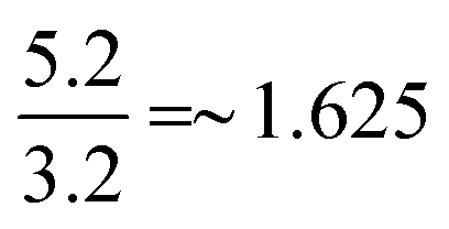 , which clearly shows that at lower current densities, the contribution from the pseudocapacitance is much larger than that of EDLC, whereas for higher current densities, EDLC plays a major role in the electrochemical supercapacitor performance of the sample. A long cyclic stability test, in terms of 5000 charge–discharge cycles (at a current density of 20 A g−1) has been performed for the ZCO sample which is shown in Fig. 5. The cycle number versus capacity retention plot is illustrated in Fig. 5a. The first 5 cycles and last 5 cycles are provided in Fig. 5b and c, respectively, which show little distortion in the shape of the charge–discharge curves even after 5000 cycles. It was found that the retention power of the ZCO sample decreased to ∼78% of its initial value. The morphology of the sample was investigated after 5000 charge–discharge cycles with the help of field emission scanning electron microscopy (see Fig. S3 in the ESI†). The supercapacitor performance of the ZCO sample, in terms of the energy density and power density, has been elucidated in Fig. 5c, through a Ragone plot.
