Polyhydroxylated GdDTPA-derivatives as high relaxivity magnetic resonance imaging contrast agents

Lorenzo Tei*a, Alessandro Bargeb, Matteo Gallic, Roberta Pinallic, Luciano Lattuada*c, Eliana Gianoliod and Silvio Aimed
aDipartimento di Scienze e Innovazione Tecnologica, Università del Piemonte Orientale “Amedeo Avogadro”, Viale T. Michel 11, 15121 Alessandria, Italy. E-mail: lorenzo.tei@uniupo.it
bDipartimento di Scienza e Tecnologia del Farmaco, Università di Torino, via P. Giuria 9, 10125 Torino, Italy
cBracco Imaging SpA, Bracco Research Centre, via Ribes 5, 10010 Colleretto Giacosa, TO, Italy. E-mail: luciano.lattuada@bracco.com
dDepartment of Molecular Biotechnology and Health Sciences, Molecular Imaging Center, Università di Torino, Via Nizza 52, 10126, Torino, Italy

Received 29th July 2015 , Accepted 28th August 2015

First published on 28th August 2015


Abstract

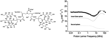
The search for new MRI agents endowed with high relaxivity at the magnetic field strength of clinical scanners (1.5–3 T) is still receiving great attention from researchers involved in the development of new probes. Such Gd(III) complexes should combine a fast inner-sphere water exchange rate (kex) with an enhanced contribution from water molecules in the second and outer coordination spheres. In the present work, dendrimeric-like structures were synthesized by coupling diethylenetriaminepentaacetic acid (DTPA) and its mono-methylphosphonic derivative (P-DTPA) with two differently branched, highly hydrophilic, gluconyl moieties. A 1H and 17O NMR relaxometric study on the corresponding Gd(III) complexes reveals that the Gd-P-DTPA-polyol complex displays very high relaxivities (around 20 mM−1 s−1 at 298 K) over the 0.5–3 T range of field strengths as a result of a fast kex and the presence of a strong second sphere contribution.

Introduction

The use of Gd(III)-chelates as contrast enhancing agents (CA) for Magnetic Resonance Imaging (MRI) has developed considerable research activity aimed at the design of effective, specific and safe CAs.1 The efficacy of a contrast agent is expressed by its relaxivity, r1, which is the enhancement of the longitudinal proton relaxation rate induced by the paramagnetic agent at 1 mM concentration. Theory predicts high relaxivity for a Gd(III) complex when the rate of water exchange between the inner sphere and the bulk solvent (kex = 1/τM) is around 106–107 s−1 and when the molecular reorientational time and electron spin relaxation time are similar and long at the selected field strength.1,2 Nowadays, clinical MR imaging is increasingly moving to higher fields since the number of installed 3 Tesla scanners is steadily growing worldwide.3 Higher fields improve the signal-to-noise ratio (SNR) and provide higher spatial resolution and/or reduced acquisition times.4 Considering this trend, there is the need of contrast agents endowed with a high relaxivity over an extended range of magnetic field strength.5 High-relaxivity CAs permit the use of lower doses in routine clinical applications and are the candidates of choice in molecular imaging procedures aimed at detecting low-concentration targets. Optimizing the three contributions that determine the observed relaxation rates of water protons, namely inner-, second- and outer-coordination sphere, could lead to highly efficient CAs.

One route followed in the search for higher relaxivities relied on the lengthening of the molecular reorientational time through the formation of polymers or of covalent and non-covalent conjugates between the paramagnetic chelate and slowly moving substrates6 (dendrimers,7 proteins,8 carbohydrates9) and the formation of supramolecular adducts like micelles or liposomes.10 In most cases, these systems display maximum r1 values at 20–40 MHz (i.e. 0.5–1 T) but their relaxation enhancement ability decreases quickly at higher fields. Therefore, medium size molecular weight Gd-based compounds have been proposed in order to obtain good relaxivity values over the 0.5–3 T magnetic field range. Examples of these systems are based either on oligomeric (2–8 units) Gd(III) complexes4,11 or on monomeric Gd-complexes with the Gd3+ ion at the baricentre of the macromolecule with a well-structured second sphere of hydration.12 Similar symmetric structures based on DOTA (DOTA = 1,4,7,10-tetraazacyclododecane-1,4,7,10-tetraacetic acid) but with higher molecular weights (5–7 kDa) were reported to have a relaxivity drop at magnetic fields higher than 1 T (40 MHz).13 Moreover, their dimensions allow only a slow diffusion in the extravascular space.

Due to their pharmacokinetic profile, the diagnostic applications of this type of contrast agents are different not only from the already marketed non-specific agents that freely and quickly distribute into the extracellular space, but also from blood pool agents that mainly distribute into the blood system. In light of these considerations, there still is the need for improved non-specific contrast agents able to combine high relaxivity with rapid extravasation, as required for angiographic- and perfusion-based diagnostic applications.

We have faced this problem by synthesizing new Gd(III) complexes based on the structure of DTPA functionalized with highly hydrophilic substituents in order to remarkably increase the second sphere contribution to the relaxivity by creating a structured network of water molecules localised around the polyhydroxyl groups. Ligands L1 and L2 are DTPA-bisamides bearing two differently branched gluconyl moieties and L3 is based on a DTPA derivative in which the central arm has been replaced by a methyl phosphonate moiety (P-DTPA) and two outer acetate moieties bear each the triply gluconyl substituted polyol. The choice of a P-DTPA derivative relies on the published observation that its Gd(III) complex shows an optimal value of τM (about 88 ns).14 Moreover, phosphonate moieties have been shown to yield good second sphere contributions to the observed relaxivity due to hydrogen bonded water molecules localised between the phosphonate group and the Gd.15 In the present work, we report on the synthesis of L1–L3 ligands and on a 1H and 17O NMR relaxometric study on the corresponding Gd(III) complexes.

Results and discussion

Ligands design

The introduction of polyhydroxylic or glycosylic groups at the periphery of a Gd(III) complex has been considered previously with different purposes. In some reports, CAs structures that incorporate glucose and galactose/mannose moieties were developed with the intent to enhance targeting in vivo.16 Others groups engineered complexes in which the Gd(III) ion lied at the barycentre of the macromolecular structure surrounded by a well-defined second hydration sphere to obtain higher relaxivities.12,13 Furthermore, a water-solubilizing polyhydroxylic dendron was incorporated to improve the poor water solubility of the parent complex.17 Our work started with the synthesis of two diamides of DTPA (L1 and L2) bearing differently branched polygluconyl groups on the outer surface. The assessment of the relaxometric properties of their Gd-complexes allowed us to determine the best hydrophilic pendant group for the attainment of a large second sphere contribution to relaxivity. Two examples of DTPA-bisamides bearing two acetylglucose units and a dendrimeric glycosidic cluster with six acetylgluconamides at the periphery were already reported with the aim to create new blood pool CAs more than to evaluate the effect of the second sphere water molecules surrounding the Gd(III) centre.18 As we were aware that DTPA-bisamides are characterized by a very slow water exchange rate (kex) that “quenches” any putative inner sphere relaxivity gain, our next task dealt with the introduction of two polyhydroxy-containing moieties on P-DTPA ligand.

Bifunctional derivatives of DTPA have been reported either with the remote functional group on the central acetate arm or on the diethylenetriamine backbone.19 Only one example of functionalization of a lateral acetate pendant arm was described20 and to the best of our knowledge a bis-functionalized DTPA-like agent has never been reported. Thus, the bifunctional chelating ligand 15 (Scheme 4) was designed having four carboxylic and the phosphonic groups tert-butyl protected and two free carboxylic groups able to react with amino functionalized moieties. Then, the ligand L3 could be prepared by reacting the bifunctional agent 15 with two dendronized polygluconyl groups with the aim to increase the amount of hydroxyl groups and to create a symmetric Gd(III) complex with lateral hydrophilic moieties and a central phosphonate group.

Synthesis

The polyol functionalised DTPA bisamide L1 (Scheme 1) was synthesized in one step with an overall 61% yield starting from DTPA bis-anhydride 4[thin space (1/6-em)]21 and N,N′-(iminodi-2,1-ethanediyl)bis-D-gluconamide 3, previously obtained by amidation of diethylenetriamine 1 with δ-gluconolactone 2 (Scheme 1). Similarly, the DTPA bisamide L2 was synthesised by reaction of DTPA bis-anhydride 4 with the tris-gluconoyl amide derivative 6 obtained by reaction between pentaerythrityl tetramine 5[thin space (1/6-em)]22 and δ-gluconolactone (Scheme 2). Aiming to use this amino-functionalised polyol for further syntheses, we decided to prepare the fully acetylated derivative 9 (Scheme 3). Thus, the amino group of 6 was protected with (Boc)2O and tetramethylammonium hydroxide in DMF to give 7. Then, all the hydroxyl groups were acetylated with Ac2O in pyridine to give 8 and, finally, 9 was obtained by removing the Boc protection with TFA in CH2Cl2.
image file: c5ra15071j-s1.tif
Scheme 1 Synthesis of the DTPA-bisamide L1.

image file: c5ra15071j-s2.tif
Scheme 2 Synthesis of the DTPA-bisamide L2.

image file: c5ra15071j-s3.tif
Scheme 3 Synthesis of the fully acetylated aminopolyol 9; (i) Boc2O, TMAOH, DMF, 48h; (ii) Ac2O, Py, 90 °C, 24 h.

The synthetic procedure to obtain L3, depicted in Scheme 4, relies on the strategy published by Williams and Rapoport who reported the N-bisalkylation of p-nitrophenylalanine benzyl ester with different bromoethylamines as a direct method for the construction of protected DTPA analogues.23 Moreover, following this procedure, modified amino acids have been bisalkylated with [N-(bromoethyl)amino]diacetic acid t-butyl ester in order to obtain functionalized DTPA pentaesters as versatile intermediates for the preparation of conjugates of metal complexes.19,24 We exploited this idea in a different way by synthesising a bromoethyl derivative of protected aspartic acid and reacting it with an aminomethyl phosphonate. By this route, after orthogonal deprotection of two carboxy benzylester groups, we were able to attach an amino-polyol molecule on both sides of the P-DTPA derivative. In particular, L-aspartic acid 4-benzyl ester 10 was esterified, using t-butyl acetate in the presence of catalytic perchloric acid, into L-aspartic acid 1-tert-butyl 4-benzyl diester 11, which was directly reacted with one equivalent of t-butyl bromoacetate under Rapoport's two-phase conditions (i.e.: MeCN/pH 8 phosphate buffer) to yield, after chromatographic purification, the mono-alkylated compound 12 in 76.5% yield. Triester 12 was then further alkylated with 2-bromoethyl-trifluoromethansulfonate in order to obtain in good yield the bromoethyl component 13 to be used for the construction of the P-DTPA derivative (Scheme 4).


image file: c5ra15071j-s4.tif
Scheme 4 Synthesis of L3.

The other component was the aminomethylphosphonic acid di-tert-butyl ester 19 (Scheme 5) obtained in two steps by Mannich reaction of dibenzylamine with paraformaldehyde and tert-butyl phosphite, followed by hydrogenolysis of the benzyl group at atmospheric pressure with 10% Pd/C catalyst. The Rapoport's conditions were also used for the alkylation of 19 with two equivalents of 13 (no excess was used) to get the P-DTPA derivative 14 in 42.4% yield after column purification. Cleavage of the benzyl ester protections of 14 by hydrogenolysis yielded the diacid pentaester 15. This compound was then coupled to the amino-protected polyol 9 in DMF at room temperature for 48 h by using HATU (O-(7-azabenzotriazol-1-yl)-N,N,N′,N′-tetramethyluroniumhexafluoro phosphate) as coupling reagent and N,N-diisopropyl ethylamine as base. Product 16, together with a small amount of the mono-substituted derivative, was collected by precipitation in cold water. Due to difficulties met during the purification procedures, it was decided to use this mixture without separating the mono-functionalized impurity. De-acetylation of the polyalcohol was afforded by bubbling ammonia in a refrigerated methanol solution of 16. This reaction was completed only after 9 days and after bubbling more ammonia every two days. In order to obtain the final ligand L3, the last step was the cleavage of the t-butyl esters by reaction with a 50% solution of trifluoroacetic acid in CH2Cl2 and precipitation by addition of diethyl ether. In order to get pure L3 a simple procedure was followed: the trifluoroacetate salt obtained after t-butyl ester deprotection was redissolved in water, brought to pH 7, lyophilized and recovered with methanol. Ligand L3 was thus obtained as a white solid in 30% yield by filtration of the MeOH solution. On the other hand, evaporation of the solvent give rise to an oil which resulted to be the mono-polyol derivative present in about 28% of the total raw material.


image file: c5ra15071j-s5.tif
Scheme 5 Synthesis of aminomethylphosphonic acid di-tert-butyl ester 19.

Relaxivity of the Gd(III) complexes

The Gd(III) complexes GdL1, GdL2 and GdL3 were prepared by 1H NMR relaxometric titration with a stock solution of GdCl3 at pH 6.5 monitoring the change in the longitudinal water proton relaxation rate (R1) at 20 MHz and 298 K as a function of the concentration of Gd3+. The slope of the straight line obtained corresponds to the relaxivity (r1) of the complex. The residual free Gd3+ ion was quantified by Orange Xylenol UV method as lower than 0.2% in all complexes.25 The exact concentration of Gd3+ ions was determined by measurement of the bulk magnetic-susceptibility shifts of the tBuOH signal. The relaxivity (r1) values at 20 MHz and 298 K are 5.0 mM−1 s−1 for GdL1, 4.3 mM−1 s−1 for GdL2 and 19.2 mM−1 s−1 for GdL3. In Fig. 1, the relaxivities of the three Gd-chelates are plotted as a function of their molecular weight and compared to those reported in the literature for monomeric Gd-complexes with one coordinated water molecule at T = 298 K and B0 = 0.5 T. While the r1 values for the two GdDTPA-bisamides GdL1 and GdL2 are significantly lower than the values expected on the basis of their molecular weight, the r1 of the Gd(III) complex with the monophosphonic DTPA derivative L3, is markedly higher than expected for a molecule of such molecular weight. Noteworthy, the relaxivity of GdL3 is more than four times higher than that of the parent GdDTPA (r1 = 4.7 mM−1 s−1). A more detailed relaxometric characterization was carried out through the registration of the 17O NMR-R2p profiles as a function of temperature (for an accurate determination of the exchange lifetime (τM) of the coordinated water molecule – Fig. 2) and of the 1H-NMRD profiles over the range of frequencies from 0.01 to 120 MHz (Fig. 3A and B).
image file: c5ra15071j-f1.tif
Fig. 1 Relaxivities (r1) values, measured at 298 K and 20 MHz, of GdL1, GdL2 and GdL3 as a function of their molecular weight compared to those reported for other q = 1 complexes. Values for Gd-DOTA, Gd-DTPA and Gd-HPDO3A come from ref. 2, for Gd-4 and Gd-5 from ref. 12a, for Gd-6b, 6c, 6d and 6e from ref. 12b and for P760 and P792 from ref. 13.

image file: c5ra15071j-f2.tif
Fig. 2 Temperature dependence of the paramagnetic contribution to the 17O transverse relaxation rate of water for GdL1, GdL2 and GdL3 (10 mM) measured at 2.1 T and pH 7.

image file: c5ra15071j-f3.tif
Fig. 3 1/T1 NMRD profiles of GdL1, GdL2 (A) and GdL3 (B) recorded at 298 K and pH 7. The data refer to a 1 mM concentration of the paramagnetic complexes. The solid curves through the data points were calculated with the parameters reported in Table 1, whereas the dotted lines in (B) refer to the different contributions to the overall relaxivity.

The analysis of the 17O NMR experimental data yielded the τM values reported in Table 1. The value found for GdL3 (93 ns) is very similar to that already reported for related GdP-DTPA derivatives14 and very close to the optimal value for the achievement of high relaxivity.1,2 On the contrary, very long τM values (5.6 μs and 7.6 μs, for GdL1 and GdL2 respectively) were found for the two bisamides derivatives. This slow water exchange rate, which is invariably found in GdDTPA-bisamides,26 is responsible for the low relaxivity shown by GdL1 and GdL2. The proton relaxivity data measured as a function of the magnetic field strength at 298 K and neutral pH (Fig. 3A and B) were well interpolated with the set of values calculated on the basis of the classical Solomon-Bloembergen-Morgan theory by fixing the τM values to those obtained by 17O NMR-R2p analysis. The main parameters obtained from the fitting of experimental to calculated data are reported in Table 1. As expected, a progressive increase in the reorientational correlation time (τR) occurs upon increasing the molecular weight.

Table 1 Best-fit parameters obtained from 1H-NMRD at 298 K and 17O-R2p versus T analysis shown in Fig. 2 and 3a
  Δ2 (×1019 s−2) τV (ps) τR (ps) τM (μs) q qss rss (Å) τss (ps)
a On carrying out the fitting procedure, some parameters were fixed to reasonable values: rGd-H (distance between Gd and protons of the innersphere water molecule) = 3.1 Å; a (distance of minimum approach of solvent water molecules to Gd ion) = 4 Å; D (solvent diffusion coefficient) = 2.24 × 10⁻⁵ cm2 s−1. rss (distance between Gd and protons of the second sphere water molecules) = 3.7 Å. [Δ2] Squared mean transient zero-field splitting (ZFS) energy. [τV] Correlation time for the collision-related modulation of the ZFS Hamiltonian. [τR] Reorientational correlation time. [τM] Exchange lifetime of the coordinated water molecule. [q] Number of inner-sphere water molecules. [qss] Number of second-sphere water molecules. [τss] correlation time associated with the motions (reorientation or exchange) of the second-sphere water molecules.
Gd-L1 2.9 ± 0.57 22.1 ± 1.8 305 ± 12.8 5.6 ± 1.2 1 0
Gd-L2 2.1 ± 0.57 25.0 ± 1.5 470 ± 68.1 7.6 ± 0.6 1 0
Gd-L3 2.2 ± 0.13 34.2 ± 0.10 560 ± 10.8 0.093 ± 0.003 1 3.5 ± 0.4 3.7 93 ± 0.15


In the case of GdL3 (Fig. 3B), a good fitting of the experimental data was possible only by taking into account a contribution from second sphere water molecules.27 3.5 second sphere water molecules (qss) were estimated to be located at an average distance of 3.7 Å from the Gd3+ ion with an overall correlation time (τss) of 93 ps (Table 1). The increase in the correlation time associated with the collision-related modulation of the zero-field splitting Hamiltonian (τV) observed in the case of GdL2 and GdL3, is likely related to the occurrence of a reduced rate of solvent collisions between the coordinated and bulk water molecules. Tentatively, one may relate the observed behavior to the enhanced shielding of the poly-hydroxylic containing moieties with respect to GdL1.

In summary, one may conclude that the high relaxivity shown by GdL3 is the result of the occurrence of three positive conditions: (i) an array of second sphere water molecules linked through hydrogen bond to the phosphonate moiety, (ii) a fast water exchange rate of the coordinated water molecule and (iii) the increased molecular weight (τR = 560 ns) determined by the introduction of the poly-hydroxylic substituents on the surface of the metal complex. Noteworthy, the relaxivity values for GdL3, measured at the Larmor frequencies more used on clinical scanners (40, 60, 120 MHz corresponding to 1, 1.5 and 3 T, respectively), are somehow constant, being 20.8 mM−1 s−1 at 40 and 60 MHz and 19.1 mM−1 s−1 at 120 MHz.

Experimental

General

All reagents and solvents were obtained from commercial suppliers and directly used without further purification. N,N′-Bis-[2-(2,6-dioxo-4-morpholinyl)ethyl]glycine (DTPA-bis-anhydride) 4,21 pentaerythrityl tetramine tetrahydrochloride 5,22 tert-butyl phosphite28 and 2-bromoethyl trifluoromethanesulfonate29 were synthesized as reported in literature. TLC was performed on Merck silica gel 60 TLC plates F254 and visualized by using UV (254 nm) or 1% KMnO4 in 1 N NaOH. Column chromatography was performed by using silica gel 60 (70–230 mesh) while flash chromatography was carried out on silica gel 60 (230–400 mesh). The 1H, 13C and 31P spectra were recorded on a Bruker Avance 400 instrument. Chemical shifts are reported in δ relative to an internal standard of residual chloroform (δ 7.27 for 1H NMR and 77.16 for 13C NMR). ESI MS spectra on novel compounds were acquired on a high resolving power mass spectrometer LTQ Orbitrap (Thermo Scientific, Rodano, Italy), equipped with an atmospheric pressure interface and an ESI ion source. Other mass spectra were recorded with a ThermoFinnigan TSQ700 triple-quadrupole instrument equipped with an electrospray ionization source. Analytical HPLC was performed on a Merck KGaA apparatus with the following method: stationary phase: Lichrospher RP-Select B 5 μm, 250 × 4 mm column packed by Merck KGaA; mobile phase: eluent A = 0.01 M KH2PO4 and 0.017 M H3PO4 in H2O, eluent B = MeCN, gradient elution: t = 0 min (5% B), t = 30 min (80%B); T = 45 °C; flow rate: 1 mL min−1; UV detection: 210 nm.
Synthesis of N,N′-(iminodi-2,1-ethanediyl)bis-D-gluconamide (3). Diethylenetriamine 1 (1 g, 9.7 mmol) was added dropwise to a suspension of δ-gluconolactone 2 (3.6 g, 20.3 mmol) in DMF (10 mL). The reaction mixture was stirred at room temperature for 1.5 h. The precipitate was filtered, washed with CH2Cl2 (500 mL), to remove DMF and the excess of δ-gluconolactone, and dried to obtain 3 as a white solid (4.06 g, 8.83 mmol), yield 91%; mp 142 °C (dec.); Rf 0.12 (eluent CHCl3/MeOH/25% NH4OH 30[thin space (1/6-em)]:[thin space (1/6-em)]60[thin space (1/6-em)]:[thin space (1/6-em)]20); 1H NMR (600 MHz, D2O) δ 4.24 (d, J = 5.0 Hz, 2H), 4.00 (d, J = 5.0 Hz, 2H), 3.72 (d, J = 16.9 Hz, 2H), 3.66 (m, 4H), 3.55 (dd, J = 11.0, 4.4 Hz, 3H), 3.31 (dt, J = 12.1, 9.2 Hz, 4H), 2.71 (t, J = 9.2 Hz, 4H); 13C NMR (151 MHz, D2O) δ 175.08 (CO), 73.81 (C-2), 72.50 (C-3), 71.45 (C-4), 70.70 (C-5), 62.96 (C-6), 47.33 (CH2N), 38.45 (CH2N).
Synthesis of N,N-bis[2-[(carboxymethyl)[2-[bis[2-[(D-gluconoyl)amino]ethyl]amino]-2-oxoethyl]amino] ethyl]glycine (L1). N,N′-Bis-[2-(2,6-dioxo-4-morpholinyl)ethyl]glycine (DTPA-bis-anhydride)21 4 (0.19 g; 0.54 mmol) was added to a solution of 3 (0.5 g; 1.09 mmol) in DMSO (5 mL) and the yellow solution was stirred at 50 °C for 8 h. After cooling at room temperature, the reaction mixture was diluted with CH2Cl2 (50 mL); the precipitate was filtered, washed with CH2Cl2 (200 mL) then lyophilized to remove the residual DMSO. L1 was obtained as white solid (0.42 g; 0.33 mmol), yield 61%; mp 128–130 °C Rf 0.30 (eluent MeOH/25% NH4OH 7[thin space (1/6-em)]:[thin space (1/6-em)]3); HPLC: retention time 2.1 min, 98% (area%); 1H NMR (600 MHz, D2O, 333 K) δ 4.70–4.58 (m, 10H), 4.37 (s, 4H), 4.18 (s, 4H), 4.12–4.05 (m, 12H), 3.98–3.85 (m, 12H), 3.78 (m, 12H), 3.49 (m, 4H); 13C NMR (151 MHz, D2O, 333 K) δ 175.68, 175.36, 174.90, 171.11, 167.40 (CO), 74.21 (C-2), 74.15 (C-2′), 73.97 (C-3), 73.07 (C-3′), 72.72 (C-4), 72.19 (C-4′), 71.30 (C-5), 62.56 (C-6), 58.16, 56.80, 56.02 (CH2CO), 53.92, 50.42 (CH2N), 47.41, 46.55 (CH2N), 37.49, 36.50 (CH2NH). ESI-HRMS (m/z): 1276.4563 (M + H+) (calcd 1276.5379).
Synthesis of N-[[2,2-bis[(D-gluconoylamino)methyl]-3-amino]propyl]-D-gluconamide (6). Pentaerythrityl tetramine tetrahydrochloride22 5 (14 g; 0.051 mol) was suspended in a mixture of DMF (170 mL) and Et3N (25.4 mL; 0.184 mmol). A solution of δ-gluconolactone 2 (32.7 g; 0.184 mol) in DMF (195 mL) was slowly added dropwise (1.5 h) at room temperature. The mixture was stirred at room temperature for 4 days. The reaction mixture was filtered and concentrated at reduce pressure. The residue was treated with MeCN to give a precipitate which was filtered and washed with MeCN. The solid was dried to give 6 as a white solid and as hydrochloride salt (30.5 g; 0.046 mmol), yield 90%; Rf 0.44 (eluent MeOH/25% NH4OH 7[thin space (1/6-em)]:[thin space (1/6-em)]3); 1H NMR (600 MHz, D2O) δ 4.35 (m, 3H), 4.06 (dt, J = 9.5, 3.2 Hz, 3H), 3.75 (m, 6H), 3.69 (m, 3H), 3.60 (dd, J = 11.8, 6.3 Hz, 3H), 3.21–3.10 (m, 6H), 3.06–2.96 (m, 2H); 13C NMR (151 MHz, D2O) δ 176.55 (CO), 175.99 (CO), 74.04 (C-2), 73.91 (C-2′), 72.95 (C-3), 72.74 (C-3′), 71.52 (C-4), 70.83 (C-5), 70.68 (C-5′), 62.96 (C-6), 62.93 (C-6′), 44.86 (C), 38.59 (CH2N), 38.47 (CH2N). ESI-HRMS (m/z): 667.2175 (M + H+) (calcd 667.2880).
Synthesis of N,N-bis[[[2,2-bis[(D-gluconoylamino)methyl]-3-amino]propyl]-D-gluconamide]-2-oxoethyl]amino] ethyl]glycine (L2). N,N′-Bis-[2-(2,6-dioxo-4-morpholinyl)ethyl]glycine (DTPA-bis-anhydride)21 4 (0.19 g; 0.54 mmol) was added to a solution of 6 (0.8 g; 1.20 mmol) in DMSO (5 mL) and stirred at 50 °C for 8 h. After cooling at room temperature, the reaction mixture was diluted with CH2Cl2 (50 mL); the precipitate was filtered, washed with CH2Cl2 (200 mL) then lyophilized to remove the residual DMSO. L2 was obtained as white solid (0.64 g; 0.38 mmol), yield 70%. 1H NMR (300 MHz, D2O) δ 4.18 (m, 4H), 3.94 (d, J = 3.2 Hz, 2H), 3.86–3.73 (m, 6H), 3.65–3.54 (m, 8H), 3.51 (m, 8H), 3.45–3.32 (m, 8H), 3.06–2.77 (m, 6H), 2.51 (s, 18H), 2.29 (m, 6H), 0.78 (t, J = 7.2 Hz, 4H). 13C NMR (75 MHz, D2O, pH = 10) δ 179.4, 173.7, 168.1, 74.8, 74.7, 73.9, 71.9, 71.4, 70.9, 70.3, 63.0, 45.2, 38.5, 9.9. ESI-HRMS (m/z): 1690.6211 (M + H+) (calcd 1690.6865).
Synthesis of (N-[[2,2-bis[D-gluconoylamino)methyl]-3-tert-butoxycarbonylamino]propyl]-D-gluconamide (7). Compound 6 (30.0 g, 45.0 mmol) was dissolved in DMF (400 mL) and a solution of (Boc)2O (17.2 g; 65.2 mmol) in DMF (100 mL) was slowly added dropwise. Then, tetramethylammonium hydroxide (8.15 g, 45.0 mmol) was added and the mixture was stirred at room temperature for 2 days. The solution was concentrated at reduced pressure and the residue was treated with CH2Cl2 to give a precipitate which was filtered and washed with H2O (100 mL) and CH3OH (100 mL). The solid so obtained was dried to give 7 (25 g, 32.6 mmol), yield: 72%. 1H NMR (600 MHz, D2O) δ 4.35 (d, J = 2.7 Hz, 3H), 4.07 (t, J = 3.1 Hz, 3H), 3.75 (m, 6H), 3.69 (m, 3H), 3.61 (dd, J = 11.8, 6.4 Hz, 3H), 3.12 (s, 15H), 3.02 (m, 6H), 2.85 (m, 2H), 1.41 (s, 9H); 13C NMR (151 MHz, D2O) δ 175.79 (CO), 158.81 (CO), 81.88 (C), 74.06 (C-2), 72.94 (C-3), 71.52 (C-4), 70.69 (C-5), 62.97 (C-6), 55.67, 45.52 (C), 39.94 (CH2N), 38.72 (CH2N), 28.07 (CH3). ESI-HRMS (m/z): 789.3003 (M + Na+) (calcd 789.3229).
Synthesis of (N-[[2,2-bis[2,3,4,5,6-penta-O-acetyl-D-gluconoylamino)methyl]-3-tert-butoxycarbonylamino]propyl]-2,3,4,5,6-penta-O-acetyl-D-gluconamide (8). To a suspension of compound 7 (10.0 g; 13.0 mmol) in Ac2O (186 mL; 1.97 mol), pyridine (25.7 g; 326 mmol) was added at room temperature. The mixture was then heated to 90 °C to dissolve the reagents, then cooled at room temperature. After 1 day at room temperature, the mixture was concentrated at reduce pressure and the residue was dissolved in EtOAc (200 mL) and washed with water (200 mL) and with 10% aqueous NaHCO3 (200 mL). The organic phase was dried (Na2SO4) and evaporated to give 8 (15 g; 10.7 mmol), yield: 82%, 1H NMR (600 MHz, CDCl3) δ 8.21 (s, 1H), 7.81 (s, 2H), 6.00 (s, 1H), 5.65 (m, 3H), 5.45 (m, 3H), 5.26 (m, 3H), 5.07 (m, 3H), 4.31 (m, 3H), 4.17 (br m, 3H), 3.37 (m, 4H), 3.22 (m, 2H), 3.04 (m, 2H), 2.29, 2.10, 2.06 (m, 45H), 1.42 (s, 9H). 13C NMR (151 MHz, CDCl3) δ 170.84 (CO), 170.11 (CO), 170.04 (CO), 158.25 (CO), 80.26 (C), 71.50 (C-2), 70.43 (C-3), 70.28 (C-4), 69.23 (C-5), 61.68 (C-6), 39.65, 28.66 (CH3), 21.09 (CH3), 21.04 (CH3), 20.92 (CH3), 20.64 (CH3); ESI-HRMS (m/z): 1419.4123 (M + Na+) (calcd 1419.4814).
Synthesis of (N-[[2,2-bis[2,3,4,5,6-penta-O-acetyl-D-gluconoylamino)methyl]-3-amino]propyl]-2,3,4,5,6-penta-O-acetyl-D-gluconamide (9). Trifluoroacetic acid (3.67 g; 32.0 mmol) was added to a solution of compound 8 (4.50 g; 3.20 mmol) in CH2Cl2 (60 mL). After 3 days at room temperature, further TFA (1.82 g; 16.0 mmol) was added to the reaction mixture. After further 4 days at room temperature, the solvents were removed under reduced pressure and the residue was dissolved in CH2Cl2 (7 mL) and TFA (5.49 g; 48.0 mmol). The mixture was stirred at room temperature for 1 day, then evaporated under reduced pressure. The residue was dissolved in CH2Cl2 (10 mL) and evaporated several times affording 9 as a white solid and as trifluoroacetic salt (3.00 g; 2.13 mmol), yield: 67%; Rf 0.59 (eluent EtOAc/n-hexane 8[thin space (1/6-em)]:[thin space (1/6-em)]2); 1H NMR (400 MHz, CDCl3) δ 8.27 (s, 1H), 7.78 (s, 2H), 7.34 (s, 2H), 5.60 (m, 3H), 5.46 (m, 3H), 5.24 (d, J = 3.3 Hz, 3H), 5.09 (td, J = 6.0, 3.2 Hz, 3H), 4.34 (m, 3H), 4.16 (dd, J = 12.4, 5.8 Hz, 3H), 3.02 (m, 6H), 2.70 (s, 2H), 2.24, 2.13, 2.09, 2.07 (s, 45H). 13C NMR (101 MHz, CDCl3) δ 170.99 (CO), 170.61 (CO), 170.47 (CO), 72.46 (C-2), 70.07 (C-3), 69.37 (C-4), 69.07 (C-5), 62.00 (C-6), 45.20 (C), 38.96 (CH2N), 38.49 (CH2N), 21.01 (CH3), 20.96 (CH3), 20.72 (CH3), 20.59 (CH3); ESI-HRMS (m/z): 1297.3996 (M + H+) (calcd 1297.4470).
Synthesis of (dibenzylamino)methylphosphonic acid di-tert-butyl ester (18). Paraformaldehyde (2.14 g; 71.2 mmol) was added to a solution of dibenzylamine (12.7 g; 64.5 mmol) in MeCN (130 mL). The suspension was heated to 80 °C for 1 h, then the resulting solution was cooled to rt. A solution of tert-butyl phosphite (71.2% of purity, 25 g; 71.2 mmol) in MeCN was added dropwise over 25 min to the reaction mixture and the solution was stirred at room temperature for 22 h. The solvent was evaporated under vacuum and 0.1 N HCl (320 mL) was added to the residue. The suspension was extracted with CH2Cl2 (3 × 150 mL) and the combined organic phases washed with water (3 × 150 ml), dried (Na2SO4) and evaporated in vacuo. The crude (27.3 g) was purified by chromatography on silica gel (15[thin space (1/6-em)]:[thin space (1/6-em)]85 EtOAc/petroleum ether; Rf 0.2) and the desired product was obtained (12.4 g, 30.8 mmol) as a colourless oil. Yield: 47.7%. 1H-NMR (CDCl3) 400 MHz δ = 7.43 (d, 4H, J = 7.2 Hz, Har), 7.32 (t, 4H, J = 7.2 Hz, Har), 7.24 (m, 2H, Har), 3.94 (s, 4H, CH2Ph), 2.79 (d, 2H, J = 10.2 Hz, CH2P), 1.49 (s, 18H, CCH3). 13C-NMR (CDCl3) 100 MHz δ = 139.6 (Car), 129.6–128.6–127.3 (CHar), 82.4 (CCH3), 59.5 (CH2Ph), 52.5 (CH2P, J = 163 Hz), 30.9 (CCH3); 31P-NMR (CDCl3) 162 MHz δ = 19.4. MS (ESI+; MeOH): m/z 404 (M + H+).
Synthesis of aminomethylphosphonic acid di-tert-butyl ester (19). (Dibenzylamino)methylphosphonic acid di-tert-butyl ester (33.3 g; 82.7 mmol) was dissolved in MeOH (500 mL) and hydrogenated at atmospheric pressure on Pd/C 10% (3.3 g). After 2 h the mixture was filtered through Millipore® apparatus (0.5 μm) and the solution evaporated under vacuum. The crude (18.4 g) was used without any further purification. Quantitative yield. 1H-NMR (CDCl3) 400 MHz δ = 2.81 (d, 2H, J = 10.2 Hz, CH2P), 1.49 (s, 18H, CCH3). 13C-NMR (CDCl3) 100 MHz δ = 83.2 (CCH3), 40.6 (CH2P, J = 153 Hz), 29.5 (CCH3); 31P-NMR (CDCl3) 162 MHz δ = 21.0. MS (ESI+; MeOH): m/z 224 (M + H+).
Synthesis of L-aspartic acid, 1-tert-butyl-4-benzylester (11). Perchloric acid (70% aq.; 19.28 g; 0.134 mol) was dropped into a suspension of L-aspartic acid 4-(phenylmethyl) ester (25 g; 0.112 mol) in tert-butyl acetate (515 mL; 3822 mol) stirred at room temperature. After 18 h at room temperature the obtained clear solution was diluted with H2O (470 mL) and the phases were separated; the aqueous phase was extracted with EtOAc (2 x 235 mL). The organic phases were collected and washed with 5% aq. NaHCO3 (2 × 250 mL) and H2O (2 × 200 mL); the new aqueous phases were collected and extracted with EtOAc (3 × 100 mL). All organic phases were collected, dried over Na2SO4 and evaporated to obtain 11 (27 g; 0.097 mol) as a pale yellow oil. Yield 86.6%. TLC (silica gel; eluent: EtOAC; Rf 0,57). 1H-NMR (CDCl3) 400 MHz δ = 7.31 (m, 5H, Har), 5.14 (s, 2H, CH2Ph), 3.68 (t, 1H, J = 6.3 Hz, CHNH2), 2.77 and 2.66 (dd, 2H, J1 = 16.3 Hz, J2 = 4.9 Hz, CH2CO2Bn), 1.76 (bs, 2H, NH2), 1.40 (s, 9H, CCH3). 13C-NMR (CDCl3) 100 MHz δ = 173.7 (CO2Bn), 171.5 (CO2tBu), 136.1 (Car), 128.7 (CHar), 81.8 (CCH3), 66.8 (CH2Ph), 52.2 (CHNH2), 39.6 (CH2CO2Bn), 28.3 (CCH3). MS (ESI+; MeOH): m/z 280.2 (M + H+); 302.2 (M + Na+).
Synthesis of N-2-(t-butoxycarbonylmethyl)-L-aspartic acid, 1-tert-butyl-4-benzylester (12). A mixture of L-aspartic acid, 1-t-butyl-4-benzylester 11 (27 g; 0.0967 mol), t-butyl bromoacetate (20.00 g; 0.102 mol), acetonitrile (160 mL) and 2 M phosphate buffer pH 8 (80 mL) was vigorously stirred at room temperature; after 18 h the phases was separated and the organic phase was evaporated. The residue thus obtained was dissolved in EtOAc (300 mL) and washed with H2O (2 × 150 mL) and brine (2 × 150 mL). After drying over Na2SO4, the organic solution was evaporated to give a crude (34 g) that was purified by flash-chromatography [silica gel; eluent: 4[thin space (1/6-em)]:[thin space (1/6-em)]1 n-hexane/EtOAc; Rf 0,34] to obtain 29.1 g of 1. (Yield: 76.5%) 1H-NMR (CDCl3) 400 MHz δ = 7.32 (m, 5H, Har), 5.14 (s, 2H, CH2Ph), 3.59 (t, 1H, J = 6.3 Hz, CHNH), 3.36 (dd, 2H, J1 = 17.0 Hz, CH2CO2tBu), 2.78 and 2.71 (dd, 2H, J1 = 16.0 Hz, J2 = 6.4 Hz, CH2CO2Bn), 1.48 and 1.44 (s, 18H, CCH3). 13C-NMR (CDCl3) 100 MHz δ = 172.3 (CO2Bn), 171.3, 171.1 (CO2tBu, 2C), 136.1 (Car), 128.9, 128.6 (CHar), 82.1, 81.6 (CCH3), 66.9 (CH2Ph), 58.0 (CHNH), 50.3 (CH2CO2tBu), 38.5 (CH2CO2Bn), 28.6, 28.3 (CCH3). ESI-HRMS (m/z): 394.2167 (M + H+) (calcd 394.2230).
Synthesis of N-2-bromoethyl-N-2-(t-butoxycarbonylmethyl)-L-aspartic acid, 1-tert-butyl-4-benzylester (13). 2-Bromoethyl trifluoromethanesulfonate29 (34.93 g; 0.136 mol, 1.7 eq.) was slowly dropped into a solution of compound 12 (31.5 g; 0.08 mol) and 2,6-lutidine (27 g; 0.25 mol, 3.1 eq.) in toluene (450 mL) stirred at −15 °C under nitrogen atmosphere. The reaction mixture was stirred at room temperature for 22 h then diluted with H2O (200 mL) and extracted with EtOAc (200 mL). The organic solution was dried over Na2SO4 and evaporated to give a crude (33 g) that was purified by chromatography [silica gel, eluent: 7[thin space (1/6-em)]:[thin space (1/6-em)]3 n-hexane/iPr2O, Rf 0,44] to obtain 13 (24,9 g; 0,05 mol) as a pale yellow oil. Yield 62.4%. 1H-NMR (CDCl3) 400 MHz δ = 7.37 (m, 5H, Har), 5.15 (s, 2H, CH2Ph), 3.87 (t, 1H, J = 7.4 Hz, CHN), 3.40, 3.15 (m, 6H, CH2CH2Br and CH2CO2tBu), 2.85 and 2.69 (dd, 2H, J1 = 16.0 Hz, J2 = 7.4 Hz, CH2CO2Bn), 1.46 and 1.44 (s, 18H, CCH3). 13C-NMR (CDCl3) 100 MHz δ = 171.0 (CO2Bn and CO2tBu, 3C), 136.1 (Car), 128.9, 128.7 (CHar), 82.4, 81.5 (CCH3), 67.0 (CH2Ph), 62.9 (CHN), 56.1, 54.9 (NCH2, 2C), 36.7 (CH2CO2Bn), 30.8 (CH2Br), 28.5 (CCH3). ESI-HRMS (m/z): 500.1206 (M + H+) (calcd 500.1648).
Synthesis of N,N′-[[[[bis(1,1-dimethylethoxy)phosphinyl]methyl]imino]di-2,1-ethanediyl]bis[N-[2-(1,1-dimethylethoxy)-2-oxoethyl]-L-aspartic acid 1,1′-bis(1,1-dimethylethyl) 4,4′-bis(phenylmethyl) ester (14). A solution of bromo-derivative 13 (4.11 g; 8.22 mmol) in acetonitrile (20 mL) was slowly dropped into an emulsion of aminomethylphosphonic acid di-tert-butyl ester 19 (916 mg; 4.11 mmol) in acetonitrile (10 mL) and 2 M phosphate buffer pH 8 (20 mL) over 8 hours under vigorous stirring. After 22 h, the phases were separated and the aqueous layer was extracted with EtOAc (2 × 30 mL). The organic layer was evaporated. The oily residue was dissolved with EtOAc (20 mL), and the solution was washed with 1[thin space (1/6-em)]:[thin space (1/6-em)]1 water/brine (2 × 30 mL) and dried over Na2SO4. The crude (4.49 g) was purified by silica gel chromatography (silica gel; EtOAc/petroleum ether 4[thin space (1/6-em)]:[thin space (1/6-em)]6; Rf 0.32). Compound 14 was obtained (1.86 g; 1.75 mmol) as a yellow oil. Yield 42.4%. 1H-NMR (CDCl3) 400 MHz δ = 7.32 (m, 10H, Har), 5.09 (s, 4H, CH2Ph), 3.80 (dd, 2H, J = 3.6 Hz, CHN), 3.35 (s, 4H, CH2CO2tBu), 2.82–2.62 (m, 14H, NCH2CH2N, NCH2P and CH2CO2Bn), 1.46 and 1.41 (s, 54H, CCH3). 13C-NMR (CDCl3) 100 MHz δ = 171.3 (CO2Bn and CO2tBu, 6C), 136.3 (Car), 128.9, 128.6 (CHar), 82.5, 81.8, 81.0 (CCH3), 66.7 (CH2Ph), 62.2 (NCH), 55.0 (CH2CO2tBu), 54.7, 51.2 (NCH2CH2N), 52.2 (CH2P), 36.5 (CH2CO2Bn), 30.9 and 28.5 (CCH3). 31P-NMR (CDCl3) 162 MHz δ = 18.6. ESI-HRMS (m/z): 1062.5503 (M + H+) (calcd 1062.6031).
Synthesis of N,N′-[[[[bis(1,1-dimethylethoxy)phosphinyl]methyl]imino]di-2,1-ethanediyl]bis[N-[2-(1,1-dimethylethoxy)-2-oxoethyl]-L-aspartic acid 1,1′-bis(1,1-dimethylethyl) ester, (15). Compound 14 (973 mg; 1.10 mmol) was dissolved in methanol (50 mL) and hydrogenated at atmospheric pressure on 10% Pd/C (50 mg). After 30 min the reaction mixture was filtered through Millipore apparatus® (0.5 m filter) and the solution evaporated under vacuum. The crude (725 mg) was used in the next step without any further purification. 1H-NMR (CDCl3) 400 MHz δδ = 3.89 (m, 2H, CHN), 3.49–3.36 (m, 4H, CH2CO2tBu), 3.25–2.95 (m, 10H, NCH2CH2N, NCH2P) 2.75 (CH2CO2H), 1.50 and 1.46 (s, 54H, CCH3). 13C-NMR (CDCl3) 100 MHz δδ = 173.0 (COOH), 170.8, 170.2 (CO2tBu, 4C), 84.2, 82.7, 82.2 (CCH3), 61.7 (NCH), 54.5 (CH2CO2tBu), 54.0, 50.1 (NCH2CH2N), 51.8 (CH2P, J = 155 Hz), 35.6 (CH2CO2H), 30.9 and 28.5 (CCH3). 31P-NMR (CDCl3) 162 MHz δ = 15.0. ESI-HRMS (m/z): 882.4855 (M + H+) (calcd 882.5092).
Synthesis of compound (16). A solution of diacid 15 (4.91 g; 5.58 mmol), polyalcohol 9 (17.40 g; 13.38 mmol, 2.4 equivalents) and HATU (5.08 g; 13.38 mmol) in 50 mL of DMF was brought to 0 °C with an ice bath in nitrogen atmosphere. To this solution, DIPEA (9.32 mL, 53.52 mmol, 9.6 eq.) was added keeping the temperature below 5 °C. The mixture was kept under stirring at room temperature for 48 h monitoring the reaction by TLC (silica gel; CHCl3/MeOH 9[thin space (1/6-em)]:[thin space (1/6-em)]1), then the reaction solution was poured in ice, stirred for 30 min and filtered. The solid was washed with water, dissolved in chloroform and dried over Na2SO4. After filtration and solvent evaporation, the desired product was obtained as pale yellow gummy solid (15.3 g, 80% yield). (Rf 0.52, silica, CHCl3/MeOH 9[thin space (1/6-em)]:[thin space (1/6-em)]1). 1H NMR (400 MHz, CDCl3) δ 7.86 (sb, 8H), 5.61 (m, 6H), 5.46 (m, 6H), 5.31 (m, 6H), 4.97 (m, 6H), 4.29 (m, 6H), 4.16 (m, 6H), 3.83 (m, 2H, CHN), 3.60–2.75 (m, 14H, CH2CO2tBu, NCH2CH2N, NCH2P), 2.28 (s, 16H, CCH2N), 2.07 and 2.02 (m, 90H, CH3COO), 1.54 and 1.46 (s, 54H, CCH3).13C NMR (101 MHz, CDCl3) δ 172.8, 170.9, 170.5, 169.8, 169.6, 168.7, 81.4, 69.9, 68.9, 61.4, 45.8, 38.4, 30.7, 30.0, 28.3, 28.2, 20.7. ESI-HRMS (m/z): 3439.2986 (M + H+) (calcd 3439.3665).
Synthesis of compound (17). Ammonia was bubbled for 45 min into a solution of the fully protected ligand 16 (19.48 g) in methanol (400 mL) refrigerated with an ice bath. After 1 h the ice bath was removed, CaCl2 valve was applied and the solution was allowed to stir at room temperature for 9 days. Every two days more ammonia was bubbled for 10–15 min into the solution. The reaction was followed by TLC (CHCl3/MeOH 9[thin space (1/6-em)]:[thin space (1/6-em)]1) and by MS until disappearance of any peaks related to partially acetylated product. Then, the solvent was evaporated in vacuum obtaining 18.1 g of the crude which was used without any further purification in the next deprotection step. 1H NMR (400 MHz, D2O) δ 4.34 (bs, 6H), 4.06 (bs, 6), 3.82–3.71 (m, 8H), 3.68 (t, J = 6.8 Hz, 4H), 3.59 (m, 4H), 2.94 (bs, 8H), 2.78 (s, 4H), 2.73 (s, 2H), 1.42 (m, 56H). 13C NMR (101 MHz, CDCl3) δ 171.9, 171.5, 170.8, 170.7, 169.7, 82.4, 78.0, 70.9, 70.0, 62.4, 46.7, 39.2, 31.7, 31.0, 29.2. ESI-HRMS (m/z): 2179.1102 (M + H+) (calcd 2179.0495).
Synthesis of compound (L3). A round-bottomed flask containing the phosphonic proligand 17 (18.1 g, 8.3 mmol) was cooled with an ice bath; then a mixture of CH2Cl2/TFA 1[thin space (1/6-em)]:[thin space (1/6-em)]1 (100 mL) was added dropwise giving a yellow-brown solution. After 20 min, the ice bath was removed and the solution was kept under stirring at room temperature for 16 h. The solvent was removed and the residue was dissolved in dichloromethane (5 mL) and evaporated; this procedure was repeated four times. The crude was treated with Et2O to obtain the precipitation of a white solid which was filtered, dissolved in water and lyophilized. The slightly yellow solid was then re-dissolved in water, brought to pH 7 by adding 2 M NaOH and lyophilized again. The solid obtained was recovered with MeOH and kept under stirring for 1 h. The white solid formed was filtered and dried in vacuum to obtain 4.6 g (2.50 mmol, 30% yield) of product. The methanolic solution was dried at the rotary evaporator and in vacuum to obtain 2.9 g of the mono-derivative as a yellow oil. 1H NMR (400 MHz, D2O) δ 4.39 (bs, 6H), 4.10 (bs, 6H), 3.82–3.76 (m, 8H), 3.76–3.70 (m, 4H), 3.64 (dd, J = 11.6, 6.3 Hz, 4H), 3.32 (s, 4H), 3.07 (s, 8H), 2.96 (s, 2H). 13C NMR (101 MHz, CDCl3) δ 171.9, 171.5, 170.8, 170.7, 169.7, 82.4, 78.0, 70.9, 70.0, 62.4, 46.7, 39.2, 31.7, 31.0, 29.2. ESI-HRMS (m/z): 1842.6012 (M + H+) (calcd 1842.6739).

Water proton relaxation measurements

The longitudinal water proton relaxation rates were measured at 25 °C by using a Stelar Spinmaster (Stelar, Mede, Pavia, Italy) spectrometer operating at 0.5 T (21.5 MHz Proton Larmor Frequency), by mean of the standard inversion-recovery technique. The temperature was controlled with a Stelar VTC-91 air-flow heater equipped with a copper constantan thermocouple (uncertainty 0.1 °C). The proton 1/T1 NMRD profiles were measured at 25 °C on a fast field-cycling Stelar relaxometer over a continuum of magnetic field strengths from 0.00024 to 0.47 T (corresponding to 0.01–20 MHz proton Larmor frequencies). The relaxometer operates under computer control with an absolute uncertainty in 1/T1 of ±1%. Additional data points in the range 21.5–70 MHz were obtained on the Stelar Spinmaster spectrometer. The concentration of the solutions used for the relaxometric characterization was determined by measuring the bulk magnetic-susceptibility shifts of the tBuOH signal.

17O-NMR measurements

Variable temperature 17O-NMR measurements were recorded at 2.1 T on a JEOL90 spectrometer, equipped with a 5 mm probe, by using a D2O external lock. The experimental settings were: spectral width 9000 Hz, 90° pulse (12 μs), acquisition time 10 ms, 1024 scans and without sample spinning. Aqueous solutions containing 2.6% of 17O isotope (Yeda, Israel) were used. The observed transverse relaxation rates (RO2obs) were calculated from the signal width at half-height (Δν1/2): RO2obs = πΔν1/2. Paramagnetic contributions to the observed transversal relaxation rate (R2p) were calculated by subtracting from RO2obs the diamagnetic contribution measured at each temperature value on pure water enriched with 2.6% 17O isotope.

Conclusions

The synthesis of three novel DTPA derivatives bearing polyhydroxylated pendant arms is reported. While the two DTPA bisamides L1 and L2 with differently branched polygluconyl groups were easily synthesised from DTPA bis-anhydride, the monophosphonic P-DTPA ligand L3 was prepared by a multistep procedure which included the synthesis of a bis-functionalized DTPA bifunctional ligand via the Rapoport reaction. The relaxivity of GdL1 and GdL2 were very low because DTPA-bisamides are characterized by slow water exchange rates of the coordinated water molecule. On the other hand, the relaxivity values measured for GdL3 over a wide range of imaging fields (0.5–3 Tesla) are among the highest till now reported for monomeric Gd(III) complexes with medium sized molecular weight (∼2000 Da). This finding makes GdL3 a very promising system for applications at the currently used magnetic fields of the clinical MRI scanners.

Notes and references

  1. The Chemistry of Contrast Agents in Medical Magnetic Resonance Imaging, ed. A. Merbach, L. Helm and É. Tóth, John Wiley & Sons Ltd, Chichester, 2nd edn, 2013 Search PubMed.
  2. (a) C. F. G. C. Geraldes and S. Laurent, Contrast Media Mol. Imaging, 2009, 4, 1 CrossRef CAS PubMed; (b) P. Hermann, J. Kotek, V. Kubíček and I. Lukeš, Dalton Trans., 2008, 3027 RSC; (c) P. Caravan, J. J. Ellison, T. J. McMurry and R. B. Lauffer, Chem. Rev., 1999, 99, 2293 CrossRef CAS PubMed.
  3. (a) Y. Lvovsky, E. W. Stautner and T. Zhang, Supercond. Sci. Technol., 2013, 26, 1 CrossRef; (b) T. C. Cosmus and M. Parizh, IEEE Trans. Appl. Supercond., 2011, 21, 2104 CrossRef CAS.
  4. (a) P. R. Luijten and D. W. J. Klomp, Drug Discovery Today: Technol., 2011, 8, e103 CrossRef PubMed; (b) C. K. Khul, F. Träber and H. H. Schild, Radiology, 2008, 246, 675 CrossRef PubMed.
  5. (a) L. Helm, Future Med. Chem., 2010, 2, 385 CrossRef CAS PubMed; (b) P. Caravan, C. T. Farrara, L. Frullano and U. Ritika, Contrast Media Mol. Imaging, 2009, 4, 89 CrossRef CAS PubMed.
  6. M. Botta and L. Tei, Eur. J. Inorg. Chem., 2012, 1945 CrossRef CAS PubMed.
  7. (a) M. R. Longmire, M. Ogawa, P. L. Choyke and H. Kobayashi, Wiley Interdiscip. Rev.: Nanomed. Nanobiotechnol., 2014, 6, 155 CrossRef CAS PubMed; (b) A. Bumb, M. W. Brechbiel and P. Choyke, Acta Radiol., 2010, 51, 751 CrossRef PubMed.
  8. P. Caravan, Acc. Chem. Res., 2009, 42, 851 CrossRef CAS PubMed.
  9. J. Martinelli, M. Fekete, L. Tei and M. Botta, Chem. Commun., 2011, 47, 3144 RSC.
  10. (a) W. J. M. Mulder, G. J. Strijkers, G. A. F. Van Tilborg, D. P. Cormode, Z. A. Fayad and K. Nicolay, Acc. Chem. Res., 2009, 42, 904 CrossRef CAS PubMed; (b) E. Terreno, D. Delli Castelli, C. Cabella, W. Dastrù, A. Sanino, J. Stancanello, L. Tei and S. Aime, Chem. Biodiversity, 2008, 5, 1901 CrossRef CAS PubMed.
  11. (a) Y. Song, E. K. Kohlmeir and T. J. Meade, J. Am. Chem. Soc., 2008, 130, 6662 CrossRef CAS PubMed; (b) J. B. Livramento, E. Toth, A. Sour, A. Borel, A. E. Merbach and R. Ruloff, Angew. Chem., Int. Ed. Engl., 2005, 44, 1480 CrossRef CAS PubMed.
  12. (a) D. A. Fulton, E. Elemento, S. Aime, L. Chaabane, M. Botta and D. Parker, Chem. Commun., 2006, 1064 RSC; (b) D. A. Fulton, M. O'Halloran, D. Parker, K. Senanayake, M. Botta and S. Aime, Chem. Commun., 2005, 474 RSC.
  13. (a) L. Van Der Elst, Y. Raynal, M. Port, P. Tisnes and R. N. Muller, Eur. J. Inorg. Chem., 2005, 6, 1142 CrossRef PubMed; (b) M. Port, C. Corot, O. Rousseaux, I. Raynal, L. Devoldere, J. M. Idée and A. Dencausse, Magn. Reson. Mater. Phys., Biol. Med., 2001, 12, 121 CrossRef CAS; (c) M. Port, D. Meyer, B. Bonnemain, C. Corot, M. Schaefer, O. Rousseaux, C. Simonot, P. Bourrinet, S. Benderbous, A. Dencausse and L. Devoldere, Magn. Reson. Mater. Phys., Biol. Med., 1999, 8, 172 CrossRef CAS.
  14. J. Kotek, P. Lebduskova, P. Hermann, L. Vander Elst, R. N. Muller, C. F. G. C. Geraldes, T. Maschmeyer, I. Lukes and J. A. Peters, Chem.–Eur. J., 2003, 9, 5899 CrossRef CAS PubMed.
  15. M. Botta, Eur. J. Inorg. Chem., 2000, 399 CrossRef CAS.
  16. (a) P. Baia, J. P. Andre, C. F. G. C. Geraldes, J. A. Martins, A. E. Merbach and E. Toth, Eur. J. Inorg. Chem., 2005, 2110 CrossRef CAS PubMed; (b) J. P. Andre, C. F. G. C. Geraldes, J. A. Martins, A. E. Merbach, M. I. M. Prata, A. C. Santos, J. J. P. de Lima and E. Toth, Chem.–Eur. J., 2004, 10, 5804 CrossRef CAS PubMed; (c) K. Luo, G. Liu, X. Zhang, W. She, B. He, Y. Nie, L. Li, Y. Wu, Z. Zhang, Q. Gong, F. Gao, B. Song, H. Ai and Z. Gu, Macromol. Biosci., 2009, 9, 1227 CrossRef CAS PubMed.
  17. V. C. Pierre, M. Botta and K. N. Raymond, J. Am. Chem. Soc., 2005, 127, 504 CrossRef CAS PubMed.
  18. (a) G. Yu, M. Yamashita, K. Aoshima, M. Takahashi, T. Oshikawa, H. Takayanagi, S. Laurent, C. Burtea, L. Vander Elst and R. N. Muller, Bioorg. Med. Chem. Lett., 2007, 17, 2246 CrossRef CAS PubMed; (b) M. Takahashi, Y. Hara, K. Aoshima, H. Kurihara, T. Oshikawa and M. Yamashita, Tetrahedron Lett., 2000, 41, 8485 CrossRef CAS.
  19. L. Lattuada, A. Barge, G. Cravotto, G. B. Giovenzana and L. Tei, Chem. Soc. Rev., 2011, 40, 3019 RSC.
  20. J. F. W. Keana and J. S. Mann, J. Org. Chem., 1990, 55, 2868 CrossRef CAS.
  21. (a) C. F. G. C. Geraldes, A. M. Urbano, M. C. Alpoim, A. D. Sherry, K.-T. Kuan, R. Rajagopalan, F. Maton and R. N. Muller, Magn. Reson. Imaging, 1995, 13, 401 CrossRef CAS; (b) W. C. Eckelman, S. M. Karesh and R. C. Reba, J. Pharm. Sci., 1975, 64, 704 CrossRef CAS PubMed.
  22. (a) S. Dutta, P. Biswas, U. Florke and K. Nag, Inorg. Chem. Commun., 2010, 13, 1074 CrossRef CAS PubMed; (b) W. Hayes, H. M. I. Osborn, S. D. Osborne, R. A. Rastall and B. Romagnoli, Tetrahedron, 2003, 59, 7983 CrossRef CAS PubMed; (c) A. McAuley, S. Subramanian and T. W. Whitcombe, Can. J. Chem., 1989, 67, 1650 CrossRef CAS.
  23. M. A. Williams and H. Rapoport, J. Org. Chem., 1993, 58, 1151 CrossRef CAS.
  24. P. L. Anelli, F. Fedeli, O. Gazzotti, L. Lattuada, G. Lux and F. Rebasti, Bioconjugate Chem., 1999, 10, 137 CrossRef CAS PubMed.
  25. A. Barge, G. Cravotto, E. Gianolio and F. Fedeli, Contrast Media Mol. Imaging, 2006, 1, 184 CrossRef.
  26. (a) S. Laurent, L. Vander Elst, F. Botteman and R. N. Muller, Eur. J. Inorg. Chem., 2008, 4369 CrossRef CAS PubMed; (b) E. Toth, F. Connac, L. Helm, K. Adzamli and A. E. Merbach, Eur. J. Inorg. Chem., 1998, 2017 CrossRef CAS.
  27. S. Aime, M. Botta, F. Fedeli, E. Gianolio, E. Terreno and P. Anelli, Chem.–Eur. J., 2001, 7, 5261 CrossRef CAS.
  28. (a) J. R. Cox Jr and M. G. Newton, J. Org. Chem., 1969, 34, 2600 CrossRef; (b) H. C. Manning, M. Bai, B. M. Anderson, R. Lisiak, L. E. Samuelson and D. J. Bornhop, Tetrahedron Lett., 2005, 46, 4707 CrossRef CAS PubMed.
  29. M. Yar, E. M. McGarrigle and V. K. Aggarwal, Angew. Chem., Int. Ed., 2008, 47, 3784 CrossRef CAS PubMed.

Footnote

Electronic supplementary information (ESI) available: 1H and 13C NMR spectra of novel compounds. See DOI: 10.1039/c5ra15071j

This journal is © The Royal Society of Chemistry 2015