Hybrid biomaterial based on porous silica nanoparticles and Pluronic F-127 for sustained release of sildenafil: in vivo study on prostate cancer

C. P. Silveiraa, L. M. Apolináriob, W. J. Fávarobce, A. J. Paula*d and N. Durán*ace
aInstitute of Chemistry, Universidade Estadual de Campinas (UNICAMP), Campinas, SP, Brazil
bDepart. Struct. Funct. Biol., Universidade Estadual de Campinas (UNICAMP), Campinas, SP, Brazil
cFarmabrasilis R&D Division, Campinas, SP, Brazil
dSolid-Biological Interface Group (SolBIN), Departamento de Física, Universidade Federal do Ceará (UFC), Fortaleza, Ceará, Brazil. E-mail: amaurijp@gmail.com
eNanoBioss, Institute of Chemistry, Universidade Estadual de Campinas (UNICAMP), Campinas, SP, Brazil. E-mail: duran@iqm.unicamp.br

Received 28th July 2015 , Accepted 17th September 2015

First published on 17th September 2015


Abstract

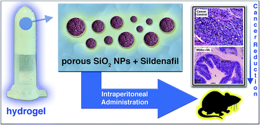
We describe here a drug depot hydrogel system comprising sildenafil (SIL; commercially available as Viagra®) incorporated in mesoporous silica nanoparticles (MSNs; around 60 nm of diameter) and conjugated with a thermosensitive poloxamer (Pluronic F-127 or PF127; possessing a liquid–gel transition over a determined temperature). The conjugation of these components results in an anticancer biomaterial with optimized physico-chemical properties: (i) MSNs are colloidally stabilized mainly through repulsive depletion forces manifested in PF127 concentrations above 5 wt%; (ii) the gelation temperature of the systems is set to about 20 °C (attained with 18 wt% of PF127) in order to result in prompt gelation when they are intraperitoneally administered (in rats) as liquids; (iii) and the hydrogel is able to induce a prolonged release of MSNs + SIL. After testing these new formulations against chemically induced prostate cancer in rats, the histopathological analyses of the prostate showed that the hybrid systems were able to largely reduce the prostate tumor level, and also highlighted the role of the MSNs in this process, since the improvement of the tumor condition was proportional to the nanoparticles concentration.


Introduction

Nanoparticles have been introduced in new and alternative therapies for cancer in the last decade. Their use has unique advantages because they can provide a sustained release and effective drug delivery.1–3 Among nanomaterials, mesoporous silica nanoparticles (MSNs) have great potential for use in drug delivery systems for cancer treatment mainly because they are biocompatible and safe,2,4,5 and have been presenting promising results.2,6 In a relevant in vivo study performed by Lu et al. to investigate the biodistribution and biocompatibility of MSNs in mice, the authors showed that MSNs administered intraperitoneally at 5 mg kg−1 did not induce inflammatory processes, and that 94.4% of the MSNs administered were excreted in the urine and feces within 4 days.7 Similarly, Hudson et al. showed that MSNs are safe when administered intraperitoneally or intravenously at 40 mg kg−1, though lethal at 1.2 g kg−1.4 In another study, Fu et al. verified that MSNs are excreted in urine and feces of mice without altering the kidney morphology.5

The high pore content of MSNs makes them capable of loading high drug volumes. Specifically in cancer nanomedicine, MSNs have shown promising results in carrying both hydrophilic and hydrophobic cancer drugs, such as doxorubicin8–10 and cisplatin,11 and camptothecin,7,12,13 respectively. Among the benefits are the improvement of the solubility of insoluble drugs,14 the higher accumulation in cancer cells,15 and the increase of bioavailability and decrease of toxicity towards healthy cells.16 It is known that most of the tumors present an acidic pH and, in this pH range, the silanol groups on the MSNs surface (negatively-charged in neutral pH) become more protonated, diminishing the electrostatic interaction between these groups and positively-charged drug molecules, leading to the release of the drug.16

Cancer drugs usually trigger a broad range of side effects that affect patients' quality of life and the treatment itself, so that alternative and/or conjugated therapies have been increasingly considered.17 For instance, sildenafil (SIL; commercially available as Viagra®) is an inhibitor of the enzyme phosphodiesterase-5 (PDE-5), which has an important role in the tumor progression and which expression was found to be increased in many carcinomas,18 reason why this enzyme has been widely studied. The PDE-5 inhibition prevents the biotransformation of cyclic guanosine monophosphate (3′,5′-cGMP) into 5′-GMP, which is associated with an increase in blood flow caused by vasodilatation.19 Although in the literature there are some studies making use of SIL to treat some kinds of cancer, they mostly confirm the anticancer effect of this molecule in vitro and without any conjugation (e.g., vesicles, nanoparticles). Sarfati et al. suggested that SIL could be used in the treatment of chronic lymphocytic leukemia, in which it induces apoptosis via caspase activation.20 In addition, Serafini et al. suggested the use of PDE-5 inhibitors as assistants to tumor-specific immune therapy.21 From these perspectives, the anticancer (and/or other beneficial) effects of SIL could be even increased by conjugating it with nanoparticles.

In the particular case of the prostate cancer, a highly aggressive tumor, so far there are very few treatment options and none is highly effective. Moreover, the treatment options depend on the tumor level, varying from hormonal blockage and chemotherapy to prostatectomy, being all very aggressive approaches.22,23 In this context, we introduce here an alternative approach for prostate cancer treatment making use of MSNs incorporated in a hydrogel of a poloxamer (i.e., Pluronic F-127; PF127), for administering SIL. PF127 was used in the formulation for attaining a long-term release of mesoporous silica nanoparticles (MSNs), which in turn act as nanocarriers for SIL against prostate cancer. PF127 was used since it is a biocompatible thermo-reversible block copolymer that has a tunable liquid–gel transition temperature near room temperature,24 thus making it suitable for pharmaceutical formulations with prolonged release and in situ gelation.25,26 The choice of the PF127 hydrogel and MSNs was also based on their biocompatibility, and proven efficiency as a drug depot system (PF127 hydrogel), and as a molecular vehicle (MSNs).

Experimental section

Synthesis of the mesoporous silica nanoparticles

Mesoporous silica nanoparticles (MSNs) were synthesized according to Paula et al. (2012)27 using an approach based on the methods introduced by Stöber28 and Bein,29,30 which allow the synthesis of spherical and monodisperse porous silica nanoparticles with high colloidal stability in aqueous medium. Firstly, 0.75 g of cetyltrimethylammonium bromide (CTAB) were added to a 0.050 mol L−1 ammonium hydroxide solution (∼pH 11). To this solution, 3.2 mL of absolute ethanol were added under magnetic stirring and, after 15 minutes, 2.5 mL of tetraethyl orthosilicate (TEOS) were added maintaining the reaction under reflux at 60 °C for two hours. The products were separated by centrifugation for 60 min at 18[thin space (1/6-em)]400 rcf and resuspended in ethanol for further extraction of CTAB. To do so, more absolute ethanol was added to the resuspended nanoparticles so that the volume of the suspension reached 90 mL, followed by the addition of 10 mL of hydrochloric acid (HCl) (thus resulting in a 1[thin space (1/6-em)]:[thin space (1/6-em)]9 HCl[thin space (1/6-em)]:[thin space (1/6-em)]ethanol-volume ratio). Finally, the resulting suspension was sonicated in an ultrasound bath for 10 minutes. A stock ethanolic suspension of the nanoparticles was obtained by (i) centrifuging for 60 minutes at 18[thin space (1/6-em)]400 rcf the MSNs[thin space (1/6-em)]:[thin space (1/6-em)]HCl[thin space (1/6-em)]:[thin space (1/6-em)]ethanol mixture after the sonication; (ii) washing the resulting pellet twice with absolute ethanol; (iii) and resuspending the MSNs in absolute ethanol. The MSNs aqueous suspensions used in all assays were obtained from this stock ethanolic suspension after its centrifugation for 60 min at 18[thin space (1/6-em)]400 rcf, and redispersion in deionized water.

The colloidal stability assays were performed through centrifugation studies, in which information on the MSNs colloidal state was obtained in situ (i.e., directly from the suspension), by quantifying the amount of MSNs dispersed in the liquid through inductively coupled plasma optical emission spectrometry (ICP-OES; see details in the ESI, A2), after varied inertial forces (i.e., centrifugal forces) were applied on the suspensions. The complete characterization of the MSNs, and also the description of the methods used in the colloidal stability assay are provided in the ESI (Sections A1 and A2).

Hybrid systems: synthesis and characterization

Two hybrid systems (PF127-hydrogel + MSNs + SIL) were developed containing MSNs in different concentrations. The first system (S1) comprises PF127, SIL and MSNs at 1.0 mg mL−1; while the second system (S2) contains five times more MSNs (5.0 mg mL−1), keeping the same amount of PF127 and SIL. The hybrids were produced by adding SIL to a PF127 solution, under magnetic stirring, and inserted in an ice bath (to keep the temperature under 4 °C therefore maintaining the PF127 solution as a liquid). MSNs were added subsequently and NaCl(aq) was added (lastly, to avoid aggregation) to reach the physiological saline condition (0.9% w/v). The preparation of the hybrid systems in NaCl 0.9% aimed to avoid pain to the animals that could be caused by a difference in the osmotic pressure between cells and the hybrids. SIL concentration in the systems was calculated to reach 5.0 mg of drug per kilogram of animal body weight,18 considering injections of 0.3 mL and an animal body weight (average) of 150 g. Along with S1 and S2, a third system was produced without MSNs (Control-SIL group). In this way, the differentiation between the samples is related to the presence and amount of MSNs (see Table 1).
Table 1 Description of the drug depot systems
System Components Concentrations Tgel (°C)
Control-SIL PF127 18% (w/w) 20.0 ± 0.5
SIL 4.16 mg mL−1
S1 PF127 18% (w/w) 20.0 ± 0.5
SIL 4.16 mg mL−1
MSNs 1.0 mg mL−1
S2 PF127 18% (w/w) 19.0 ± 0.5
SIL 4.16 mg mL−1
MSNs 5.0 mg mL−1


PF127 concentration was chosen in regard to its gelation temperature (Tgel). The final concentration of PF127 ensured a Tgel in a temperature range (around 20 °C) that would allow a suitable handling of the hybrid system: intraperitoneal administration in the animal as a liquid (T < Tgel) – and minimizing the loss of product due to gelation inside the syringe, and then gelation in the peritoneum as a result of a thermal equilibrium with the animal body temperature (T > Tgel), forming a drug depot-like system. In this way, to evaluate the Tgel of each system, the assay was accomplished by adding 5 mL of the PF127 solution in a beaker in an ice bath containing a thermometer. The solution was heated under magnetic stirring and the gelation temperature was considered the temperature at which the bar stopped moving (see Table 1).

To assess the influence of MSNs on the hydrogel disruption that results in the long-term release of the components (MSNs + SIL), their in vitro release was studied through a membrane-less dissolution method using NaCl 0.9% (w/v) as the release medium. For this, 0.5 mL of the hybrid systems (PF127-hydrogel + MSNs + SIL) at low temperatures (present as liquids) was added to a weighted vial and incubated at 35 °C using a dry bath. This temperature corresponds to the rats' body temperature (Fischer 344 – used in the in vivo assay). After achieving thermal equilibrium, the vials were weighted and 0.5 mL of the release medium (also at 35 °C) was carefully put on the surface of the hydrogel, avoiding mixing the system. At pre-determined intervals, the release medium was removed and the vial was weighed. Then, a new release medium (NaCl 0.9% solution) was put on the surface of the hydrogel to avoid the saturation of the solution. The disruption rate was calculated through the hydrogel weight loss. Due to the fact that the MSNs and SIL absorb light in the same wavelength range (Fig. SM-1; ESI), it was not possible to precisely quantify the SIL amount in the release medium through UV-Vis spectrometry. Considering the biological context of application of the hybrid system, it must be simulated in the release assay at least the concentration gradient that manifests in vivo, which will prevent the hybrid system of attaining a thermodynamical equilibrium and thus will rule over the hydrogel disruption and the release of MSNs and SIL. This is the reason why it was performed a careful and progressive replacement of the supernatant above the gelified hybrid system, in order to provide a concentration gradient between the hydrogel and the supernatant (i.e., NaCl 0.9% solution). Thus, it was analyzed if the presence of MSNs inside the hydrogel has altered the hydrogel disruption, or even prevented it.

In vivo assays

To evaluate the anticancer activity of the hybrid systems, they were tested in rats with chemically induced prostate cancer, and the effects of the treatments were assessed through histopathological analyses. For this, seventeen seven-week-old male Fischer 344 rats were used in the study. They were obtained from the Multidisciplinary Center for Biological Investigation (CEMIB) at University of Campinas (UNICAMP). Four of them comprised the healthy control group (HC, n = 4), composed of healthy animals. The prostate cancer induction was then performed in 13 animals according to a new protocol,31 which consisted firstly of daily subcutaneous injections of testosterone cypionate (100 mg kg−1) diluted in 0.5 mL of peanut oil for three days. Then, the animals were anesthetized with xylazine hydrochloride 2% (5 mg kg−1) and ketamine hydrochloride 10% (60 mg kg−1), for further performance of a 0.5 cm suprapubic incision and inoculation of 0.2 mL of n-methyl-n-nitrosourea (MNU) dissolved in 0.3 mL of sodium citrate (1 M, pH 6.0) and PF127 25%, which allows the in situ gelation of the solution. The final dose of MNU was of 15 mg kg−1. After one week from the MNU inoculation, the animals received subcutaneous injections of testosterone cypionate (5 mg kg−1) diluted in 5 mL of peanut oil on alternate days for 120 days. After this period, animals with prostate cancer were divided in 4 groups, one of them being the cancer control group (CC, n = 4, untreated animals with prostate cancer). The other 3 groups were treated with each of the hybrid systems produced here (see Table 1). Each group of animals was named after the respective system received as treatment, i.e. the animals treated with system Control-SIL (SIL in the PF127 hydrogel, absence of MSNs) was named Control-SIL group (n = 3); the group treated with the system S1 was named S1 group (n = 3); and the group treated with the system S2 was named S2 group (n = 3). All groups received a weekly dose of 0.3 mL of the respective treatment via intraperitoneal for 30 days. Both CC and HC groups received physiological saline solution (NaCl 0.9%), while groups Control-SIL, S1 and S2 received the treatments as described in Table 1. To prevent gelation of the systems inside the needles and consequent loss of product, the systems were kept in ice bath before each application. This allows a better flow of the hybrid systems through the needle and that gelation occurs only inside the animal body (and not in the syringe or needle). The animals received water and the same solid diet ad libitum (Nuvilab), and were allocated in single solid-bottom boxes lined with wood shavings in a room with controlled light and temperature (12 hours with light and 12 in the dark; 20–25 °C). The research was approved by the Animal Experimentation Ethics Commission (CEUA-IB-UNICAMP), according to the ethical principles adopted by SBCAL-Brazilian Laboratory Animal Science Association (former COBEA-Brazilian College of Animal Experimentation).

After one week from the last dose-application, the animals were euthanized and samples of the prostatic ventral lobe of each animal underwent histopathological analyses. The samples were collected and fixed in Bouin for 12 hours. After fixing, the tissues were washed with ethanol 70% and dehydrated. In addition, the fragments were diafanized in xylol and embedded in a plastic polymer (Paraplast Plus, ST. Louis, MO, EUA). The fragments were cut into 5 mm-thick pieces using a microtome Biocut 1130 (Reichert-Jung), stained with hematoxylin–eosin and photographed using a photomicroscope Nikon Eclipse Ni-U (equipped with a camera Nikon DS-RI-1). The diagnoses of prostatic lesions were based on morphological features evaluations that are already established in the literature.32 Thus, the information drawn was correlated with histopathological changes, which were qualitatively evaluated. Therefore, the values represented in Table 2 are related to an absolute number of animals (expressed in percentage) that presented the respective histopathological change, without standard deviations.

Table 2 Percentage of histopathological changes in the ventral prostate from five experimental groupsa
Histopathology Groups
HC (n = 4) CC (n = 4) Ctrl-SIL (n = 3) S1 (n = 3) S2 (n = 3)
a HGPIN = high-grade prostate intraepithelial neoplasia; PHN = prostatic nodular hyperplasia; ctrl = control.
Normal 100%
Prostatic atrophy 66.6%
PNH 25% 33.3% 66.6%
HGPIN 75% 66.6% 100%
Low grade prostatic adeno-carcinoma 25% 66.6% 66.6%
Intermediate prostatic adeno-carcinoma 50% 100% 33.3%
High-grade prostatic adeno-carcinoma 25%


Evaluation of silicon amount in prostatic tissues

The accumulation of MSNs in prostatic tissues was evaluated by measuring the silicon amount. Tissue samples from all animals were digested with hydrofluoric acid (HF) 40% and the resultant solution was neutralized with potassium hydroxide (KOH) prior to its injection into an inductively coupled plasma optical emission spectrometer (ICP-OES). Silicon was quantified by meanings of the silicon concentration per unit-mass of prostatic tissue.

Results and discussion

Mesoporous silica nanoparticles (MSNs)

Transmission electron microscopy (TEM) images of MSNs reveal nanoparticles with spherical morphology and a size distribution varying from 45 to 75 nm (Fig. 1a). Besides having a spherical morphology, the MSNs contain near-cylindrical pores. The conclusion of the existence of cylindrical-shaped pores was drawn from the N2-sorption isotherm (Fig. SM-2a; ESI), which indicated the manifestation of an adsorption pattern that resembles a type IV-reversible isotherm (IUPAC), with a subtle stepwise behavior from 0.2 to 0.6 P/P0.27,33 This step is characteristic of well-ordered mesopores such as those present in MCM-41-like structures, and the height of the step reflects on the pores ordering degree. The surface area of the nanoparticles, calculated from the adsorption branch of the isotherm through the BET method resulted in a value of about 850 m2 g−1. Furthermore, the pore size distribution was calculated through the BJH method by using the adsorption branch of the isotherm, and it indicated the majority of pores with about 2 nm of diameter (Fig. SM-2b; ESI).
image file: c5ra15006j-f1.tif
Fig. 1 (a) TEM image of MSNs (inset: size distribution histogram of nanoparticles obtained by measuring at least 100 nanoparticles in several images). (b) A photograph of a hybrid system produced (MSNs + PF127 + SIL) at 25 °C (T > Tgel; Tgel = ∼20 °C).

The colloidal stability of the nanoparticles is imperative for nanomedicine applications such as imaging and cancer therapy. In these contexts, nanoparticles need to be individualized in the appropriate medium for their administration, in order to provide homogeneity similar to that of a soluble molecule. The homogeneity allows an efficient circulation of the nanoparticle in the organism, and prevents side effects from aggregation. In addition, it is known that nanoparticles (and macromolecules) with sizes up to about 100 nm are subjected to the EPR effect (i.e., enhanced permeability and retention), which enables these nanoparticles to accumulate in the tumor interstitial cavities. This effect is a result of (i) the high vascularization and the presence of well-spaced endothelial cells in tumorous blood vessels, which increase the nanoparticle permeability in the tumor; along with the result of (ii) a deficient lymphatic system, which leads to the nanoparticle retention inside the tumor cavities.34–37 Thus, it is important to maintain the colloidal stability of nanoparticles throughout their cycle of action.

Unfunctionalized nanoparticles (i.e., without surface chemical modifications) are commonly not stable in physiological media (e.g., NaCl 0.9%) due to their high ionic strength, which prevents charge-based mechanisms of colloidal stabilization. However, it was observed through centrifugation assays a high colloidal stability for MSNs dispersed in NaCl 0.9% (Fig. SM-3; ESI). This stabilization is a result of the negatively charged surface of MSNs in the NaCl 0.9% solution, manifesting from the deprotonation of silanol groups at the nanoparticle surface (i.e., an electric-based colloidal stabilization, with a zeta potential of −10 mV; see Table SM-1; ESI). Furthermore, size measurements from dynamic light scattering (DLS) also indicated that MSNs have essentially the same size and polydispersity index (PDI) when they are dispersed in both deionized water and in the NaCl 0.9% solution; a result that confirms their colloidal stability in both conditions (see Table SM-1; ESI). Considering the MSNs colloidal stability when they are dispersed in NaCl 0.9% under normal conditions (i.e., shelf life), the suspension is visually stable over weeks when conserved at room temperatures (25 °C). The same occurs with the MSNs suspensions in the presence of PF127 (see below).

On the other hand, nonionic copolymers such as PF127 have the ability to induce or prevent nanoparticles aggregation through entropy-based mechanisms, i.e., depletion forces. The manifestation of attractive or repulsive depletion forces depends mostly on the polymer concentration.38–40 When mixed with MSNs in the NaCl 0.9% solution, it was observed that at 3 wt% PF127 leads to a destabilization of the MSNs sols (i.e., colloidal suspension), whereas at 5 wt% (or above) the same polymer is able to stabilize MSN (Fig. SM-3; ESI). This result indicates the existence of a threshold for the manifestation of attractive or repulsive depletion forces (5 wt% ≥ threshold ≥ 3 wt%). It is import to mention that the long-range interaction manifesting between MSNs and PF127 that is responsible for the depletion forces occurs along with the adsorption (i.e., short-range interaction) of PF127 on the MSNs surface (see the decrease in the zeta potentials as a function of the PF127 concentration; Table SM-1; ESI). The manifestation of both kinds of interactions (short- and long-range; i.e., adsorption and depletion forces) was previously observed for PF127 (ref. 40) and also for polyethylene glycol.39 Therefore, as in the hybrid systems the PF127 concentration is 18 wt% (to form hydrogels), much higher than the observed threshold for repulsive depletions forces to occur, this polymer will act as an initial stabilizing agent for MSNs from the time of injection up to a possible formation of the protein capping (i.e., corona effect), resulted from the adsorption of proteins on the MSNs surface,41–43 which are also able to provide colloidal stabilization for MSNs.13,44,45

Hybrid systems

The hybrid systems produced here make use of a thermosensitive drug depot system for the controlled release of MSNs and SIL. In this way, it was developed a drug depot formulation comprising a poloxamer hydrogel (i.e., Pluronic F-127; PF127) incorporated with MSNs and SIL. In order to obtain preliminary information on the role of the MSNs in the systems, we developed two formulations (S1 and S2) differing only in the MSNs concentration (Table 1). Along with these two formulations, it was produced another without MSNs, thus comprising just of PF127 and SIL (Control-SIL; Table 1).

PF127 was used as a gelling agent to achieve in situ gelation, so that the liquid formulation could be administered in a minimally invasive manner and form a drug depot inside the animal, ensuring the controlled release of the components. PF127 is a biocompatible, FDA-approved, thermo-reversible block copolymer comprising poly(ethylene oxide)–poly(propylene oxide)–poly(ethylene oxide) monomers (PEO–PPO–PEO), distributed in a PPO[thin space (1/6-em)]:[thin space (1/6-em)]PEO ratio of approximately 1[thin space (1/6-em)]:[thin space (1/6-em)]2 (PEO100–PPO65–PEO100; MW ∼ 12[thin space (1/6-em)]500).24,46 The presence of hydrophobic PPO chains (MW ∼ 4000) leads to the formation of PF127 micelles. The micellization process occurs via an endothermic process with entropic gains, in which hydrophobic PPO chains cluster themselves, resulting in the dehydration of the hydrophobic domains.47,48 The PF127 gelation occurs with the temperature rise, from which critical micellar concentration (CMC) drops and micelles cluster themselves in a three-dimensional arrangement maintained by the interaction of PEO domains.24,47

The PF127 liquid–gel transition temperature can be adjusted, so that it can be a liquid at low temperatures and a gel at room temperature. By adjusting the PF127 concentration to 18% (w/w), it was possible to attain a liquid–gel transition temperature (Tgel) at ∼20 °C, a range over which the liquefied system (at T < Tgel) can be easy handled towards its administration into the animals (for instance, with the aid of an ice bath). Furthermore, with Tgel over this range of temperature (of about 20 °C), after the intraperitoneal administration the drug depot system (i.e., hybrid system) promptly gelifies in contact with the peritoneal cavity, which is at about 35 °C (T > Tgel).

The potential of the hybrid systems for controlled release was confirmed through a membraneless dissolution method, by measuring the rate of gel disruption over time. In this method, the release medium (NaCl 0.9% w/w) is put carefully on the surface of the hydrogel and the disruption rate is obtained by measuring the weight loss over time and by changing the release medium at pre-determined intervals to avoid saturation. The results (Fig. 2) confirm the potential for controlled released. Furthermore, it was also observed that MSNs alter the disruption process of PF127 hydrogels (see blue and red curves in Fig. 2), decreasing its rate after 200 min. This is possibly due to their interaction with the micelles chains present in the hydrogel macrostructure. Considering the permanent concentration gradient induced by cells on the hydrogel (when it is inside the peritoneal cavity), which was roughly simulated in this assay, it is expected that the hybrid system containing MSNs will delay the release of SIL after administration.


image file: c5ra15006j-f2.tif
Fig. 2 Gel disruption profile of hybrid systems.

As previously mentioned, besides the supramolecular assembly of Pluronic F-127 that leads to the formation of the hydrogel, there are also secondary short- and long-range interactions of the polymer with the MSNs, which provide their colloidal stabilization mainly through repulsive depletion forces (at PF127 concentrations above 5 wt%). This means that MSNs incorporated in the PF127 hydrogel are present in a colloidal state. In this way, the hydrogel disruption consequently leads to the diffusion of the cargo (MSNs + SIL) to the medium, which means that the SIL and MSNs release will obey the same profile observed for the hydrogel. From thermodynamical considerations, the same phenomena will occur in the peritoneal cavity, although with a different kinetics, considering that the concentration gradients provided by cell and tissues as well as the releasing medium (i.e., peritoneal fluid) are very distinct.

In vivo application of the hybrid systems

After their administration in the animals, the evaluation of the effects of the hybrid systems on the prostate was performed through histopathological analyses (Fig. 3). Diagnoses of prostatic lesions were based on Shappell et al.,32 and the morphological changes in the ventral prostatic lobe of the rats were evaluated. The results (Table 2) confirm the antitumor potential of the systems when compared to Cancer Control (CC; rats with cancer; not treated) and Control-SIL (rats treated with SIL incorporated in the PF127 hydrogel) groups. Groups S1 and S2 (containing MSNs) induced a decrease in the frequency of more aggressive tumors (intermediate and high grade), and a higher incidence of low-grade tumors. The improvement in the tumor conditions was proportional to the MSNs concentration, thus confirming the role of MSNs in this process. There are few studies that propose the use of SIL for cancer treatment in vivo, and there is still no consensus about its role in tumor progression. For example, Das et al. studied the effect of the association of SIL and doxorubicin in two prostate cancer cell lines (PC-3 and DU145), and compared the results with the effects of doxorubicin and SIL alone.18 The authors reported that SIL alone had no effect in relation to the cell death, production of reactive oxygen species (ROS) or apoptosis. In the same work, they also studied the effects of the association of doxorubicin and SIL in tumor reduction in mice carrying PC-3 flank tumors. The authors verified that SIL alone had no effect in relation to the tumor volume and weight. Using other cell lines, Sarfati et al. studied the effects of SIL and other PDE-5 inhibitors on chronic lymphocytic leukemia cells (B-CLL),20 and they concluded that SIL induces cell apoptosis via caspase. Other studies have also found that the PDE-5 inhibition results in an increase of the anticancer immune response, such as the in vivo study conducted by Serafini et al.21 However, unlike the work conducted here, the authors did not use prostate models. In the specific case of the effects of the hybrid system (MSNs + SIL + PF127) against prostate cancer cells, although these in vivo results shown here represent capital results in order to introduce perspectives of using these systems for prostate cancer treatment, further detailed studies (e.g., in vitro) must be carried out in order to reveal the mechanisms of action of the systems.
image file: c5ra15006j-f3.tif
Fig. 3 Photomicrographs of the ventral prostate from (A and B) HC, (C and D) CC, (E) Control-SIL, (F–H) S1 (I–L) S2 groups. (A and B) Acini with different sizes, pleated borders and epithelium composed of two cell types, one layer of basal cells (Bc) and one layer of columnar secretory cells (or luminal cells) (Lc). (C and D) High-grade adenocarcinoma was characterized by rare acini, neoplastic cells arranged in cords or nests (Na) through the stroma and the absence of basal cells. (E) Intermediate degree adenocarcinoma was characterized by neoplastic sharp acini (Na) that began to merge and an absent basal cells layer. (F) In prostatic atrophy, the acini showed no papillary projections and were mainly composed of cuboidal shape luminal cells (Lc) that presented a reduction in the nuclei/cytoplasm relation and the basal cell (Bc) layer was maintained. (G and K) High-grade prostatic intraepithelial neoplasia (HGPIN) was characterized by focal proliferation of the epithelium, cytologic atypia (arrows), identified by large nuclei and prominent nucleoli. (H and L) Low-grade adenocarcinoma consisting of well-formed acini (Na) showing discrete neoplasia and cytologic atypia and that resembled normal acini but with the absence of basal cells. (I and J) Prostatic Nodular Hyperplasia (PNH) was characterized by well-defined hyperplastic nodules, which were characterized by acini displaying epithelial papillomatosis and rounded borders. The acini epithelium was composed of columnar secretory cells and basal cells. (A–L) Epithelium – Ep; lumen – L; stroma – St.

Due to its thermoreversibility and biocompatibility, Pluronic F-127 can be used as a depot system for most routes of administration, including oral, topical, intranasal and rectal.49–51 Considering its characteristics, currently there is not a premise to believe that its administration would not be safe. Furthermore, it is worth mentioning that it was not observed here any macroscopic evidence of alteration on the abdominal wall or peritoneal cavity of the animals after the treatment. Regarding the route of administration, it was chosen the intraperitoneal rather than intratumoral in order to perform a systemic treatment. Caudal vein injection was discarded due to its volume limitation and difficulty when compared to intraperitoneal. Furthermore, we did not use intratumoral administration due to the mechanical limitation of the prostatic capsule, which was already under a tension caused by the prostate swelling.

The accumulation of MSNs in prostatic tissues was evaluated by measuring the silicon amount. Tissue samples from all animals were digested with hydrofluoric acid (HF) 40%, and the resultant solution was neutralized with potassium hydroxide (KOH) before injection into an inductively coupled plasma optical emission spectrometer (ICP-OES). Silicon was quantified by means of the silicon concentration per unit-mass of prostatic tissue. The results (Fig. 4) provide information about the accumulation of MSNs after one week from the last dose-application. It was observed that MSNs accumulated in prostatic tissues, and that the accumulation was proportional to the MSNs concentration. S2 group present a silicon amount almost five times greater than S1 group.


image file: c5ra15006j-f4.tif
Fig. 4 Silicon amount (Si-weight/prostatic tissue-weight) in prostatic tissues of rats from the groups Control-SIL, S1 and S2 after one week from the last dose-administration.

Conclusions

In summary, we developed a hydrogel-based formulation (based on Pluronic F-127; PF127) that acts as a drug depot system for the delivery of mesoporous silica nanoparticles (MSNs) incorporated with sildenafil (SIL). This conjugation (PF127 + MSNs + SIL) was able to largely reduce the tumor level in rats with chemically induced prostate cancer, but only when MSNs are present. The hybrid system was intraperitoneally administered in the rats as a liquid (at T < 20 °C; below the hydrogel gelation temperature Tgel), and it was gelified when in contact with the peritoneal cavity (at around 35 °C; T > Tgel). The progressive release of the components (MSNs + SIL) from the hydrogel was also confirmed through in situ assays. Results reveal that silica nanoparticles incorporated with SIL play a determinant role in the improvement of tumor conditions. Finally, the analysis of the silicon (Si) amount in prostatic tissues after one week from the last injection of the hybrid system indicates that the Si accumulation in the organ is proportional to the initial concentration of MSNs administered. To our knowledge, this is the first in vivo study that considers the association of SIL and MSNs for prostate cancer treatment, using SIL as the main drug (not as a conjugate with doxorubicin, for instance). SIL has been commonly used as co-adjuvant, usually to overcome multidrug resistance of cancer drugs. Thus, this paper opens a new perspective of using SIL and MSNs as the main therapy in prostate cancer treatments. Furthermore, these important findings strongly encourage future studies for exploring the association of SIL with other cancer drugs loaded in the same nanostructure, and also conjugated with drug depot systems (e.g. hydrogels), which can possibly achieve even a higher effectiveness.

Acknowledgements

Support from FAPESP, CNPq, INOMAT (MCTI/CNPq), FUNCAP, NanoBioss (MCTI) and the Brazilian Network of Nanotoxicology (MCTI/CNPq) are acknowledged.

References

  1. T. M. Allen and P. R. Cullis, Science, 2004, 303, 1818 CrossRef CAS PubMed.
  2. W. X. Mai and H. Meng, Integr. Biol., 2013, 5, 19 RSC.
  3. F. Chen, T. R. Nayak, S. Goel, H. F. Valdovinos, H. Hong, C. P. Theuer, T. E. Barnhart and W. Cai, Mol. Pharm., 2014, 11, 4007 CrossRef CAS PubMed.
  4. S. P. Hudson, R. F. Padera, R. Langer and D. S. Kohane, Biomaterials, 2008, 29, 4045 CrossRef CAS PubMed.
  5. C. Fu, T. Liu, L. Li, H. Liu, D. Chen and F. Tang, Biomaterials, 2013, 34, 2565 CrossRef CAS PubMed.
  6. F. Chen, H. Hong, S. Shi, S. Goel, H. F. Valdovinos, R. Hernandez, C. P. Theuer, T. E. Barnhart and W. Cai, Sci. Rep., 2014, 4, 5080 CAS.
  7. J. Lu, M. Liong, Z. X. Li, J. I. Zink and F. Tamanoi, Small, 2010, 6, 1794 CrossRef CAS PubMed.
  8. L. Yuan, Q. Tang, D. Yang, J. Z. Zhang, F. Zhang and J. Hu, J. Phys. Chem. C, 2011, 115, 9926 CAS.
  9. H. Meng, M. Xue, T. Xia, Z. Ji, D. Y. Tarn, J. I. Zink and A. E. Nel, ACS Nano, 2011, 5, 4131 CrossRef CAS PubMed.
  10. Q. Zhang, X. Wang, P. Z. Li, K. T. Nguyen, X. J. Wang and Z. Luo, Adv. Funct. Mater., 2014, 24, 2450 CrossRef CAS PubMed.
  11. Z. Tao, B. Toms, J. Goodisman and T. Asefa, ACS Nano, 2010, 4, 789 CrossRef CAS PubMed.
  12. J. Lu, Z. Li, J. I. Zink and F. Tamanoi, Nanomedicine, 2010, 8, 212 CrossRef PubMed.
  13. A. J. Paula, R. T. Araujo Júnior, D. S. T. Martinez, E. J. Paredes-Gamero, H. B. Nader, N. Durán, G. Z. Justo and O. L. Alves, ACS Appl. Mater. Interfaces, 2013, 5, 8387 CAS.
  14. J. Lu, M. Liong, J. I. Zink and F. Tamanoi, Small, 2007, 3, 1341 CrossRef CAS PubMed.
  15. M. Liong, J. Lu, M. Kovochich, T. Xia, S. G. Ruehm, A. E. Nel, F. Tamanoi and J. I. Zink, ACS Nano, 2008, 2, 889 CrossRef CAS PubMed.
  16. M. Xie, Y. Xu, H. Shen, S. Shen, Y. Ge and J. Xie, Int. J. Pharm., 2014, 474, 223 CAS.
  17. G. Minotti, P. Menna, E. Salvatorelli, G. Cairo and L. Gianni, Pharmacol. Rev., 2004, 56, 185 CrossRef CAS PubMed.
  18. A. Das, D. Durrant, C. Mitchell, E. Mayton, N. N. Hoke, F. N. Salloum, M. A. Park, I. Qureshi, R. Lee, P. Dent and R. C. Kukreja, Proc. Natl. Acad. Sci. U. S. A., 2010, 107, 18202 CrossRef CAS PubMed.
  19. Z. Shi, A. K. Tiwari, A. S. Patel, L. W. Fu and Z. S. Chen, Cancer Res., 2011, 71, 3735 CrossRef CAS PubMed.
  20. M. Sarfati, V. Mateo, S. Baudet, M. Rubio, C. Fernandez, F. Davi, J. L. Binet, J. Delic and H. Merle-Beral, Blood, 2003, 101, 265 CrossRef CAS PubMed.
  21. P. Serafini, K. Meckel, M. Kelso, K. Noonan, J. Califano, W. Koch, L. Dolcetti, V. Bronte and I. Borrello, J. Exp. Med., 2006, 203, 2691 CrossRef CAS PubMed.
  22. B. Djavan, V. Ravery, A. Zlotta, P. Dobronski, M. Dobrovits, M. Fakhari, C. Seitz, M. Susani, A. Borkowski, L. Boccon-Gibod, C. C. Schulman and M. Marberger, J. Urol., 2001, 166, 1679 CrossRef CAS.
  23. S. A. Boorjian, R. J. Karnes, L. J. Rangel, E. J. Bergstralh and M. L. Blute, J. Urol., 2008, 179, 1354 CrossRef PubMed.
  24. G. Dumortier, J. L. Grossiord, F. Agnely and J. Chaumeil, Pharm. Res., 2006, 23, 2709 CrossRef CAS PubMed.
  25. E. A. Ibrahim, S. Ismail, G. Fetih, O. Shaaban, K. Hassanein and N. H. Abdellah, Acta Pharm., 2012, 62, 59 CrossRef CAS PubMed.
  26. S. Heilmann, S. Küchler, C. Wischke, A. Lendlein, C. Stein and M. Schäfer-Korting, Int. J. Pharm., 2013, 444, 96 CrossRef CAS PubMed.
  27. A. J. Paula, L. A. Montoro, A. G. S. Filho and O. L. Alves, Chem. Commun., 2012, 48, 591 RSC.
  28. W. Stöber, A. Fink and E. Bohn, J. Colloid Interface Sci., 1968, 26, 62 CrossRef.
  29. J. Kecht, A. Schlossbauer and T. Bein, Chem. Mater., 2008, 20, 7207 CrossRef CAS.
  30. V. Cauda, A. Schlossbauer, J. Kecht, A. Zurner and T. Bein, J. Am. Chem. Soc., 2009, 131, 11361 CrossRef CAS PubMed.
  31. W. J. Fávaro, C. P. Silveira Bueno, N. E. Durán Caballero, L. M. Apolinário and P. V. Garcia, Br. Pat. 10 2014 023118 8, 2014.
  32. S. B. Shappell, G. V. Thomas, R. L. Roberts, R. Herbert, M. M. Ittmann, M. A. Rubin, P. A. Humphrey, J. P. Sundberg, N. Rozengurt, R. Barrios, J. M. Ward and R. D. Cardiff, Cancer Res., 2004, 64, 2270 CrossRef CAS.
  33. K. Nakanishi, in Handbook of Sol–Gel Science and Technology: Processing, Characterization, and Applications, ed. S. Sakka, Kluwer Academic Publishers, New York, 2005, p. 191 Search PubMed.
  34. H. Maeda, J. Wu, T. Sawa, Y. Matsumura and K. Hori, J. Controlled Release, 2000, 65, 271 CrossRef CAS.
  35. H. Maeda, in Biomedical Aspects of Drug Targeting, ed. V. Muzykantov and V. Tochilin, Springer Science and Business Media, New York, 2002, p. 211 Search PubMed.
  36. L. Tang, N. P. Gabrielson, F. M. Uckun, T. M. Fan and J. Cheng, Mol. Pharm., 2013, 10, 883 CrossRef CAS PubMed.
  37. D. Peer, J. M. Karp, S. Hong, O. C. FaroKhzad, R. Margalit and R. Langer, Nat. Nanotechnol., 2007, 2, 751 CrossRef CAS PubMed.
  38. J. Crocker, J. Matteo, A. Dinsmore and A. Yodh, Phys. Rev. Lett., 1999, 82, 4352 CrossRef CAS.
  39. X. Zhang, M. R. Servos and J. Liu, J. Am. Chem. Soc., 2012, 134, 9910 CrossRef CAS PubMed.
  40. G. C. Padovani, R. Petry, C. A. Holanda, F. A. Sousa, V. M. Saboia, C. A. Silva, A. R. Paschoal, A. G. Souza Filho and A. J. Paula, J. Phys. Chem. C, 2015, 119, 18741 CAS.
  41. M. P. Monopoli, F. B. Bombelli and K. A. Dawson, Nat. Nanotechnol., 2011, 6, 11 CrossRef CAS PubMed.
  42. M. Lundqvist, J. Stigler, G. Elia, I. Lynch, T. Cedervall and K. A. Dawson, Proc. Natl. Acad. Sci. U. S. A., 2008, 105, 14265 CrossRef CAS PubMed.
  43. M. P. Monopoli, C. Aberg, A. Salvati and K. A. Dawson, Nat. Nanotechnol., 2012, 7, 779 CrossRef CAS PubMed.
  44. A. J. Paula, D. S. T. Martinez, R. T. Araujo Jr, A. G. Souza Filho and O. L. Alves, J. Braz. Chem. Soc., 2012, 23, 1807 CrossRef CAS.
  45. A. J. Paula, C. P. Silveira, D. S. T. Martinez, A. G. Souza Filho, F. V. Romero, L. C. Fonseca, L. Tasic, O. L. Alves and N. Durán, ACS Appl. Mater. Interfaces, 2014, 6, 3437 CAS.
  46. S. Li, J. Guo, R. A. Patel, A. L. Dadlani and R. M. Leblanc, Langmuir, 2013, 29, 5742 CrossRef CAS PubMed.
  47. P. Alexandridis and T. A. A. Hatton, Colloids Surf., A, 1995, 96, 1 CrossRef CAS.
  48. A. M. Pragatheeswaran and S. B. Chen, Langmuir, 2013, 29, 9694 CrossRef CAS PubMed.
  49. S. Miyazaki, C. Yokouchi, T. Nakamura, N. Hashiguchi, W.-M. Hou and M. Takada, Chem. Pharm. Bull., 1986, 34, 1801 CrossRef CAS.
  50. K. Morikawa, F. Okada, M. Hosokawa and H. Kobayashi, Cancer Res., 1987, 47, 37 CAS.
  51. M. Padilla, G. T. Clark and R. L. Merrill, J. Am. Dent. Assoc., 2000, 131, 184 CrossRef CAS.

Footnote

Electronic supplementary information (ESI) available: Experimental details and characterization data. See DOI: 10.1039/c5ra15006j

This journal is © The Royal Society of Chemistry 2015