A renewable resource-derived thixotropic self-assembled supramolecular gel: magnetic stimuli responsive and real-time self-healing behaviour

Krishnamoorthy Lalithaa, Y. Siva Prasada, Vellaisamy Sridharana, C. Uma Maheswaria, George Johnb and Subbiah Nagarajan*a
aOrganic Synthesis Group, Department of Chemistry & The Centre for Nanotechnology and Advanced Biomaterials, School of Chemical and Biotechnology, SASTRA University, Thanjavur - 613401, Tamil Nadu, India. E-mail: nagarajan@scbt.sastra.edu
bDepartment of Chemistry, The City College of New York, 160 Convent Ave., New York, NY 10031, USA

Received 25th July 2015 , Accepted 7th September 2015

First published on 8th September 2015


Abstract

We designed and synthesized a simple fluorescent self-assembled molecular gel from a renewable resource and characterized it well using various techniques. The self-assembly mechanism was studied relative to the molecular structure. We also demonstrate that Fe3O4 nanoparticles were encapsulated in situ into the organogel by a simple process, which generated a magnetic gel with remarkable self-healing features. More importantly, stimuli responsive self-healing properties of the magnetic gel have been characterized by visual inspection, morphological and rheological analyses.
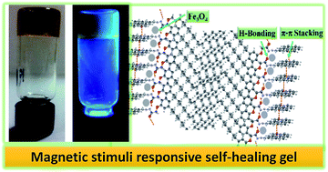

Plants and animals have the unique capability to alter their physiological functions in response to environmental conditions such as temperature, light, pH, electromagnetic fields, mechanical stress, and chemical stimuli. The basic science behind nature's activity has always been an interesting source of knowledge for scientists to develop new technologies for advanced “smart” materials.1,2 Among these, magnetic responsive gel materials have attracted a great deal of research interest in the field of actuators, image enhancement, separation science and drug delivery, to name a few.3 Generally polymeric gels possesses covalently cross-linked irreversible structures with certain limitations in the real time field applications, whereas physical gels, are self-assembled structures based on reversible intermolecular interactions, which overcome the limitations of polymeric gel systems.4 Recently, Weiss presented a brief perspective on the status quo of molecular gels with inputs from scientists across a broad range of interests such as rheology, structural characterization, soft matter, thermodynamics, etc.4a Generally self-assembled gels are continuous in structure and own solid-like rheological behaviour due to the entrapment of solvent in a self-assembled 3D solid matrix through surface tension and capillary forces.5 We report herein the ability of small amphiphilic π-gelators derived from cashew nut shell liquid (CNSL), a renewable resource, and pyrene, to generate a stimuli responsive self-healing magnetic gel. Establishing and optimizing efficient materials from renewable resources has been a focus that substantially addresses the needs of the 21st century.6 Cardanol, a bio-based non-isoprene unit derived from CNSL, is comprised of a rich mixture of phenolic lipids: 5% of 3-(pentadecyl) phenol (3-PDP), 50% of 3-(8Z-pentadecenyl) phenol, 16% of 3-(8Z,11Z-pentadecadienyl) phenol and 29% of 3-(8Z,11Z,14-pentadecatrienyl) phenol.7 The uniqueness of cardanol stems from its structural features and easily accessible saturated and unsaturated hydrocarbon chains. Although few stimuli responsive self-healing molecular gels and polymer gels were reported in the literature,8 the preparation of supramolecular gel through non-covalent interactions with special properties, such as self-healability, stimuli responsive character are becoming an important strategy to develop advanced materials. Recently our research group has explored the self-assembled molecular gels and its application in medicine and biology.9 In the present study, we demonstrate the design, synthesis and stimuli responsive self-healing behaviour of super gel and magnetic gel derived from renewable resource.

The π-gelators were constructed from renewable resources in two steps. First step involves the nucleophilic substitution reaction of cardanol, 1a and 3-n-pentadecylphenol, 1b with methyl bromoacetate resulted in the formation of corresponding methylesters, 2a and 2b in good yield. Reaction of compound 2a and 2b with hydrazine followed by Schiff base formation with a fluorescent signaling unit, 1-pyrenecarboxaldehyde resulted in the formation of π-gelators, 3a and 3b respectively in good yield (Scheme 1). Here we have adapted a simple and economical synthetic pathway that could be used directly for practical applications. The gelation ability of compounds 3a and 3b were consistently studied for different solvents by the “stable to inversion” method.2 Compound 3a forms supramolecular organogels in a number of solvents at very low critical gelation concentrations (CGCs). The gelation ability of π-gelators in various solvents and oils is summarized in Table S1. Among the various solvents studied, gelator 3a showed excellent organogelation ability with lowest CGC of 0.18% (wt/v) in heavy paraffin oil. Gels thus formed are thermos reversible in nature, and stable at ambient conditions.


image file: c5ra14744a-s1.tif
Scheme 1 Synthesis of π-gelators, 3a and 3b.

In the case of 3b, a remarkable change in gelation ability was observed; 3b formed a weak gel in heavy paraffin oil with CGC of 0.75 wt/v%. From this result, we presume that the existence of a kink in the hydrophobic part of 3a, which stabilizes on self-assembly, might be the driving force for the formation of a strong gel (SG).10 We also report a straightforward method to prepare a magnetic gel (MG) by mixing Fe3O4 nanoparticles into SG, followed by sonication for 5 min. A detailed procedure for the preparation of MG has been given in the experimental section. FTIR is very useful tool for investigating hydrogen bonding interactions or non-covalent interactions between molecular building blocks and interacting component. More specifically, stretching frequencies of –OH, –NH and –C[double bond, length as m-dash]O are more distinctive, shift in wave number can be observed as a result of non-covalent interactions.11 The clustering of Fe3O4 on gel was observed mainly due to the interaction between the imine nitrogen (–N[double bond, length as m-dash]CH–) and carbonyl carbon (–NH–C[double bond, length as m-dash]O) with Fe3O4 nanoparticles, as inferred from FTIR studies (see ESI). A plot of Tgel vs. gelator concentration (Fig. 1a) clearly depicts a steady increase in Tgel with respect to the concentration of the gelator. The positive influence of Fe3O4 nanoparticles in the gelation of SG could also be directly ascertained from Fig. 1a. The fluorescent nature of SG and MG was studied using emission spectral technique. In a self-assembled state, SGs in heavy paraffin oil (1 × 10⁻⁵M) show emission peaks at 408, 428, 453 and 483 nm, which upon further titration with heavy paraffin oil, show increased peak intensity near 408 nm with an observed blue shift. This result clearly demonstrates the disassembly of self-assembled SG. Further, the addition of Fe3O4 nanoparticles into SG dissolved in heavy paraffin oil (1 × 10⁻⁵M) leads to the hypochromic shift in emission peak centred at 428 nm. The introduction of Fe3O4 cause little distortion in self-assembly, thus this effect intends that the intensity of the absorption maximum is lowered (Fig. 1b).


image file: c5ra14744a-f1.tif
Fig. 1 (a) A plot of Tgel vs. gelator. (b) Fluorescence titration of SG with heavy paraffin oil [λex = 350 nm]. In titration experiments, direction of arrow shows the response of emission intensity with piecemeal addition of 100 μL of heavy paraffin oil. 2 mL of initial volume of solution (1 × 10⁻⁵ M) was taken for titration experiments. Black line pointed by arrow is for MG in paraffin oil (1 × 10⁻⁵ M solution).

The morphology of Fe3O4 nanoparticles, SG and MG, were obtained through high-resolution transmission electron microscopy (HRTEM). HRTEM analysis of Fe3O4 clearly depicts the existence of particle sizes of 20–40 nm (Fig. 2a). The formation of 3D entangled fibrous network with a diameter ranging from 100–200 nm for SG can be consistently observed by HRTEM analysis. (Fig. 2b). In the case of MG, an attachment of Fe3O4 nanoparticles on the fibrous network was observed.12 Li et al. clearly explain the morphology of magnetic nanoparticles doped gel, where non-covalent interaction between gelator and magnetic nanoparticles plays a vital role.12c We also believe that the binding of Fe3O4 with SG by means of noncovalent interaction might be the driving force for such morphological changes. After incorporation of appropriate magnetic nanoparticles into the 3D-fiberous network of gel, their response to the external magnetic stimuli gel may alter gel network structure, thereby mechanical property of the system changes and becomes magnetic responsive.


image file: c5ra14744a-f2.tif
Fig. 2 HRTEM image of (a) Fe3O4 nanoparticles, (b) SG and (c) MG. For morphological analysis, 100 mL of self-assembled fibers of SG/MG in heavy paraffin oil was dispersed in 1 mL of acetone and drop casted the resultant solution on HRTEM grid.

Small angle X-ray diffraction (SAXD) was employed to gain the additional insight into the molecular packing of gelator molecule in supramolecular self-assembled state. SAXD of the SG gave a Bragg's reflection at 3.1 nm indicating an ordered arrangement of the molecule in gel state, obtained from the packing of hydrophobic unit due to the van der Waals interaction. The peaks observed at 1.5 nm, 0.81 nm and 0.54 nm articulates the stacking of phenyl moiety, H-bonding and π–π stacking. The observed d-spacing value is greater than the molecular length of SG, determined by molecular modelling studies using energy minimized calculations, suggest that during the course of self-assembly process, hydrophobic part intercalates to form a supramolecular structure. Typically, crystalline plane diffraction pattern of Fe3O4 is observed at 21°, 30°, 35°, 41°, 51°, 63° and 67° respectively.13 In the case of MG, in addition to the peaks corresponding to SG, moderately intense peak at 30.5°, 35.7°, 43.4°, 57.1° and 62.5° was also observed (Fig. 3a). Based on spectroscopic, microscopic and SAXD results, one can easily predict the mechanism for the formation of self-assembled supramolecular structure by utilizing H-bonding, π–π stacking and weak van der Waals interaction. A schematic representation of gelator arrangement in MG is given in Fig. 3b.


image file: c5ra14744a-f3.tif
Fig. 3 (a) SAXD pattern of SG and MG. (b) Schematic representation of possible molecular packing model for MG.

The elastic behaviour of a gel could be directly keyed by measuring the difference between storage moduli (G′) and loss moduli (G′′).14 In SG and MG, the value of G′ was found to be much higher than that of G′′ in the entire range of frequency sweep, and this result clearly suggests the formation of strong gel with good tolerance to external forces (Fig. 4a). In the strain sweep experiment, G′ and G′′ remain constant up to a certain level. Afterwards, a gradual drop was observed and a crossover occurred between G′ and G′′. This crossover point is considered as the critical strain (γc) of the gel (Fig. 4b).15 The γc values for SG and MG are 13.61 (G′ = G′′ = 312 Pa) and 4.95% (G′ = G′′ = 819 Pa) respectively (Fig. 4b).


image file: c5ra14744a-f4.tif
Fig. 4 (a) Angular frequency dependence of G′ and G′′ and (b) strain amplitude dependence of G′ and G′′. SG-red line and MG-blue line.

Thermo-responsive behaviour and processability of SG and MG, for various practical applications at different temperatures was studied by continuous temperature ramp up and ramp down experiments (Fig. 5a and b). These results clearly depict the structural and mechanical integrity of both SG and MG even after continuous ramp up and ramp down in temperature for more than three cycles. After looking at the exceptional mechanical behaviour of these gels, we planned to analyse the thixotropic or self-healing nature of SG and MG by performing the step strain experiment of G′ and G′′ (Fig. 5c and d). Generally thixotropic materials that change their physical property in response with mechanical action, such systems were well known in natural systems. Functions of protoplasm, red blood cells, nerve regeneration and muscular activities were specific examples.16 Thixotropic gels are dynamic self-assembled system constitutes a challenging and unique self-healing property. A detailed investigation on the behaviour of these self-assembled system under mechanical stress and resting time is required. Shinkai et al. clearly demonstrate the requirements of thixotropic gelator, which are as follows; (a) it should an efficient super gelator, (2) gel should display the thixotropic property even at a very low concentration, and (c) gels should not lost its property during the cycles of breaking and regeneration process.17 Thixotropic or self-healing nature of SG and MG was identified using step strain experiment. By applying high magnitude of strain (100%), both G′ and G′′ values were apparently decreased because of the broken network structure.17 Recovery of G′ and G′′ was observed within 5 seconds by simultaneous decline in strain to 0.5% in the case of SG, and 0.01% in case of MG. This result clearly argues the self-healing nature by reconstruction of network structure and fast recovery of the mechanical property (Fig. 5c and d). It is interesting to note that even after 5 cycles of breaking and regeneration process, there is no significant loss in mechanical behaviour. Most of the self-healing materials reported in literature were polymer gels and only little is known related to the self-healing of gels.8 Recently Shi and co-workers reported the spontaneous, macroscopic supramolecular assembly of building blocks into an advanced assembled structures, the assembly process joined the hydrophobic surfaces, thereby minimizing the overall interfacial free energy of the system.18 In this report we present a low molecular weight gel, SG and MG displaying remarkable self-healing property. In particular, owing to increasing interest of magnetic gels in the fields of controlled drug-delivery, separation science, artificial muscle and smart biomaterials,19 we have investigated the macroscopic supramolecular assembly of building blocks prepared from SG and MG. In order to differentiate SG from MG, SG has been loaded with rhodamine dye.


image file: c5ra14744a-f5.tif
Fig. 5 (a and b) Storage (G′) and loss modulus (G′′) of SG and MG in temperature ramp experiment respectively. Ramp up and ramp down temperature for SG and MG are 25 to 55 °C and 15 to 60 °C. (c & d) Time course change of G′ and G′′ of SG and MG in step strain experiment respectively. For SG, 100% strain (4.5 min) and 0.05% strain (4.5 min) were applied alternatively and for MG 100% strain (4.5 min) and 0.01% strain (4.5 min) were applied alternatively. Concentration of SG and MG: 1 wt/v% in heavy paraffin oil.

Self-healing of SG to MG started immediately after connecting the two pieces of gels one over the other, and within 5 min, the two pieces of gels start healing themselves via non-covalent interactions and were strong enough to hold vertically by using tweezer (Fig. 6a). By utilizing self-healing process, non-magnetic SG has been connected with magnetic responsive MG, and the entire system attains magnetic stimuli responsive character. Owing to the interesting self-healing behaviour displayed by SG and MG, we were more curious in investigating self-healing behaviour in water medium. In the absence of external magnetic stimuli, hydrophobic SG and MG come close to each other by mere shaking and self-healing occurred, in order to minimizing the overall interfacial free energy. The resultant self-healed gel could be picked by tweezer (Fig. 6b, see ESI video). To understand the mechanism of self-healing process, we designed a gel fusion experiment. Fusion of SG with gel prepared from 3b (moiety without kink) was not proceeded well because of absence of kink in 3b, whereas fusion or self-healing of SG with SG or SG with MG worked very well. From this result, we presume that the cooperative effect of hydrophobic interaction and kink present in it were responsible for self-healing mechanism.18,20


image file: c5ra14744a-f6.tif
Fig. 6 (a) Macroscopic level self-healing of SG and MG and picking up of the resultant by tweezer. (b) Self-healing of SG and MG in water with the evolution of time, 5 s to 300 s. After 300 s, self-healed gel were scooped by spatula and picked up using tweezer. To show the colour difference between SG and MG, pieces of SG was tagged with red colour using rhodamine dye.

Thus the magnetism cooperates well with the self-healing process and induces the stimuli responsive behaviour. Small pieces of MG in water could be systematically aligned and self-healed under the guidance of external magnetic field (see ESI video). This phenomenon can be potentially used for the development of advanced materials operated from remote place. The bulk conveyance path guided under an external magnetic field strength of 0.556 T in both water and air medium is shown in ESI video.

In summary, a fluorescent, self-healing and magnetic stimuli responsive gel was obtained from renewable resources by a simple process utilizing self-assembly phenomenon. Hydrogen bonding and π–π stacking interactions that exist in the SG and MG stabilize the self-assembled structures, which was identified by various techniques. Morphological analysis clearly depicted the formation of a highly entwined fibrous network with attached Fe3O4 nanoparticles. Stability and mechanical strength of SG and MG has been identified by rheological measurements. The magnetic properties and self-healing behaviour of this system have been demonstrated, and work well on the macroscopic level. SG and MG were both compatible for practical applications since they retained their inherent mechanical properties, even after processing the gels several times under different magnitudes of strain and temperature. We envision that this self-healing and magnetic stimuli responsive gel might have potential applications in the medical, biological, environmental, and electronics fields. Further detailed investigation of these gel materials are in progress.

Acknowledgements

This work was financially supported by the Department of Science and Technology (IFA-CH-04 and #SB/FT/CS-024/2013), India and Board of Research in Nuclear Science (#37(1)/20/47/2014), Department of Atomic Energy, India. GJ acknowledges the support from the Gulf of Mexico Research Initiative (GoMRI) through the Consortium for the Molecular Engineering of Dispersant Systems (C-MEDS) subcontract (TUL-626-11/12).

Notes and references

  1. Recent reviews on stimuli responsive materials: (a) E.-K. Lim, T. Kim, S. Paik, S. Haam, Y.-M. Huh and K. Y. Lee, Chem. Rev., 2015, 115, 327–394 CrossRef CAS PubMed; (b) L. Cheng, C. Wang, L. Feng, K. Yang and Z. Liu, Chem. Rev., 2014, 114, 10869–10939 CrossRef CAS PubMed; (c) J. Yao, M. Yang and Y. Duan, Chem. Rev., 2014, 114, 6130–6178 CrossRef CAS PubMed; (d) X. Zhang, J. Yin and J. Yoon, Chem. Rev., 2014, 114, 4918–4959 CrossRef CAS PubMed; (e) S. S. Babu, V. K. Praveen and A. Ajayaghosh, Chem. Rev., 2014, 114, 1973–2129 CrossRef CAS PubMed; (f) J. E. Stumpel, D. J. Broer and A. P. H. J. Schenning, Chem. Commun., 2014, 50, 15839–15848 RSC; (g) S. Mura, J. Nicolas and P. Couvreur, Nat. Mater., 2013, 12, 991–1003 CrossRef CAS PubMed; (h) O. Kuksenok, D. Deb, P. Dayal and A. C. Balazs, Annu. Rev. Chem. Biomol. Eng., 2014, 5, 35–54 CrossRef CAS PubMed; (i) A. S. Hoffman, Adv. Drug Delivery Rev., 2013, 65, 10–16 CrossRef CAS PubMed; (j) X. Hou, W. Guo and L. Jiang, Chem. Soc. Rev., 2011, 40, 2385–2401 RSC; (k) Y. Chen, H. Chen and J. Shi, Adv. Mater., 2013, 25, 3144–3176 CrossRef CAS PubMed; (l) C.-H. Lu, B. Willner and I. Willner, ACS Nano, 2013, 7, 8320–8332 CrossRef CAS PubMed; (m) N. M. Sangeetha and U. Maitra, Chem. Soc. Rev., 2005, 34, 821–836 RSC; (n) M. Suzuki and K. Hanabusa, Chem. Soc. Rev., 2009, 38, 967–975 RSC.
  2. For book chapters: (a) R. G. Weiss and P. Terech, Molecular Gels, Materials with Self-Assembled Fibrillar Networks, Springer, New York, 2006 Search PubMed; (b) Functional Molecular Gels, ed. B. Escuder and J. F. Miravet, RSC Soft Matter Series, 2014, pp. 1–319 Search PubMed.
  3. (a) V. N. Rao, M. N. Ganivada, S. Sarkar, H. Dinda, K. Chatterjee, T. Dalui, J. D. Sarma and R. Shunmugam, Bioconjugate Chem., 2014, 25, 276–285 CrossRef PubMed; (b) R. Libanori, F. B. Reusch, R. M. Erb and A. R. Studart, Langmuir, 2013, 29, 14674–14680 CrossRef CAS PubMed; (c) S. Bolisetty, J. J. Vallooran, J. Adamcik and R. Mezzenga, ACS Nano, 2013, 7, 6146–6155 CrossRef CAS PubMed; (d) W.-H. Chiang, V. T. Ho, H.-H. Chen, W.-C. Huang, Y.-F. Huang, S.-C. Lin, C.-S. Chern and H.-C. Chiu, Langmuir, 2013, 29, 6434–6443 CrossRef CAS PubMed.
  4. (a) R. G. Weiss, J. Am. Chem. Soc., 2014, 136, 7519–7530 CrossRef CAS PubMed; (b) S. Dong, Y. Luo, X. Yan, B. Zheng, X. Ding, Y. Yu, Z. Ma, Q. Zhao and F. Huang, Angew. Chem., Int. Ed. Engl., 2011, 50, 1905–1909 CrossRef CAS PubMed; (c) X. Yan, D. Xu, X. Chi, J. Chen, S. Dong, X. Ding, Y. Yu and F. Huang, Adv. Mater., 2012, 24, 362–369 CrossRef CAS PubMed; (d) M. Zhang, D. Xu, X. Yan, J. Chen, S. Dong, B. Zheng and F. Huang, Angew. Chem., Int. Ed. Engl., 2012, 51, 7011–7015 CrossRef CAS PubMed; (e) S. Dong, B. Zheng, D. Xu, X. Yan, M. Zhang and F. Huang, Adv. Mater., 2012, 24, 3191–3195 CrossRef CAS PubMed.
  5. (a) T. G. Barclay, K. Constantopoulos and J. Matisons, Chem. Rev., 2014, 114, 10217–10291 CrossRef CAS PubMed; (b) S. I. Stupp and L. C. Palmer, Chem. Mater., 2014, 26, 507–518 CrossRef CAS; (c) D. Ivnitski, M. Amit, B. Rubinov, R. Cohen-Luria, N. Ashkenasy and G. Ashkenasy, Chem. Commun., 2014, 50, 6733–6736 RSC; (d) J. D. Tovar, Acc. Chem. Res., 2013, 46, 1527–1537 CrossRef CAS PubMed; (e) R. Marty, R. Nigon, D. Leite and H. Frauenrath, J. Am. Chem. Soc., 2014, 136, 3919–3927 CrossRef CAS PubMed.
  6. V. S. Balachandran, S. R. Jadhav, P. K. Vemula and G. John, Chem. Soc. Rev., 2013, 42, 427–438 RSC.
  7. (a) K. Lalitha, P. Jenifer, Y. S. Prasad, K. Muthusamy, G. John and S. Nagarajan, RSC Adv., 2014, 4, 48433–48437 RSC; (b) L. Faure, S. Nagarajan, H. Hwang, C. L. Montgomery, B. R. Khan, G. John, P. Koulen, E. B. Blancaflor and K. D. Chapman, J. Biol. Chem., 2014, 289, 9340–9351 CrossRef CAS PubMed.
  8. (a) W. Miao, D. Yang and M. Liu, Chemistry, 2015, 21, 7562–7570 CrossRef CAS PubMed; (b) M. J. Gao, J. L. Wang, Q. Cong, B. Zhang, X. C. He, X. F. Ma and G. Li, Mater. Sci. Forum, 2015, 815, 675–683 CrossRef; (c) K. Miyamae, M. Nakahata, Y. Takashima and A. Harada, Angew. Chem., Int. Ed. Engl., 2015, 54, 8984–8987 CrossRef CAS PubMed; (d) T. Rossow and S. Seiffert, Supramolecular Polymer Networks and Gels, Springer International Publishing, Cham, 2015, vol. 268 Search PubMed; (e) L. Peng, H. Zhang, A. Feng, M. Huo, Z. Wang, J. Hu, W. Gao and J. Yuan, Polym. Chem., 2015, 6, 3652–3659 RSC; (f) X. Yu, L. Chen, M. Zhang and T. Yi, Chem. Soc. Rev., 2014, 43, 5346–5371 RSC; (g) K. Imato, T. Ohishi, M. Nishihara, A. Takahara and H. Otsuka, J. Am. Chem. Soc., 2014, 136, 11839–11845 CrossRef CAS PubMed; (h) Z. Qi and C. A. Schalley, Acc. Chem. Res., 2014, 47, 2222–2233 CrossRef CAS PubMed; (i) A. Harada, Y. Takashima and M. Nakahata, Acc. Chem. Res., 2014, 47, 2128–2140 CrossRef CAS PubMed; (j) S. Dong, B. Zheng, F. Wang and F. Huang, Acc. Chem. Res., 2014, 47, 1982–1994 CrossRef CAS PubMed; (k) P. Xing, X. Chu, M. Ma, S. Li and A. Hao, Phys. Chem. Chem. Phys., 2014, 16, 8346–8359 RSC; (l) R. Du, J. Wu, L. Chen, H. Huang, X. Zhang and J. Zhang, Small, 2014, 10, 1387–1393 CrossRef CAS PubMed; (m) S. Basak, J. Nanda and A. Banerjee, Chem. Commun., 2014, 50, 2356–2359 RSC; (n) M. Vatankhah-Varnoosfaderani, S. Hashmi, A. GhavamiNejad and F. J. Stadler, Polym. Chem., 2014, 5, 512–523 RSC.
  9. (a) K. Lalitha and S. Nagarajan, J. Mater. Chem. B, 2015, 3, 5690–5701 RSC; (b) K. Lalitha, Y. S. Prasad, C. U. Maheswari, V. Sridharan, G. John and S. Nagarajan, J. Mater. Chem. B, 2015, 3, 5560–5568 RSC.
  10. D. Chandler, Nature, 2005, 437, 640–647 CrossRef CAS PubMed.
  11. (a) Y. Zhang, B. Yang, X. Zhang, L. Xu, L. Tao, S. Li and Y. Wei, Chem. Commun., 2012, 48, 9305–9307 RSC; (b) Y. Zhang, L. Tao, S. Li and Y. Wei, Biomacromolecules, 2011, 12, 2894–2901 CrossRef CAS PubMed.
  12. (a) E. Taboada, L. N. Feldborg, A. Pérez del Pino, A. Roig, D. B. Amabilino and J. Puigmartí-Luis, Soft Matter, 2011, 7, 2755 RSC; (b) I. O. Shklyarevskiy, P. Jonkheijm, P. C. M. Christianen, A. P. H. J. Schenning, A. Del Guerzo, J.-P. Desvergne, E. W. Meijer and J. C. Maan, Langmuir, 2005, 21, 2108–2112 CrossRef CAS PubMed; (c) S. Li, V. T. John, G. C. Irvin, S. H. Rachakohda, G. L. McPherson and C. J. O’Connor, J. Appl. Phys., 1999, 85, 5965 CrossRef CAS PubMed.
  13. M. R. Guilherme, R. S. Oliveira, M. R. Mauricio, T. S. P. Cellet, G. M. Pereira, M. H. Kunita, E. C. Muniz and A. F. Rubira, Soft Matter, 2012, 8, 6629–6637 RSC.
  14. Y. Zhang, B. Yang, X. Zhang, L. Xu, L. Tao, S. Li and Y. Wei, Chem. Commun., 2012, 48, 9305–9307 RSC.
  15. D. Liu, D. Wang, M. Wang, Y. Zheng, K. Koynov, G. K. Auernhammer, H.-J. Butt and T. Ikeda, Macromolecules, 2013, 46, 4617–4625 CrossRef CAS.
  16. (a) U. Proske, D. L. Morgan and J. E. Gregory, Prog. Neurobiol., 1993, 41, 705–721 CrossRef CAS; (b) R. W. Briehl, Nature, 1980, 288, 622–624 CrossRef CAS PubMed; (c) K. E. Hagbarth, J. Hugglund, M. Nordin and E. U. Wallin, J. Physiol., 1985, 368, 323–342 CrossRef CAS.
  17. P. Mukhopadhyay, N. Fujita, A. Takada, T. Kishida, M. Shirakawa and S. Shinkai, Angew. Chem., Int. Ed., 2010, 49, 6338–6342 CrossRef CAS PubMed.
  18. M. Cheng, G. Yu, Y. Zhang, M. Song, Y. Zhang and F. Shi, Small, 2014, 10, 3907–3911 CrossRef CAS PubMed.
  19. (a) C. He, S. W. Kim and D. S. Lee, J. Controlled Release, 2008, 127, 189–207 CrossRef CAS PubMed; (b) A. Fernández-Barbero, I. J. Suárez, B. Sierra-Martín, A. Fernández-Nieves, F. J. de Las Nieves, M. Marquez, J. Rubio-Retama and E. López-Cabarcos, Adv. Colloid Interface Sci., 2009, 147–148, 88–108 CrossRef PubMed; (c) L. Frkanec and M. Zinić, Chem. Commun., 2010, 46, 522–537 RSC; (d) J. Ramos, A. Imaz, J. Callejas-Fernández, L. Barbosa-Barros, J. Estelrich, M. Quesada-Pérez and J. Forcada, Soft Matter, 2011, 7, 5067 RSC; (e) C. Aimé and T. Coradin, J. Polym. Sci., Part B: Polym. Phys., 2012, 50, 669–680 CrossRef PubMed; (f) J. Estelrich, E. Escribano, J. Queralt and M. A. Busquets, Int. J. Mol. Sci., 2015, 16, 8070–8101 CrossRef CAS PubMed.
  20. Z. Wei, J. H. Yang, J. Zhou, F. Xu, M. Zrinyi, P. H. Dussault, Y. Osada and Y. M. Chen, Chem. Soc. Rev., 2014, 43, 8114–8131 RSC and references cited therein.

Footnote

Electronic supplementary information (ESI) available: Experimental details, figures, tables and copy of NMR spectra. Videos demonstrating the self-healing behavior in water, magnetic stimuli responsiveness in water and air medium. See DOI: 10.1039/c5ra14744a

This journal is © The Royal Society of Chemistry 2015
Click here to see how this site uses Cookies. View our privacy policy here.