A. Galluda,
D. Wartherb,
M. Maynadierc,
M. Seftad,
F. Poyerefg,
C. D. Thomasefg,
C. Rouxelh,
O. Mongini,
M. Blanchard-Descejg,
A. Morèrea,
L. Raehmb,
P. Maillard*efg,
J. O. Durand*bg,
M. Garciaa and
M. Gary-Bobo*a
aInstitut des Biomolécules Max Mousseron de Montpellier, UMR 5247 CNRS-Université Montpellier-ENSCM, Bâtiment (E), Faculté de Pharmacie, 15 avenue Charles Flahault BP14491, 34093 Montpellier, France. E-mail: magali.gary-bobo@inserm.fr
bInstitut Charles Gerhardt Montpellier, UMR5253, CNRS-UM2-ENSCM-UM1, CC1701 Place Eugène Bataillon, 34095 Montpellier Cedex 05, France. E-mail: durand@univ-montp2.fr
cNanoMedSyn, 15, avenue Charles Flahault BP14491, 34093 Montpellier, France
dInstitut Curie, UMR144, Hôpital, 26 rue d'Ulm, 75248 Paris Cedex 05, France
eInstitut Curie, Research Center, PSL Research University, Chemistry, Modelisation and Imaging for Biology (CMIB), Bât 110-112, Centre Universitaire, F-91405 Orsay, France
fCNRS UMR 9187, INSERM U 1196, Université Paris-Saclay Université Paris Sud 11, Bât 110-112, Centre Universitaire, Rue Henri Becquerel, F-91405 Orsay Cedex, France. E-mail: philippe.maillard@curie.fr
gCNRS GDR 3049 PHOTOMED, UMR5623, Université Paul Sabatier, F 31062 Toulouse, France
hChimie et Photonique Moléculaires, CNRS UMR6510, Campus de Beaulieu, Université Rennes 1, 35042 Rennes, France
iInstitut des Sciences Chimiques de Rennes (CNRS, UMR 6226), Université de Rennes 1, F-35042 Rennes Cedex, France
jUniversité de Bordeaux, ISM, UMR 5255 CNRS, F-33400 Talence, France
First published on 26th August 2015
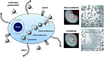
Research in nanomedicine has grown rapidly over the past few years and is playing a key role in the development of effective treatments for ophthalmological purpose. Retinoblastoma is an intraocular tumor triggered by genetic mutation in young children and photodynamic therapy (PDT) is currently developed as a promising non-mutagenic approach to treat this cancer. In this work, we have first identified two receptors, MRC2 and CD209, which are highly expressed by retinoblastoma. Then, we developed mesoporous silica nanoparticles (MSN) grafted with the antibodies anti-MRC2 and/or anti-CD209 for retinoblastoma PDT and imaging.
The use of lectins for direct therapeutic targeting has drawn attention in various research areas including cancers.6–11 In this way, glycoconjugated-porphyrins were synthesized to obtain efficient photosensitizers against retinoblastoma cells for one-photon photodynamic therapy (PDT).12–14 To develop more effective therapies for ophthalmological purposes, nanomedicine could play a key role. There are a number of nanomedicine tools offering innovative solutions to ophthalmological issues and targeted therapy is the clue for therapeutic efficiency improvement.15 In our previous work, mannose or galactose functionalized-mesoporous silica nanoparticles (MSN) were designed for PDT and drug delivery for retinoblastoma treatment.16,17 In continuation to this work and to enhance the specific targeting of retinoblastoma, we investigated four members of the mannose receptor family which belong to the group VI of C-type lectin superfamily. Gene expression of MRC2, PLA2R, LY75 and CD209 were examined by transcriptomic analysis on 37 human retinoblastoma tumors. Then, expression levels of these receptors were investigated by immunohistochemistry and Western blot on Y-79 and WERI-Rb1 retinoblastoma cells and on tumor tissues (Rb102, Rb111 and Rb200) derived from xenografted mice models. Finally, we evaluated the targeting of retinoblastoma cells by antibody-grafted MSN and their therapeutic efficiency using two-photon PDT. For the study of mannose receptors as targets for retinoblastoma treatment, the expression of macrophage mannose receptor 1 (MRC1), C-type mannose receptor 2 (MRC2), lymphocyte antigen 75 (LY75), phospholipase A2 receptor (PLA2R) and CD209 receptor (see ESI, Fig. S1†) were analyzed.
First, a transcriptomic analysis was conducted on the gene expression of PLA2R, LY75, CD209 and MRC2 in retinoblastoma using Affymetrix U133 plus 2.0 arrays (MRC1 is not probed on these arrays). Transcriptomic profiling was thus performed on a total of 37 primary human retinoblastoma tumors (Fig. 1). For PLA2R, LY75 and CD209, mean sample gene expression values were of 2.6, 2.8 and 3.5, respectively (log-scaled). The mean gene expression of MRC2 was 6.6. In addition, the analysis of the open dataset from McEvoy et al. confirms these expression profiles (see ESI, Fig. S2†). This is in accordance with previous obtained results in 4 retinoblastoma cell lines (WERI-Rb1, Y-79, RB113 and RB355) and 55 additional primary human tumors.3 These results highlight the significant MRC2 gene expression in retinoblastoma cell lines and tumors.
![]() | ||
Fig. 1 Box plots of normalized relative scores for CD209, LY75, MRC2 and PLA2R type-specific gene signatures for 37 retinoblastoma human tumors. |
To evaluate protein expression and to confirm results obtained by transcriptomic analysis, immunohistochemistry experiments were performed on retinoblastoma cells and human tumors obtained from xenografted mouse (Fig. 2A). Y-79 and WERI-Rb1 cells were fixed, permeabilized and immunostained. Anti-PLA2R, anti-LY75,18 anti-CD209,19,20 anti-MRC2,21,22 and anti-MRC123,24 antibodies were used for immunocytochemistry experiments after a validation of their specificity in stringent processes. Anti-IgG rabbit and anti-IgG mouse antibodies were used as negative control of unspecific staining.
In Y-79 cells, immunostaining showed no difference between PLA2R and control while for WERI-Rb1 a moderate staining of cytoplasmic and membrane granular pattern was obtained. Considering CD209, MRC2 and MRC1, an important granular membrane and cytoplasmic staining was observed for both retinoblastoma cell lines. Then, expression of mannose receptors was evaluated on human retinoblastoma tumors. Rb102, Rb111 and Rb200 tissues were obtained from xenografted mouse with primary cancer cells of three patients affected by retinoblastoma (Fig. 2B). Granular membrane and cytoplasmic localization of LY75, CD209, MRC2 and MRC1 were observed with different immunostaining intensities in tissues. In Rb102, tumor area LY75, CD209 and MCR2 were strongly expressed while in Rb111 tumor expression of LY75 appears higher than CD209, MRC2 or MRC1. Similarly, tumor area in Rb200 is tightly immunostained and particularly for LY75 and CD209. By these experiments we highlighted the overexpression of LY75, CD209, MRC2 and MRC1 on retinoblastoma and point out their potential interest for targeted therapy.
Protein expression was also performed by Western blot analysis. We evaluated expression of LY75, CD209, MRC2 and MRC1 in retinoblastoma cells and tissues (Fig. 2C). Rabbit polyclonal antibody anti-actin was used to control the protein loading. The results obtained confirm the high expression of LY75, CD209, MRC2 and MRC1, in vitro (except that MRC2 expression is less pronounced for WERI-Rb1) and in vivo for the samples tested. However, Western blot expression profile was also performed on other human cancer cell lines (see ESI Fig. S19†) and a moderate expression of LY75 is found in prostate (LNCaP), breast (MDA-MB-231 and MCF-7), colon (HCT-116) and osteosarcoma (Saos-2) cancer cells. This excludes the use of LY75 as a specific target of retinoblastoma. MRC1 and CD209 proteins are particularly overexpressed in retinoblastoma compared to the other cancer cell lines. By contrast, MRC2 is also found in Saos-2 cells but not in the other cancer cells studied. Thus, CD209 and MRC1 could be specific targets for retinoblastoma as shown by Western blot analyses.
The results obtained by these different analysis methods were summarized and arbitrarily semi-quantified (Table 1). The data indicate a correlation between the different analytical approaches. Indeed, CD209 and MRC2 mannose receptors are mainly overexpressed in retinoblastoma cell lines and tumors.
PLA2R | LY75 | CD209 | MRC2 | MRC1 | ||
---|---|---|---|---|---|---|
a N.D.; none determined.3 | ||||||
Transcriptomic | Y-79a | −/+ | −/+ | +++ | +++ | N.D. |
WERI-Rb1a | −/+ | −/+ | +++ | +++ | N.D. | |
Tumors (n = 37) | −/+ | −/+ | +++ | + | N.D. | |
IHC | Y79 | −/+ | ++ | +++ | +++ | + |
WERI-Rb1 | ++ | ++ | +++ | +++ | + | |
Rb102 | N.D. | ++ | +++ | +++ | −/+ | |
Rb111 | N.D. | +++ | +++ | +++ | +++ | |
Rb200 | N.D. | ++ | ++ | + | −/+ | |
WB | Y-79 | N.D. | + | ++ | ++ | +++ |
WERI-Rb1 | N.D. | ++ | ++ | −/+ | + | |
Rb111 | N.D. | ++ | +++ | +++ | ++ | |
Rb200 | N.D. | + | +++ | +++ | +++ |
In order to study the targeting of MRC2 and CD209 mannose receptors, MSN nanoparticles covalently grafted with commercial anti-MRC2 or/and anti-CD209 antibodies were synthesized (Fig. 3). The ligation of the antibodies to the nanoparticles was performed through a specific semi-carbazide/aldehyde reaction. The semi-carbazide function was first grafted on the nanoparticles. The aldehyde function was obtained through mild oxidation of the carbohydrate chains of the Fc part of the antibodies. This strategy leads to an end-on orientation of the antibodies at the surface of the nanoparticles and thus maintains a maximal activity.25,26 Characterizations of nanoparticles: size, repartition, surface grafting and morphology were performed by DLS, TEM, UV-visible absorption and FTIR spectra analysis (see ESI Fig. S3–S16†).
![]() | ||
Fig. 3 Synthesis of MSN loaded with two-photon photosensitizer (2hνPS) (A) or rhodamine. (rho) (B) and grafted with or without anti-IgGRb, anti-CD209 or anti-MRC2 antibodies. |
The antibodies-grafted MSN were designed to improve therapeutic targeting in two-photon PDT.
Firstly, confocal microscopy was performed on retinoblastoma cells using rhodamine-loaded MSN (Fig. 4). The cellular uptake was assessed by fluorescence imaging on living Y-79 and WERI-Rb1 incubated 5 h with antibodies-grafted MSN-(rho)-IgGRb, MSN-(rho)-CD209, MSN-(rho)-MRC2 or the non-grafted one MSN-(rho)–NHCO–NHNH2 at 20 μg mL−1. MSN-(rho)-IgGRb and MSN-(rho)–NHCO–NHNH2 were introduced as unspecific and untargeted controls, respectively. Cells were co-stained with a lysosomal marker. The emission of rhodamine was localized within the cells for MSN-(rho)-CD209 and MSN-(rho)-MRC2, while MSN-(rho)-IgGRb and MSN-(rho)–NHCO–NHNH2 (not shown) remain outside the cells. MSN-(rho)-CD209 and MSN-(rho)-MRC2 are co-localized with the lysosomes thus demonstrating the successful internalization through the endolysosomal pathway.
The MSNs loaded with the photosensitizers were then screened under two-photon excitation in retinoblastoma cells (Fig. 5). Y-79 and WERI-Rb1 cells were seeded in a poly-ornithine coated 384 multi-well plate and incubated 5 h with a single-dose of 40 μg mL−1 of MSN-(2hνPS)–NHCO–NHNH2, MSN-(2hνPS)-CD209, MSN-(2hνPS)-MRC2 or MSN-(2hνPS)-(CD209+MRC2). Irradiation was performed with a Carl Zeiss confocal microscope with a focused laser beam and at maximum laser power (laser input 3 W, laser output before the objective 900 mW cm−2). The well was irradiated with three scans of 1.57 s each with a 10× magnification objective. The MTS assay was performed two days after irradiation. The safety of these nanoparticles without irradiation was verified following the same treatment conditions (see ESI, Fig. S20†). The photodynamic efficiency obtained indicates that MSN-(2hνPS)-CD209 and MSN-(2hνPS)-MRC2 were able to induce significant toxicity with around 20% to 25% of cell death for Y-79 and WERI-Rb1 cells, respectively. Cells treated with MSN-(2hνPS)-(CD209+MRC2) showed an increased toxicity with 30% of retinoblastoma cell death. In contrast, irradiated MSN-(2hνPS)–NHCO–NHNH2 did not induce any toxicity to the cells. These data provide new evidences of the potential of targeted mesoporous silica nanoparticles for retinoblastoma treatment by a non-invasive method with reduced side effects.
The primary focus of this study was to further improve the specificity in the treatment of retinoblastoma. It was already known that some mannose receptors were involved in the active transport of nanoparticles in Y-79 retinoblastoma cells.17 In this work, mannose receptors expressions were evaluated in immortalized cell lines and in human retinoblastoma tumors. Combining several biological analyzes, we demonstrated for the first time that CD209 and MRC2 could be specific targets for retinoblastoma. Due to their overexpression in cells and tissues, we focused on the synthesis of antibodies-grafted MSN designed for two-photon photodynamic therapy. Herein, commercial specific antibodies were grafted on MSN using an oriented strategy. The cellular uptake of these MSNs mediated by the endocytosis pathway and the co-localization into the lysosomes was successfully monitored by confocal imaging on living retinoblastoma cell lines with rhodamine-loaded MSNs and a co-localization into the lysosomes. The potential of biphotonic PDT, to control in time and space the cytotoxic effect, induced by using mannose receptors antibodies-grafted nanoparticles was also demonstrated. Using two-photon excitation in the near infra-red region exhibits strong advantages such as deeper penetration in tissues (down to 2 cm), lower scattering losses and three-dimensional spatial resolution (operative for an efficient and safe treatment of retinoblastoma). Work is in progress to provide a local administration in an orthotopic retinoblastoma mouse model newly developed at Institut Curie. In parallel, current efforts are made on downsizing the MSN diameter in order to cross the blood retinal barrier.27 Indeed, eye drops are easy to use and ensure high levels of patient compliance and in this context, innovative formulations of nanomaterials as eye drops are promising systems for ophthalmological drug delivery.
Footnote |
† Electronic supplementary information (ESI) available. See DOI: 10.1039/c5ra14640b |
This journal is © The Royal Society of Chemistry 2015 |