Template free synthesis of graphitic carbon nitride/titania mesoflowers

Reny Thankam Thomas and N. Sandhyarani*
Nanoscience Research Laboratory, School of Nano Science and Technology, National Institute of Technology Calicut, Kerala Postcode 673601, India. E-mail: sandhya@nitc.ac.in

Received 22nd July 2015 , Accepted 21st August 2015

First published on 21st August 2015


Abstract

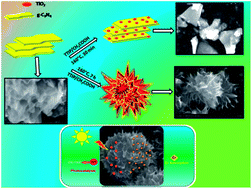
An efficient solar active catalyst is in immense demand for water splitting, waste water treatment and environmental remediation applications due to the growing energy crisis. The key to improving the activity of these materials is to increase the surface area, use a dopant and reduce the charge carrier recombination. This growing concern can be addressed by graphitic carbon nitride (g-C3N4) which has excellent visible light activity. In the present work a mesoporous flower like arrangement of carbon nitride–titania nanocomposites obtained through a solvothermal method without the use of any templates at ambient temperature is reported. A detailed investigation on the formation of these mesoflowers has been studied. The properties of the nanocomposite such as efficient absorption in the visible regime and delayed recombination of charge carriers make it an apt material for solar light photocatalysis for the degradation of organic contaminants. The high surface area of 147 m2 g−1 of the mesoflowers has been utilized for heavy metal removal such as Cr(VI) with a sorption percentage of 81% within 2 minutes of contact time in simulated water samples. This report paves the way for a new class of mesoflower nanocomposites for various environmental applications.


Introduction

Researchers around the globe are in search of a versatile material for various applications such as in energy conversion, batteries, hydrogen production and water treatment. Numerous metal oxide nanocomposites including titania, ceria and zinc oxide have been explored throughout the years to tailor its structure so as to develop new properties. Research has been focussed on the nitrogen doping of graphene and titania to enhance its visible light activity for various energy efficient technologies.1,2 Carbonaceous nanomaterials have gained great deal of interest such as carbon nanotubes, graphene, boron nitride due to its promising properties and enhanced surface area. The development of mesostructures for energy efficient application is addressed here. A facile method of synthesis of titania–carbon nitride mesostructures have been developed in this manuscript. Carbon nitride has a low band gap of 2.7 eV and excellent visible light activity.3 Graphitic carbon nitride (g-C3N4) is considered to be the most stable allotrope of carbon nitride.4 The commonly used precursors for chemical synthesis of g-C3N4 are nitrogen-rich compounds containing pre-bonded C–N core structures, such as triazine and heptazine derivatives, cyanamide, dicyanamide, urea, melamine, etc. Several methods have been employed to synthesize g-C3N4 which include liquid phase pulsed laser ablation of graphite target in the presence of ammonia,5 pyrolysis of urea,6 melamine7,8 and liquid phase exfoliation of bulk g-C3N4.9 Nanoporous graphitic carbon nitride has been synthesized using Triton X-100 and ionic liquids as templates through the self-polymerization reaction of dicyandiamide.10 All these methods require either high temperature or presence of a template for the synthesis of g-C3N4. Being a metal free photocatalyst g-C3N4 has emerged as a promising candidate for various applications such as hydrogen evolution,11 artificial photosynthesis, photocatalysis,12 fuel cells,13 and heavy metal removal.14 Titania being an environmental friendly photocatalyst has gained a lot of attention over a decade for decontamination,15 degradation of organic contaminants,16 hydrogen evolution,17 solar cells, etc. Studies are in progress to improve the solar activity of TiO2 using metals, non metals, carbon nanostructures, etc. Recently, TiO2–carbon nitride composites have gained significant interest due to the enhancement in the visible light activity. Zou et al. reported the synthesis of mesoporous TiO2 spheres which convert urea to carbon nitride at 300 °C.18 Recently, Zhou et al. has reported the synthesis of a series of composites of g-C3N4 and TiO2 with in situ doping of nitrogen in TiO2 by calcination with Ti(SO4)2 and urea as precursors for the photoreduction of CO2 under simulated light radiation.19 Reports on the development of carbon nitride–TiO2 nanocomposites are very limited in literature. As discussed earlier several steps are followed for the synthesis of these nanocomposites in literature using high temperature treatment, templates, nitrogen doping, etc. The synthesis route we present here is a one pot synthesis without the use of any templates. The sample obtained can be used as such without any further treatment.

The urge for developing an advanced material to satisfy the energy and environmental needs is addressed here. It is in this context, the synthetic approach we report here for the synthesis of g-C3N4–TiO2 mesoflowers leading to the large-scale production of 3D architectures is significant. Graphitic carbon nitride is synthesized from melamine. TiO2 and g-C3N4 is assembled into flower like morphology by a simple solvothermal method. The resulting 3D mesoflower structures provided a high surface area of ∼147 m2 g−1, and efficient photocatalytic activity under sunlight. Consequently, the 3D mesoflower composite shows excellent heavy metal adsorption rate and enhanced photocatalytic degradation of organic dye under solar light irradiation. A simple and efficient synthetic protocol for carbon nitride–TiO2 mesoflower (g-C3N4–TiO2) is developed in this work.

Experimental

Materials

All chemicals used were analytical-grade reagents without further purification. Melamine (99%, Sigma Aldrich), titanium(IV) isopropoxide (TTIP) purity 98% (Fischer scientific), acetic acid (Merck), tetraethyl orthosilicate (TEOS, Aldrich, 98%), ammonia solution 25% (Merck), 1,5-diphenylcarbazide (Merck) and potassium dichromate (Fischer scientific) were used in the experiments.

Synthesis of g-C3N4

g-C3N4 was prepared by calcining 1 g of melamine taken in a crucible covered with aluminum foil in a muffle furnace at 520 °C for 5 h.7 The white powder changes to light yellow colored powder with a yield of ∼350 mg.

Synthesis of g-C3N4/TiO2 nanocomposites

g-C3N4/TiO2 nanocomposite was prepared by one pot solvothermal method. Briefly, 0.1 wt% of g-C3N4 was added into 50 mL of concentrated acetic acid (Merck) and sonicated for 5 minutes. Then 750 μL of TTIP was added drop wise into the mixture under stirring. The mixture was stirred vigorously at room temperature for 30 minutes, then transferred into a Teflon beaker and sealed in an autoclave reactor. The reactor was heated in a muffle furnace at a temperature of 160 °C for 7 hours. Once the reactor cools down the product was collected by centrifuging the reaction mixture at 8000 rpm in water, washed thrice and dried at 60 °C. The obtained solids were denoted as g-C3N4–TiO2. The reaction was carried for different time intervals to study the evolution of these structures. The effect of concentration of g-C3N4 was also studied by varying its concentration. The samples calcined at 400 °C for 3 hours showed no change in the mesoflower structure and hence all the characterization was done for the material without any calcination.

Characterization

FE-SEM images were taken using a Hitachi SU6600 field emission gun scanning electron microscope (FE-SEM). Specimens were mounted on black carbon tape. TEM images were taken using Hitachi, H-7650 electron microscope working at an accelerating voltage of 80 kV. XRD measurements were conducted on a Rigaku Miniflex 600 X-ray diffractometer using a monochromatized Cu Kα radiation source (30 kV, 10 mA) with a wavelength of 0.1542 nm and analyzed in the range 10° ≤ 2θ ≤ 80°. UV-Vis absorption spectra were measured using a Shimadzu UV-1800 UV-Visible spectrophotometer. Functional group analysis was done using FTIR (Jasco FTIR 4108). The specific surface area was measured using BET surface area measurement (Micromeritics Gemini 2375 Surface area analyser) via multi point method after degassing the powder in flowing N2 at 200 °C for 2 h. The photoluminescence (PL) spectra were obtained by using fluorescence spectrometer (LS 55 Perkin Elmer).

Chromium adsorption

The sorption of Cr(VI) was investigated by batch experiments under ambient conditions. To 10 ppm of Cr(VI) solution, 1 g L−1 adsorbent was added. The reaction was done at a pH of 5. The solution was mixed by magnetic stirring. Then, 1 mL of the solution was centrifuged at 8000 rpm for 3 min and supernatant was collected at regular time intervals. The spectrophotometric estimation of Cr(VI) species was done using diphenylcarbazide as a complexation reagent.20 Cr(VI) form a colored compound Cr(III)-diphenyl dicarbazone which has a maximum absorption at 540 nm. The concentrations of the samples were determined by carrying out identical measurements to those of the standard using the calibration graph.

Photocatalytic degradation of Rhodamine B

In a typical process, 100 μL aqueous solution of Rhd B (1 mM) was added to 20 mL suspension of photocatalyst (0.1 mg mL−1) in water. The photocatalyst suspension was ultrasonicated for 5 minutes for better dispersion before adding the dye. The reaction vessel was exposed to sunlight with measured light intensity between 70[thin space (1/6-em)]000 and 80[thin space (1/6-em)]000 luxes. Light intensity was measured using a Lutron light meter in lux. At regular time intervals, 3 mL of the photoreacted solution was withdrawn and analyzed by the variations of the absorption maximum of Rhd B (553 nm) in the UV-Visible spectrum.

Results and discussion

Synthesis of g-C3N4–TiO2 mesoflowers

Graphitic carbon nitride was synthesized by the calcination of melamine. These nanosheets were further used for the synthesis of the nanocomposites. g-C3N4–TiO2 mesoflowers were synthesized by a facile solvothermal method at ambient temperature. Morphology of the nanocomposites was studied using FE-SEM and TEM. The sheet like nature of g-C3N4 was confirmed from the FE-SEM image (Fig. 1a). The FE-SEM images confirmed the formation of g-C3N4 as well as mesoflowers (Fig. 1b–d). It could be observed that the mesoflowers have a uniform size distribution with an average size of <2 μm (Fig. 1b). The TEM images in Fig. 1d shows the magnified region of a petal. In order to elucidate the mechanism of formation of these structures, role of various parameters such as solvents, precursors, weight ratio of g-C3N4 were investigated.
image file: c5ra14547c-f1.tif
Fig. 1 FE-SEM images of (a) graphitic carbon nitride (b) g-C3N4–TiO2 mesoflowers (c) magnified region of a mesoflower (d) TEM images of g-C3N4–TiO2, a magnified region of the petal (scale bar 100 nm) and mesoflower (scale bar 500 nm).

First a series of control experiments by varying the reaction time was carried out. The structural evolution of the flower like structures was studied using FE-SEM. Fig. 2 show the FE-SEM images of the samples synthesized at different reaction time. Initially the reaction was carried out for 30 minutes and it was found that g-C3N4 remained as sheets and titania as very small nanoparticles. When the reaction was done for 4 hours, evolution of the flower structures initiated though they were not well defined. Whereas, when the reaction time was extended to 7 hours, perfect flower like structures with petals originating from the centre were observed in SEM images. The mesoflowers were approximately of 2 μm with defined petal like structures arranged in a flower like pattern. Aggregation of structures and thereby alteration of flower like morphology were observed when the reaction time prolonged to 12 h. From EDS (Table S1, ESI) a qualitative analysis of the formation of TiO2 was done. The weight percentage of TiO2 was found to increase as the reaction time increases.


image file: c5ra14547c-f2.tif
Fig. 2 FE-SEM images of g-C3N4–TiO2 at different time intervals.

The concentration of g-C3N4 also had a major role in the formation of the flower structures. To study the effect of concentration of g-C3N4 on the structure formation 0.025, 0.05, 0.1, 0.2 wt% of g-C3N4 were used as the precursor in the reaction without changing the concentration of titania precursor. SEM images of the products are shown in Fig. 3. On addition of 0.025 wt% g-C3N4 lead to the formation of less dense flowers as evident from the SEM image. The concentrations, 0.1 and 0.05 wt% yielded almost similar flower like morphology but the evolution of petals was prominent in 0.1 wt%. There is a possibility of g-C3N4 wrapping over titania mesoflowers also. When the amount of g-C3N4 increased to 0.2 wt% the morphology was not well defined and the resulting structures were very dense. The reaction without the addition of g-C3N4 was performed which lead to the formation of rice grain like TiO2 (ESI, Fig. S1).


image file: c5ra14547c-f3.tif
Fig. 3 FE-SEM images of g-C3N4–TiO2 with varying weight ratios of g-C3N4.

The effect of other parameters such as solvent, precursor and route of synthesis was also investigated. In order to study the role of acetic acid in the evolution of flower structure, ammonia was used as the solvent under the same reaction conditions. It was found that the flower morphology was not obtained showing that acetic acid is essential for the formation of flower morphology (Fig. 4a). In order to study whether acetic acid medium itself facilitated the formation of bare g-C3N4 flowers, the reaction was carried out in acetic acid alone in the absence of titanium tetraisopropoxide. From Fig. 4b it was found that no flower structures were formed either. In order to investigate the role of titania, tetraethyl orthosilicate was introduced instead of TTIP under same conditions using acetic acid as solvent. It led to the formation of silica nanoparticles on the surface of g-C3N4. It was found that the flower like structures were not formed (Fig. 4c). The precursor for the synthesis of g-C3N4, melamine was used in the synthesis instead of g-C3N4 directly under the same conditions and the obtained product was calcined at 520 °C for 5 h. From the FE-SEM analysis, it was found that flower structures of larger sizes were obtained but the exact petal-like morphology was not obtained (Fig. 4d). Different routes of synthesis were investigated to find out their effect on the formation of mesoflowers. Initially sol–gel method of synthesis was adopted where in TTIP and g-C3N4 was allowed to react in an ethanol water mixture (1[thin space (1/6-em)]:[thin space (1/6-em)]1) for 3 h at 60 °C. The nanocomposites were washed and collected through centrifugation. The SEM images show the distribution of spherical shaped TiO2 nanoparticles on the surface of g-C3N4. It was evident that the flower shaped structures were not formed in this route of synthesis (Fig. 4e). Then sonochemical route of synthesis was also investigated using 1[thin space (1/6-em)]:[thin space (1/6-em)]2 weight ratio of g-C3N4[thin space (1/6-em)]:[thin space (1/6-em)]TiO2(rice grain shaped). The mixture was dispersed in water and sonicated for 1 h. This route of synthesis also did not yield flower-like morphology. It can be concluded that the route of synthesis also had a prominent role in the formation of these mesostructures (Fig. 4f). These experiments suggest that the flower structure formation is mediated through the formation of titanium–acetic acid complex21 leading to the formation of g-C3N4–TiO2 composite. In order to elucidate the evolution of the g-C3N4 mesoflower structures in the presence of TiO2 further characterization were done.


image file: c5ra14547c-f4.tif
Fig. 4 FE-SEM images of g-C3N4 (a) reaction with TTIP in ammonia solution (b) in acetic acid alone, solvothermal synthesis using (c) TEOS as precursor (d) melamine as the precursor for g-C3N4, using different routes of synthesis of g-C3N4–TiO2 (e) sol–gel route (f) sonochemical route.

Characterization of g-C3N4–TiO2 mesoflowers

In order to elucidate the structural evolution of the g-C3N4–TiO2 mesoflower structures XRD analysis was performed. Fig. 5 shows the XRD patterns of g-C3N4 and g-C3N4–TiO2 mesoflowers. The XRD pattern of g-C3N4 nanosheets exhibited a small peak at 13.1° which is indexed to the characteristic in-planar structural packing (100) and a prominent peak at 27.4° assigned to inter-planar stacking peaks of the aromatic systems in graphite-like g-C3N4 (002).9,22 The interplanar spacing d(002) of 3.254 Å indicated that aromatic units in g-C3N4 are packed tighter than the packing in graphene units (3.61 Å).23 The XRD patterns of the materials synthesised at different reaction time were analyzed. In the samples after the reaction with TTIP, intensity of the major peak at 27.4° has significantly reduced and the peaks of TiO2 evolved. Upon 30 minutes of reaction time, the peaks corresponding to anatase phase have slightly evolved but not prominent. As the reaction time increased the peaks of anatase TiO2 become prominent and the intensity of g-C3N4 (002) diminished. The size of TiO2 nanoparticles calculated from XRD patterns using Scherrer equation was 5.5 nm. This correlated well with the TEM images (Fig. 1d) where formation of TiO2 nanoparticles of 5–10 nm were observed which self-assemble to form the petal like morphology. It was observed that the low angle peak at 13.1° is less prominent in flowers which in turn confirm the formation of g-C3N4.24 The characteristic peaks (2θ) of anatase TiO2 was well resolved at 25.2°, 37.6°, 47.8°, 53.9°, 62.3°, and 75.0° for the samples synthesised with reaction time 4 h, 7 h and 12 h.
image file: c5ra14547c-f5.tif
Fig. 5 XRD patterns of g-C3N4 and g-C3N4–TiO2 at different time intervals. ‘*’ shows the peaks due to carbon nitride and ‘A’ corresponds to the anatase phase of titania.

The chemical characterization of the nanocomposite was studied using FTIR spectroscopy. The FTIR spectra confirmed the existence of graphite like sp2 bonding in g-C3N4 (Fig. 6a). In the spectrum of g-C3N4, the peaks between 1200–1650 cm−1 corresponds to the C–N and C[double bond, length as m-dash]N stretching in carbon nitride which are associated with the skeletal stretching vibrations of s-triazine/tri-s-triazine rings. These peaks were observed in bare g-C3N4 as well as TiO2 incorporated samples. The peaks were sharper in the composite probably due to the weak coupling with the skeletal vibrations. The peak at 1566 cm−1 correspond to the stretching mode of g-C3N4 heterocycle was shifted to 1530 cm−1 on incorporation of TiO2 indicating that TiO2 forms bond with the triazine ring. Additionally, on reaction with TTIP for 2 h a peak at 1024 cm−1 evolved which is attributed to the C–O bond stretching. The broad peaks between 3000 and 3600 cm−1 in g-C3N4 is due to the N–H stretching and O–H stretching due to the adsorbed water in between the g-C3N4 layers. It was found to be broader on incorporation of TiO2 due to O–H stretching. The sharp peak at 809 cm−1 was attributed to the out of plane ring bending modes of s-triazine rings which is in accordance with previous studies.7,19 This peak is found to broaden upon incorporation of TiO2. The broad peak at low frequency (500–700 cm−1) was attributed to the stretching modes of Ti–O in the spectrum after 30 minute reaction time which is in accordance with the XRD patterns. It was observed that the molecular structure of g-C3N4 did not alter due to the incorporation of titania.


image file: c5ra14547c-f6.tif
Fig. 6 (a) FTIR spectra of g-C3N4 and g-C3N4–TiO2 at different reaction time (b) UV-Vis absorption spectra of g-C3N4 and g-C3N4–TiO2 (c) photoluminescence spectra of g-C3N4 and g-C3N4–TiO2 at different reaction time.

The UV-Vis absorption spectrum of g-C3N4 exhibited an absorption band centered at 235–250 nm which is attributed to π → π* electronic transition in the aromatic 1,3,5-triazine compounds and sharp absorption ∼300 nm may be attributed to the n–π* electronic transitions involving lone pairs of nitrogen atoms in the aromatic heterocyclic ring systems. The absorption edge of g-C3N4–TiO2 around 310 nm is due to the incorporated TiO2 (Fig. 6b).

To understand the extent of generation, separation, and recombination rate of the photogenerated charge carriers of g-C3N4–TiO2 mesoflower, the room temperature photoluminescence (PL) was analyzed (Fig. 6c). It was observed that the bare g-C3N4 exhibited intense photoluminescence intensity, which reduces upon 30 minutes of reaction with TTIP. Further, on increasing the reaction time the PL intensity enormously reduced. This gradual decrease confirms that on incorporation of TiO2 the recombination rate of the charge carriers have been considerably reduced which is a favorable factor for the photocatalytic activity.

Surface area analysis is an important criterion for any material in energy and environmental application. The surface area, pore volumes, and average pore sizes were determined from Brunauer–Emmett–Teller (BET) isotherms. Since the flower like morphology was well evolved upon 7 h of the reaction time, this sample was further characterized for its porous nature. Fig. 7a shows the nitrogen adsorption and desorption isotherms of g-C3N4–TiO2 after 7 h of reaction time. The isotherms showed characteristic H3 type loop which are usually seen for aggregates of plate-like particles or adsorbents containing slit-shaped pores.25 The BET surface area and pore volume (Fig. 7b) are 147 m2 g−1 and 0.001722 cm3 g−1, respectively.


image file: c5ra14547c-f7.tif
Fig. 7 (a) Nitrogen adsorption and desorption isotherms (b) pore diameter distribution curve of g-C3N4–TiO2 (7 h).

Environmental application of g-C3N4–TiO2 mesoflowers

The g-C3N4–TiO2 flower structures due to its enhanced surface area and excellent electron–hole recombination delay they were evaluated for its potential in photocatalytic degradation of organic pollutants under solar radiation and adsorption of heavy metals such as Cr(VI).

Adsorption of heavy metals – Cr(VI)

In order to evaluate the sorption property of g-C3N4–TiO2 mesoflowers for the removal of heavy metal ions, the sorption processes of hexavalent chromium was studied. Sorption of Cr(VI) on g-C3N4–TiO2 as a function of contact time is shown in Fig. 8a. A rapid adsorption of 81% was achieved in the first 2 minutes of stirring time after which equilibrium was attained. It was observed that the large surface area of the meso structures aided in the rapid removal of Cr(VI) within 2 minutes. In order to investigate the sorption kinetics in detail, the experimental data were fitted by the pseudo-second-order rate equation (Fig. 8b). From the linear plot of t/qt versus t, the value of k2 calculated from the slope and intercept is 0.0006 g mg−1 min−1. The regression coefficients (R2 [Cr(VI)] = 0.9989) suggests that the kinetics of Cr(VI) on g-C3N4–TiO2 follow the pseudo-second-order model. ESI Fig. S2 shows the variation of qt with time. In order to understand the sorption mechanism better, the experimental data were simulated by Langmuir and Freundlich isotherms (ESI, Fig. S3). The sorption capacity at equilibrium time qe (mg g−1) is compared with the experimental and calculated values as shown in Fig. 8c. It was observed that adsorption followed Freundlich model and it showed best fit with the experimental values. All the parameters and regression coefficients obtained from the equilibrium plots of Langmuir and Freundlich isotherms models are listed in Table 1. From the isotherms it is observed that Freundlich model suits better. This result indicates the heterogeneous adsorption of Cr(VI) on g-C3N4–TiO2 mesoflowers suggesting the efficient use of this structure in the heavy metal removal from contaminated water.
image file: c5ra14547c-f8.tif
Fig. 8 (a) Sorption of Cr(VI) on g-C3N4–TiO2 as a function of contact time (b) pseudo second-order kinetic plot fitting of t/qt versus t (c) comparison of experimental qe with calculated qe from Langmuir and Freundlich isotherm model.
Table 1 Regression coefficients and constants of isotherm models of adsorption of Cr(VI)
Isotherm model KL [L mg−1] KF [mg g−1 (L mg−1)1/n] R2 RL 1/n
Langmuir 3.25 0.976 0.01
Freundlich 6.48 0.988 0.324


Photocatalytic activity of g-C3N4–TiO2 mesoflowers

The photocatalytic performance of g-C3N4–TiO2 mesoflowers was evaluated using a cationic dye, Rhodamine B as a model dye pollutant under direct sunlight. Rhodamine B has an absorption maximum at 553 nm, which is directly proportional to its concentration. The absorption at λmax is monitored and the degradation efficiency of the nanocomposite was evaluated using eqn (1).
 
image file: c5ra14547c-t1.tif(1)
where A0 represents the initial absorbance, and At represents the absorbance after a reaction time (t).

The UV-Vis absorption spectra showing the degradation profile of the dye under solar radiation is shown in Fig. 9a. The enhanced surface area of mesoflowers enabled better adsorption of the dye molecules on the surface of the mesoflowers. There is possibility of wrapping of g-C3N4 over the titania mesoflowers which in turn aided the solar light activity to the nanocomposite. The reduced charge carrier recombination enhanced the photocatalytic activity of the composite under solar radiation. The kinetics of degradation of the dye was studied. It was observed that the reaction is first order with a rate constant of 49.73 × 10⁻³ min−1 (Fig. 9b). It was observed that under solar irradiation the degradation efficiency was found to be 100% within 50 minutes of exposure (Fig. 9c). A comparison table on the photocatalytic degradation of dyes using carbon nitride/titania nanocomposites as well as carbon nitride and titania is shown in Table 2. The degradation efficiency obtained in this work is higher than the previous literature wherein high power lamps and high amount of catalyst has been used in the degradation process. The mesoflower structures showed good solar photocatalysis for the degradation of organic contaminants such as dyes which can be further extended to other organic pollutants such as pesticides, phenols, etc.


image file: c5ra14547c-f9.tif
Fig. 9 (a) UV-Vis absorption spectra of photocatalytic degradation of Rhodamine B under solar radiation (b) the kinetic plot of degradation (c) photocatalytic degradation efficiency of g-C3N4–TiO2 mesoflowers over Rhodamine B under solar radiation.
Table 2 A comparison table on the photocatalytic degradation of dyes using carbon nitride/titania nanocomposites
No. Material Light source/lamp Surface area m2 g−1 Catalyst conc. (mg mL−1) Pollutant selected Time Ref.
1 SiO2/C3N4 12 W LED @ 420 nm 70 1 Rhodamine B 120 min 26
2 g-C3N4/SmVO4 350 W Xe 51 1 Rhodamine B 2 h 8
3 g-C3N4/TiO2 (B) 108 W Sulforhodamine B >5 h 27
4 Ag/g-C3N4 500 W Xe 3 Methyl orange 3 h 28
5 Porous g-C3N4 500 W Xe 5.4–60 0.5 Methylene blue 29
6 N-doped TiO2 500 W Xe 171.7 1 Methylene blue 100 min 30
7 g-C3N4–TiO2 100 W halogen 10.67 0.125 Methylene blue 100 min 31
8 Graphene-like C3N4 with 300 μL H2O2 300 W Xe 32.54 0.5 Methylene blue 25 min 32
9 g-C3N4/TiO2 mesoflowers Solar radiation 147 0.1 Rhodamine B 50 min This work


Conclusions

In summary, we have developed a facile solvothermal route of synthesis of mesoporous flower-like arrangement of g-C3N4 and titania. A detailed investigation on the formation of the flower-like structures is discussed in this work. A possible mechanism of formation of mesoflowers is discussed. The formation of flower structures strongly depended on the concentration of g-C3N4, reaction time and the solvent used. Owing to their high surface area (147 m2 g−1) they prove to be an apt candidate for removal of heavy metals with a sorption percentage of 81% within 2 minutes. The sorption processes for Cr(VI) followed the pseudo second-order kinetics, and the sorption isotherm was simulated well by the Freundlich model. They also have proved to be an efficient solar active photocatalyst. These properties of the mesostructures make them promising materials for wide range of energy and environmental applications such as heavy metal removal, photocatalysis, hydrogen evolution under solar radiation, lithium ion batteries and fuel cells. The synthetic protocol can be extended to the more general fabrication of 3D composites of carbon nitride with appropriate metal or metal oxide nanomaterials for the specific applications.

Acknowledgements

The authors thank Department of Science and Technology and Department of Biotechnology, India for the financial support to the nanoscience research laboratory. The authors thank Mrs Sabna V and Dr S. Bhuvaneshwari for their valuable discussions in the adsorption experiments. Department of Physics, Calicut University is greatly acknowledged for the XRD facility. We thank Dr C. Arunkumar and Mr Rahul Soman, Department of Chemistry, NITC for providing the fluorescence measurement facility.

Notes and references

  1. V. K. Etacheri, M. K. Seery, S. J. Hinder and S. C. Pillai, Chem. Mater., 2010, 22, 3843 CrossRef CAS .
  2. N. R. Khalid, E. Ahmed, Z. Hong, Y. Zhang and M. Ahmad, Curr. Appl. Phy., 2012, 12, 1485 CrossRef PubMed .
  3. Y. Wang, X. Wang and M. Antonietti, Angew. Chem., Int. Ed., 2012, 51, 68 CrossRef CAS PubMed .
  4. A. Thomas, A. Fischer, F. Goettmann, M. Antonietti, J. Muller, S. Robert and J. M. Carlsson, J. Mater. Chem., 2008, 18, 4893 RSC .
  5. L. Yang, P. W. May, Y. Huang and L. Yin, J. Mater. Chem., 2007, 17, 1255 RSC .
  6. J. Liu, T. Zhang, Z. Wang, G. Dawson and W. Chen, J. Matter. Chem., 2011, 21, 14398 RSC .
  7. X. Li, J. Zhang, L. Shen, Y. Ma, W. Lei, Q. Cui and G. Zou, Appl. Phys. A, 2009, 94, 387 CrossRef CAS .
  8. T. Li, L. Zhao, Y. He, J. Cai, M. Luo and J. Lin, Appl. Catal., B, 2013, 129, 255 CrossRef CAS PubMed .
  9. S. Yang, Y. Gong, J. Zhang, L. Zhan, L. Ma, Z. Fang, R. Vajtai, X. Wang and P. M. Ajayan, Adv. Mater., 2013, 25, 2452 CrossRef CAS PubMed .
  10. Y. Wang, X. Wang, M. Antonietti and Y. Zhang, ChemSusChem, 2010, 3, 435 CrossRef CAS PubMed .
  11. P. Niu, L. Zhang, G. Liu and H. M. Cheng, Adv. Funct. Mater., 2012, 22, 4763 CrossRef CAS PubMed .
  12. X. Bai, L. Wang, R. Zong and Y. Zhu, J. Phys. Chem. C, 2013, 117, 9952 CAS .
  13. Y. Zheng, J. Liu, J. Liang, M. Jaroniec and S. Z. Qiao, Energy Environ. Sci., 2012, 5, 6717 CAS .
  14. R. Hu, X. Wang, D. Shao, T. Hayat and A. Alsaedi, Chem. Eng. J., 2015, 260, 469 CrossRef CAS PubMed .
  15. J. M. Herrmann, Catal. Today, 1999, 53, 115 CrossRef CAS .
  16. R. J. Tayade, R. G. Kulkarni and R. V. Jasra, Ind. Eng. Chem. Res., 2006, 45, 922 CrossRef CAS .
  17. J. Nowotny, T. Bak, M. K. Nowotny and L. R. Sheppard, Int. J. Hydrogen Energy, 2007, 32, 2609 CrossRef CAS PubMed .
  18. X. X. Zou, G. D. Li, Y. N. Wang, J. Zhao, C. Yan, M. Y. Guo, L. Li and J. S. Chen, Chem. Commun., 2011, 47, 1066 RSC .
  19. S. Zhou, Y. Liu, J. Li, Y. Wang, G. Jiang, Z. Zhao, D. Wang, A. Duan, J. Liu and Y. Wei, Appl. Catal., B, 2014, 158, 20 CrossRef PubMed .
  20. L. Khezami and R. Capart, J. Hazard. Mater., 2005, 123, 223 CrossRef CAS PubMed .
  21. J. Ye, W. Liu, J. Cai, S. Chen, X. Zhao, H. Zhou and L. Qi, J. Am. Chem. Soc., 2011, 133, 933 CrossRef CAS PubMed .
  22. K. Maeda, X. Wang, Y. Nishihara, D. Lu, M. Antonietti and K. sDomen, J. Phys. Chem. C, 2009, 113, 4940 CAS .
  23. R. T. Thomas, P. A. Rasheed and N. Sandhyarani, J. Colloid Interface Sci., 2014, 428, 214 CrossRef CAS PubMed .
  24. B. V. Lotsch, M. Dçblinger, J. Sehnert, L. Seyfarth, J. Senker, O. Oeckler and W. Schnick, Chem.–Eur. J., 2007, 13, 4969 CrossRef CAS .
  25. M. Thommes, Chem. Ing. Tech., 2010, 82, 1059 CrossRef CAS PubMed .
  26. M. Shalom, S. Inal, D. Neher and M. Antonietti, Catal. Today, 2014, 225, 185 CrossRef CAS PubMed .
  27. L. Zhang, D. Jing, X. She, H. Liu, D. Yang, Y. Lu, J. Li, Z. Zheng and L. Guo, J. Mater. Chem. A, 2014, 2, 2071 CAS .
  28. L. Ge, C. Han, J. Liu and Y. Li, Appl. Catal., A, 2011, 409–410, 215 CrossRef CAS PubMed .
  29. M. Zhang, J. Xu, R. Zong and Y. Zhu, Appl. Catal., B, 2014, 147, 229 CrossRef CAS PubMed .
  30. G. Yang, Z. Jiang, H. Shi, T. Xiao and Z. Yan, J. Mater. Chem., 2010, 20, 5301 RSC .
  31. K. Sridharan, E. Jang and T. J. Park, Appl. Catal., B, 2013, 142–143, 718 CrossRef CAS PubMed .
  32. X. She, H. Xu, Y. Xu, J. Yan, J. Xia, L. Xu, Y. Song, Y. Jiang, Q. Zhang and H. Li, J. Mater. Chem. A, 2014, 2, 2563 CAS .

Footnote

Electronic supplementary information (ESI) available: TEM images of TiO2, amount of Cr(VI) adsorbed on g-C3N4–TiO2 at various time intervals and initial concentration, table showing the EDS analysis of g-C3N4–TiO2 at different reaction time intervals. See DOI: 10.1039/c5ra14547c

This journal is © The Royal Society of Chemistry 2015
Click here to see how this site uses Cookies. View our privacy policy here.