Amanpreet Singha,
Ajnesh Singhb,
Narinder Singh*a and
Doo Ok Jang*c
aDepartment of Chemistry, Indian Institute of Technology Ropar, Rupnagar, Punjab 140001, India. E-mail: nsingh@iitrpr.ac.in
bDepartment of Applied Science & Humanities, Jawaharlal Nehru Govt. Engineering College, Sundernagar, India
cDepartment of Chemistry, Yonsei University, Wonju 220-710, Republic of Korea. E-mail: dojang@yonsei.ac.kr
First published on 20th August 2015
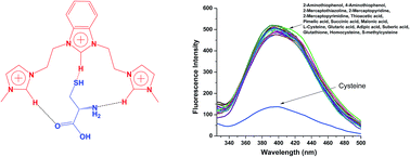
A benzimidazolium- and imidazolium-based trication was developed as a fluorescence probe for cysteine (Cys). The probe exhibits fluorescence responses to Cys in water with high selectivity and a nano-molar detection limit. Such specificity towards Cys is based on differences in the hydrogen bonding of R1 with Cys, and it provides a method for detecting Cys in the presence of other analytes in a real biological system such as human serum. Density functional theory (DFT) calculations and 1H NMR titration confirmed the cavity-based recognition of Cys.
In a biological system, the detection of Cys requires highly sensitive probes due to its low intercellular concentration (30–200 μM). Some analytical techniques for Cys determination have been reported, including high-performance liquid chromatography, electrochemical detection, and mass spectroscopy.11–14 However, these methods have some limitations in that they are time-consuming, require sophisticated instruments, and have high detection limits. As such, several recent efforts have been devoted to developing fluorescent probes for Cys determination. Zhong and co-workers prepared a colorimetric probe for the detection of biothiols using the Hg2+-mediated aggregation of gold nanoparticles.15 Li and co-workers described the self-assembling property of cysteine on gold nanoparticles, which allows for the determination of cysteine via a change in the color of gold nanoparticles due to aggregation.16 Chen modified a glassy carbon electrode surface using one-dimensional caterpillar-like manganese dioxide–carbon (MnO2–C) nanocomposites for the selective detection of cysteine.17 Zhu employed cyclic voltammetry and a carbon fiber clustered electrode modified with silver nanoparticles for the selective determination of cysteine.18
Fluorescence methods for Cys detection are more anticipated due to the simple handling and high sensitivity associated with the techniques. For example, Tan and co-workers developed a europium tetracycline-based fluorescent probe for determining the total amount of biothiols, including Cys, glutathione (GSH), and homocysteine (Hcys) in urine samples.19 Qu and co-workers employed a silver metallization-controlled conformational switch of G-quadruplex DNA as a turn-on fluorescent sensor for biothiols,2b while Yang prepared two fluorescent probes using two noble coumarin-derived products for the detection of Cys, Hcys, and GSH.20 Chu utilized fluorescent copper nano-clusters synthesized by a double-stranded DNA template (DNA-CuNCs) as a probe for biothiol determination.21 Xie and co-workers developed a graphene oxide-based fluorescent DNA sensor for sensing Cys.22 Pu and co-workers developed a fluorescence turn-on assay for detection of cysteine and histidine using a DNA/ligand/ion ensemble.23 Shang and co-workers synthesized polymer-stabilized gold nanoparticles for detection of Cys.24 Zhang and co-workers developed a probe based on the conjugate addition–cyclization reaction of a compound with a hemicyanine skeleton.25
While the use of nanoparticle or organo dye-based fluorescence probes has previously been reported for the detection of Cys, such approaches are limited by various factors, including aggregation of the probes in water and physiological conditions due to their low solubility in water.26–29 Therefore, there is great demand for the development of fluorescent Cys probes that are highly soluble in water.
A Cys molecule has several hydrogen bonding sites that can interact with a host molecule through hydrogen bonding. In this context, benzimidazolium and imidazolium cations are aptly suited to act as receptors.30–32 These organic cations have acidic hydrogens that are readily available for hydrogen bonding. Furthermore, benzimidazolium derivatives have high fluorescence quantum yields, lower cytotoxicity, high aqueous solubility, and can be easily synthesized. It has also been reported that imidazolium cations have a high affinity for various biomolecules.33–36 Consequently, it is expected that a combination of benzimidazolium and imidazolium cations may provide a cavity for hydrogen bonding with a guest molecule, thereby allowing for high solubility in water. In this work, we report on a selective and sensitive fluorescent benzimidazolium- and imidazolium-based trication probe for the detection of Cys in water.
Changes in the photophysical properties of R1 were observed so as to study its binding affinity with various anions. In particular, emission spectra were examined after the addition of F−, Cl−, Br−, I−, ClO4−, NO3−, CO32−, PO42−, CH3CO2−, and HSO42− anions (Fig. S2†). No significant changes were observed upon adding the different anions. The binding behavior of R1 was also examined with thiols and some acid derivatives such as 2-aminothiophenol, 4-aminothiophenol, 2-mercaptothiazoline, 2-mercaptopyridine, 2-mercaptopyrimidine, thioacetic acid, L-cysteine, pimelic acid, succinic acid, malonic acid, glutaric acid, adipic acid, suberic acid, glutathione, homocysteine and S-methylcysteine (Fig. 1). With one exception, no changes in the emission profile of R1 were observed after adding the analytes to the R1 solution. However, when Cys was added to a solution of R1, the fluorescence intensity of R1 decreased significantly.
![]() | ||
Fig. 1 Changes in the emission profile of R1 (10 μM) upon the addition of different analytes (1.5 equiv.) in a HEPES buffer (10 mM, pH = 7.4) solution during excitation at λex = 290 nm. |
To confirm the interaction of R1 with Cys molecules, a titration experiment was performed with successive additions of Cys to an R1 solution, as shown in Fig. 2A. As the concentration of Cys increased, the fluorescence intensity at 390 nm gradually decreased. A calibration curve was plotted based on changes observed in the spectral profile of R1 with the successive addition of Cys (Fig. 2B). The calibration plot for Cys showed excellent linear regression up to 14 μM with an R2 value of 0.9983. The limit of detection (LOD) calculated using the 3σ method38 was 48 nM, which is comparable to reported values (4–2000 nM) for fluorescent probes.2b,19–25 Quenching of the fluorescence intensity can be explained on the basis of photoinduced electron transfer (PET) caused by a lone pair electron of the Cys molecule. Upon interacting with Cys, the fluorescence of R1 is quenched due to the PET phenomenon.
The fluorescence quantum yield of R1 was determined relative to a reference compound of known quantum yield. 2-Aminopyridine was chosen as reference compound because it has an emission profile between 320–480 nm similar to probe R1.39 As shown in Table 1, the quantum yield of R1 decreased upon addition of Cys. The fluorescence quenching factor was calculated to quantify the sensing of Cys. Almost 5-fold fluorescence intensity decreased upon addition of Cys.
To ascertain the stoichiometry, fluorescence quenching data were utilized to construct Job's plots.40 The x-axis of the Job's plot, labeled as the mole fraction, is the ratio of the Cys concentration to the total concentration of Cys and R1 (Fig. 3A). The intersection of the two linear portions of the Job's plot gives the mole ratio corresponding to the binding stoichiometry between Cys and R1. From Fig. 3A, it can be confirmed that R1 interacts with Cys in a 1:
1 stoichiometry. The binding constant (Ka) of R1 for Cys was also determined by plotting 1/(F − Fo) vs. 1/[Cys]; a Ka value of 2.55 (±0.09) × 104 M−1 was obtained from the slope and intercept of the line (Fig. 3B).41
![]() | ||
Fig. 3 (A) Job's plot with a 1![]() ![]() |
To investigate the interference caused by other analytes in the detection of Cys, fluorescence spectra were recorded for Cys in the presence of different analytes. A bar diagram was subsequently generated for Io/I at 390 nm. Here, Io is the fluorescence intensity of R1, and I is the fluorescence intensity of R1 upon the addition of different analytes (Fig. 4). From the bar diagram, it is clear that none of the analytes interfere in the detection of Cys when using fluorescence spectroscopy. In addition, interference studies were performed with other amino acids such as alanine, histidine, isoleucine, leucine, lysine, methionine, aspartic acid, glutamic acid, glutamine and glycine (Fig. S3†). These analytes did not show any interference in the determination of Cys.
To evaluate the utility of R1, fluorescence spectra were recorded at different pH values in the absence or presence of Cys. The fluorescence intensity of R1 at 390 nm was low in the pH range of 4.5–6.5 and high over a pH range of 6.5–9.5 (Fig. S4†). Therefore, a HEPES buffer solution was used to maintain a pH value of 7.4 in all experiments. In the presence of Cys, the fluorescence intensity decreased and remained relatively constant over a pH range of 4.5 to 8.5. However, at higher pH values (more than 9.0), the fluorescence intensity increased due to reduced interactions of Cys with R1 in the basic pH range.
1H NMR titration of probe R1 with Cys was performed in DMSO-d6 by varying the equivalent amount of Cys with respect to R1 (Fig. 5). Probe R1 has two deshielded protons at 9.18 and 9.91 ppm that correspond to acidic imidazolium and benzimidazolium protons, respectively. The addition of 4.0 equiv. of Cys induced a shift of Δδ = 0.25 ppm in the signal of C–H (C–H of benzimidazolium cation at 9.91 ppm) and Δδ = 0.19 ppm in the signal of C–H (C–H of imidazolium cation at 9.18 ppm). The downfield shifts observed for acidic imidazolium and benzimidazolium protons imply that Cys is interacting with R1 via hydrogen bonding. Considerable shifts in the aromatic protons of R1 were also noted.
![]() | ||
Fig. 5 1H NMR spectral changes observed in the aromatic region of probe R1 in DMSO-d6 upon the addition of up to 6 equiv. of Cys. |
Density functional theory (DFT) calculations for probe R1 were performed using the GGA-DFT package of DMol3. The structure of R1 was optimized in the presence of Cys. As shown in Fig. 6, electronegative atoms such as S, N, and O form hydrogen bonds with the acidic protons of imidazolium and benzimidazolium, whereas one oxygen atom is free. This induces PET, which in turn causes fluorescence quenching. The length of C–H bonds in imidazolium and benzimidazolium rings indicates the interaction of R1 with Cys via hydrogen bonding (Fig. S6†). The bond length of C–H bonds in imidazolium and benzimidazolium rings increases during the interaction of R1 with Cys (Table S4†). The DFT calculation was also performed to study the integration of R1 with Hcys, observing no hydrogen bonding between R1 and Hcys.
To examine the use of probe R1 in real competitive environment, spike/recovery assays of Cys were performed in human serum samples. In spike/recovery assays, a known amount of an analyte is spiked into a sample. The recovery of the spiked analyte indicates whether the analyte in the sample interferes.42 The results are presented in Table 2. The recovery of added Cys was a range of 99.6–100.5. These results indicate that probe R1 can be used for estimating Cys in a real biological system without interference.
Footnote |
† Electronic supplementary information (ESI) available: Crystallographic data for compound 1, copies of NMR and mass spectra of compounds. CCDC 1038708. For ESI and crystallographic data in CIF or other electronic format see DOI: 10.1039/c5ra14501e |
This journal is © The Royal Society of Chemistry 2015 |