DOI:
10.1039/C5RA13985F
(Paper)
RSC Adv., 2015,
5, 74977-74985
L-Asparagine-assisted synthesis of flower-like β-Bi2O3 and its photocatalytic performance for the degradation of 4-phenylphenol under visible-light irradiation†
Received
16th July 2015
, Accepted 20th August 2015
First published on 20th August 2015
Abstract
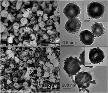
Large-scale synthesis of nanosized β-Bi2O3 is a significant challenge due to its metastable state. A facile L-asparagine-assisted reflux–calcination route was successfully developed for the large-scale preparation of β-Bi2O3 micro/nanostructures under mild conditions (low temperature, atmospheric pressure, and wide temperature windows). The composition, phase structure, morphology, surface area, and photoabsorption properties of as-synthesized β-Bi2O3 and its precursor were systematically characterized. The phase transformation conditions and possible formation mechanism of flower-like β-Bi2O3 were discussed. It is found that with a simple reflux process under atmospheric pressure at 100 °C, uniform monodisperse bismuth–asparagine complex microspheres with average diameters of ∼500 nm were produced and flower-like β-Bi2O3 micro/nanostructures were then conveniently obtained after precursor calcination at temperatures ranging from 340 °C to 420 °C. A surface CO32− coordination effect introduced from L-asparagine explained the formation of stabilized β-Bi2O3 at low temperatures (up to 420 °C). The as-synthesized β-Bi2O3 shows excellent photocatalytic activity toward the degradation of 4-phenylphenol under visible-light irradiation, which is 3.7 and 21.4 times faster than the removal rates of β-Bi2O3 nanospheres and a commercial β-Bi2O3, respectively, and allows for the elimination of 93.2% total organic carbon after 60 min of irradiation. In addition, the photogenerated reactive species were identified by radical scavenger experiments and electron paramagnetic resonance spectroscopy, and a possible visible-light-induced photocatalytic mechanism was then proposed.
1. Introduction
In the past few decades, semiconductor photocatalysis has attracted global interest because of its potential applications in solar-energy conversion and environmental purification.1 As an important Bi-based semiconductor, bismuth trioxide possesses excellent properties, such as abundant polymorphism (α-, β-, γ-, δ-, ε-, and ω-), wide band-gap range (2.0–3.96 eV), high refractive index, good electrochemical response, and high oxygen-ion conductivity, resulting in its numerous potential applications in photocatalysis, photovoltaic cell, industrial catalysis, and gas sensors.2,3 Among Bi2O3 polymorphic forms, α- and δ-phases are stable at low and high temperatures, respectively, whereas the other phases are metastable. However, according to previous studies,4 β-Bi2O3 holds a more narrow band gap and more dispersive band structures, resulting in its strong absorption of solar light, high charge-separation efficiency, and strong oxidative ability, thus demonstrating superior photocatalytic performance than other phases under visible-light irradiation. Nevertheless, compared with the widely studied α-Bi2O3, research on β-Bi2O3 is very limited due to its metastable state and synthetic challenges; moreover, the low-temperature formation mechanism of the metastable β phase is still unclear. There are two difficulties faced for large-scale nanoscale β-Bi2O3 synthesis, which greatly hinder its practical applications. First, the precursors of β-Bi2O3, such as Bi2O2CO3,5 Bi2(C2O4)3·7H2O,6 and (H2O)0.75Bi2(CH3COO)(NO3)1.12,7 are usually synthesized by hydrothermal or solvothermal processes, which restricts their large-scale production because these methods only take place under high-temperature and high-pressure conditions. Second, the calcined precursor temperatures are highly strict with a narrow temperature range to obtain a pure β phase because of its ease of conversion to a more stable α phase. In a previous study, we introduced a new synthetic route to obtain β-Bi2O3 by the direct calcination of nanometal bismuth under air atmosphere.8 Through this method, β-Bi2O3 with desired structures and morphologies can easily be obtained simply by controlling its metallic bismuth precursors.9 In addition, it has been experimentally observed in our lab that metallic bismuth precursors can be prepared in large quantities under atmospheric pressure using suitable reducing agents and temperature conditions. However, the calcination temperature to obtain a pure β-phase Bi2O3 is still restricted, otherwise a certain amount of impurity will be produced. Therefore, further development of new synthetic β-Bi2O3 routes are necessary, and the β-phase stabilization mechanism must be further identified to solve the aforementioned problems.10 Recently, by analyzing the differences of the surface chemical states of α-Bi2O3 and β-Bi2O3, Jiang et al. proposed a surface CO32− coordination mechanism to understand the stability of room-temperature β-Bi2O3, which reveals a strategy for low-temperature synthesis of β-Bi2O3, i.e. introducing organic ligand groups to the β-Bi2O3 surface to reduce its surface energy11 and thus opening a new gate for obtaining stable β-Bi2O3 in a wider temperature range.
Phenylphenols (ortho-, meta-, and para-phenylphenol), alkylphenolic compounds that consist of two linked benzene rings and a phenolic hydroxyl group have been widely used as antimicrobial agents, household pesticides, and additives in industrial materials.12 As a result, phenylphenol residues can be easily detected in riverine sediments, sewage sludge, surface water, and even in canned beer or soft drinks.13,14 However, studies have indicated that the presence of phenylphenols can cause harm to humans and other living organisms, as they might exhibit estrogenic activities, androgenic potencies, and chronic toxicity.15 It can be noted that phenylphenols often are not biodegradable, which leads to their inefficient elimination by conventional treatment methods.16 In addition, studies have shown that the hormone activity of phenylphenol isomers is highly dependent on the hydroxyl group position in the aromatic ring, and 4-OH phenylphenol (4-PP) exhibits the highest levels of estrogenic and androgenic activities.17 Thus, there is increasing concern about the presence of phenylphenols in the environment, and the removal of 4-PP from wastewaters, in particular, is urgently required. It is well known that heterogeneous photocatalysis is one of the efficient techniques for decomposing phenols,18,19 thus it can be used as a potential technology for the treatment of 4-PP. In fact, Christine et al. succeeded in photocatalytically removing 4-PP using TiO2 as a catalyst.20 However, the main drawback of TiO2 is that it can only be excited by UV light (accounting for only ∼4% of solar energy).21 To achieve higher photocatalytic efficiency and more effective utilization of the solar energy, great efforts should be made for the development of new visible-light-driven photocatalysts in the treatment of 4-PP wastewaters.
Herein, we report a facile synthesis route for the preparation of flower-like β-Bi2O3 micro/nanostructures through a reflux–calcination process assisted by L-asparagine (one of the 20 most-common natural amino acids on Earth). Compared with other methods, the proposed route allows for easy and large-scale synthesis of pure-phase β-Bi2O3 under mild conditions. The structure, morphology, photoabsorption, surface area, and formation process of the as-synthesized β-Bi2O3 were studied. The photocatalytic degradation and mineralization of 4-PP under visible-light irradiation were then reported for the first time using the as-synthesized β-Bi2O3 catalysts. In addition, the role of the primary oxidative species of the photoreaction system was examined, and a possible photocatalytic mechanism was proposed.
2. Experimental section
2.1. Materials
4-PP was supplied by Aladdin Reagent Co., Ltd. Bi(NO3)3·5H2O was purchased from Tianjin Kermel Chemical Reagent Co., Ltd. L-Asparagine was obtained from Shanghai Bo'ao Biological Technology Co., Ltd. Commercially available β-Bi2O3 was purchased from Aladdin Reagent Co., Ltd. All other chemicals were of analytical grade and used as received without further purification.
2.2. Synthesis of flower-like β-Bi2O3
In a typical synthesis, 1.8 g L-asparagine was dissolved in 50 mL of distilled water at room temperature, and 0.97 g Bi(NO3)3 was then added under continuous magnetic stirring until a homogeneous solution was formed. The solution was then transferred into a 150 mL round-bottomed flask and oil-bath heated to a temperature of 100 °C under reflux conditions. After reacting for 4 h, the resulting white precipitates were collected by centrifugation, thoroughly washed with distilled water and absolute ethanol, and then dried in an oven at 60 °C. Finally, the resultant product was calcined in a covered alumina crucible for 2 h in a muffle furnace at temperatures ranging from 260 °C to 580 °C. For comparison, β-Bi2O3 nanospheres were prepared by a solvothermal–calcining route according to the method described in our previous report.8
2.3. Characterization
The crystalline structure and phase composition of the as-synthesized samples were determined by X-ray diffraction (XRD) using a D8 Advance X-ray diffractometer (Bruker AXS, Germany) with a Cu Kα radiation source. The morphology and microstructure were observed under a field emission scanning electron microscope (FE-SEM, S-4800, Hitachi) and a field emission transmission electron microscope (FE-TEM, Tecnai G2 F20, FEI). X-ray photoelectron spectroscopy (XPS) was carried out on an electron spectrometer (Thermo Scientific ESCALAB 250Xi, USA) equipped with an Al Kα source. The ultraviolet-visible (UV-vis) diffuse reflectance spectra (DRS) of the samples were obtained on a UV-vis spectrophotometer (U-4100, Hitachi) using an integrating sphere accessory and were converted to absorption spectra by the Kubelka–Munk method. The Brunauer–Emmett–Teller (BET) surface areas and average pore volume were measured by adsorption–desorption isotherms through BET analysis (ASAP 2460, Micromeritics, USA). A desorption isotherm was applied to calculate pore-size distribution using the Barrett–Joyner–Halenda (BJH) method. Electron paramagnetic resonance (EPR, JES FA-200, JEOL, Japan) in combination with spin-trapping techniques was utilized to detect free radicals, and 5,5-dimethyl-1-pyrroline-N-oxide (DMPO) was used as a spin trap.
2.4. Photocatalytic measurements
The photocatalytic activities of the samples were evaluated for the photocatalytic degradation of an aqueous solution of 4-PP using a photochemical reactor (XPA-VII, Nanjing Xujiang Machine-electronic Plant, China), which was equipped with a 1000 W Xe lamp and a 420 nm cut-off filter as the visible-light source. In detail, 50 mg catalyst was dispersed in 50 mL 4-PP solution with an initial concentration of 20 mg L−1. Prior to irradiation, the mixture was stirred for 90 min under darkness to allow the system to reach adsorption/desorption equilibrium. During the photocatalytic procedure, approximately 3 mL of the suspension was collected at a specified time. Furthermore, the solid was separated from the solution using a 0.45 μm nitrocellulose filter, and the filtrate was then analyzed using a UV-vis spectrophotometer (UV-1800, Shimadzu, Japan, λmax = 259 nm) to determine the concentration of 4-PP. Data regarding the total organic carbon (TOC) were estimated by an automatic total organic carbon analyzer (TOC-V, Shimadzu, Japan).
3. Results and discussion
3.1. Morphology and structure
The morphology and microstructures of the as-prepared samples were explored by SEM and TEM. As shown in Fig. 1A, the products obtained in high yields before calcination (the precursor) were uniform in size and shape and displayed a dispersed, flower-like micro/nanostructure with an average diameter of approximately 500 nm. The high-magnification SEM image (Fig. 1B) shows that these 3D flower-like structures were composed of numerous nanosheets measuring less than 10 nm in thickness and approximately 400 nm in width. The TEM image (Fig. 1C) provides further details of the precursor's structure and reveals that the samples were hollow hierarchical microspheres made up of numerous radially oriented ultrathin nanosheets. The findings indicate that the precursor, which exhibited uniform flower-like hierarchitectures, was conveniently obtained by the reaction of Bi(NO3)3 with L-asparagine at a low temperature (100 °C) under an atmospheric pressure. A comparison of Fig. 1A with Fig. 1D reveals that the morphology of the sample after calcination at 340 °C still presented flower-like structures; however, parts of these structures were destroyed with their diameters being somewhat smaller than those before calcination. Moreover, Fig. 1E clearly confirms that the products were transformed from nanosheets into nanocrystal-based micro/nanostructures after the heat treatment with a mean nanocrystal size of approximately 50 nm. Furthermore, the various brightness levels in the TEM image of the sample obtained after calcination (Fig. 1F) indicate that the hollow structures of the sample were retained, which further confirms the SEM results. In addition, the high-resolution transmission electron microscopy (HRTEM) images of as-synthesized samples before and after calcination (Fig. S1, ESI†) presented clear lattice fringes with uniform interplanar spacing of 0.28 and 0.32 nm, respectively, indicating that these samples are well crystallized.
 |
| Fig. 1 SEM and TEM images of samples before (A–C) and after (D–F) calcination for 2 h at 340 °C. | |
The crystalline properties and phase structures of the as-synthesized samples before and after calcination were analyzed by XRD. As shown in Fig. 2A, it is clear that all of the diffraction peaks of the sample after calcination for 2 h at 340 °C corresponded to pure tetragonal Bi2O3, in agreement with the corresponding standard card (JCPDS no. 78-1793), with lattice constants of a = b = 7.734 Å and c = 5.639 Å. The result can also be confirmed by the HRTEM image displayed in Fig. S1 (ESI†), which reveals an interplanar spacing of 0.32 nm corresponding to the (201) plane of β-Bi2O3. Nevertheless, the sample before calcination (the precursor) could not be indexed to any XRD pattern among those featured in the powder-diffraction standard cards with relatively poor intensity of the diffraction peaks.
 |
| Fig. 2 (A) XRD patterns and (B) XPS spectra of the samples before and after calcination at 340 °C; (C–D) high-resolution XPS spectra of O 1s and C 1s. | |
To determine the chemical composition of the precursor, the samples before and after calcination were further investigated by XPS. The full-survey spectra (Fig. 2B) of the samples reveal that all of them contained Bi, O, and C, whereas before calcination, the sample also contained N. Fig. 2C and D show the high-resolution XPS spectra of O 1s and C 1s for the samples. The peak of O 1s at 529.7 eV is the characteristic peak of the Bi–O bond, and the other peak at 531.2 eV may be ascribed to O–H and chemisorbed H2O or OH− on the sample surface.22 However, the binding energy (BE) peak observed at 531.2 eV for the precursor is stronger than that observed for the sample after calcination, indicating the presence of O–H bonds in the former. With respect to the C 1s spectra, the peak located at 284.9 eV assigned to C–C bonds is attributable to the presence of adventitious carbon species during the XPS measurement,23 whereas the strong 288.8 eV peak assigned to C atoms of carbonate species can be also observed for the sample prior to calcination.24 It can be noted that a small amount of carbonate species residue could also be observed on the sample after calcination, which is an important factor for the stable formation of β-Bi2O3 at low temperatures.11 According to the XPS results, although the exact coordination number is not currently clear, the precursor should be identified as a complex of bismuth and asparagine.25,26
3.2. Possible formation process
To clarify the process by which the flower-like micro/nanostructures were formed, the precursors obtained after various reflux-heating times were characterized by SEM. Fig. 3A shows that a large number of random nanosheets were produced when the system was heated from room temperature to the target temperature (100 °C). After an additional reaction period, the random nanosheets were steadily reduced while flower-like products grew simultaneously (Fig. 3B and C). After 4 h of reaction time, high-yield and uniform hierarchical flower-like structures were produced (Fig. 1A–C). By further prolonging the reaction time (more than 4 h), the flower-like structures remained unchanged (Fig. 3D).
 |
| Fig. 3 SEM images of the samples obtained after reflux heating at 100 °C for (A) 0 h, (B) 1 h, (C) 2 h, and (D) 8 h. | |
According to the XRD, XPS, and SEM results, the growth of flower-like architectures in the as-synthesized sample before calcination can be described as a coordination–hydrolysis following an aggregation/self-assembly mechanism (Scheme 1). In the first stage, asparagine acts as a ligand to react with Bi3+ to form a uniform solution and prevent the rapid hydrolysis of Bi(NO3)3 in water. When the reaction system reaches a certain temperature by oil-bath heating, the bismuth–asparagine complex can further react with H2O (hydrolysis) to form basic bismuth–asparagine precipitates.26 These newly formed nucleus then develop into uniform nanosheets through a dissolution–renucleation process. Subsequently, the numerous ultrathin nanosheets tend to aggregate into larger particles under the effects of an electrostatic multipole field and gradually self-assemble into 3D flower-like micro/nanostructures to reduce their surface energy. Finally, the flower-like precursors decompose and are transferred to Bi2O3 after calcination.
 |
| Scheme 1 Schematic of the possible formation process of flower-like precursor and Bi2O3. | |
3.3. Effect of calcination temperature
The effects of the calcination temperature on the as-synthesized products were investigated by XRD. As shown in Fig. 4, when the temperature was lower than 340 °C, the diffraction intensity of the samples was poor and could not be assigned to any XRD pattern among the standard cards. However, after calcination at temperatures ranging from 340 °C to 420 °C, the crystallinity of the samples was significantly enhanced and β-Bi2O3 products were formed. By applying the Scherrer formula, the grain sizes of the samples obtained at 340, 380, and 420 °C were determined to be 41.4, 50.5, and 55.7 nm, respectively, which can be explained by the increase in calcination temperature. The results indicate that the calcination conditions required to obtain pure β-Bi2O3 from the as-synthesized precursors are very mild, and no strict temperature control is required during the operation, which facilitates its preparation. The pure beta phase of Bi2O3 stabilized at low temperature is likely due to the surface-coordination effects of CO32− derived from the precursor brought by L-asparagine (Fig. 2D). Because there are numerous CO32− retained on the surfaces of the Bi2O3 precursors, which can effectively reduce the surface energy of β-Bi2O3 and prevent its crystal structure from altering from the β phase to that of the thermally stable α, β-Bi2O3 can be stabilized even after calcination for 2 h at 420 °C. However, when the calcination temperature increases (more than 440 °C), the remaining surface-coordinated CO32− is insufficient to preserve the β-Bi2O3 structure, thus α-Bi2O3 can also be produced. Moreover, pure α-Bi2O3 (JCPDS no. 71-2274) was obtained after heat treatment at temperatures of more than 500 °C.
 |
| Fig. 4 XRD patterns of the samples after calcination at various temperatures. | |
3.4. Photoabsorption properties
UV-vis DRS was used to study the photoabsorption properties of the as-synthesized samples. As shown in Fig. 5, the maximum absorption wavelengths of the samples before (the precursor) and after calcination at 340 (pure β-Bi2O3) and 540 °C (pure α-Bi2O3) were approximately 378, 551, and 459 nm, respectively, suggesting that the β-Bi2O3 sample exhibited the best absorption from the UV to visible-light region. Moreover, corresponding digital images of the samples (inset of Fig. 5) show different colors ranging from white to yellow for the precursor and the sample received at 340 °C, respectively; however, the color turned to pale yellow after heat treatment at 540 °C for the α phase.
 |
| Fig. 5 UV-vis diffuse reflection spectra (DRS) of samples before (the precursor) and after calcination at 340 and 540 °C. The inset shows the plots of (αhν)2 vs. photon energy (hν) and corresponding images of the samples. | |
The band-gap energies of the samples were evaluated using the following equation (eqn (1)):27
|
α(hν) = A(hν − Eg)n/2
| (1) |
where
α,
ν,
Eg and
A are the absorption coefficient, light frequency, band-gap energy, and constant, respectively; and
n is determined by the type of optical transition that occurs in a given semiconductor.
28 According to the literature,
29 the value of
n is 1 for the direct optical transition of Bi
2O
3; therefore, the band-gap energies (
Eg values) of these samples can be calculated from a plot of (
αhν)
2 vs. photon energy (
hν). As shown in the inset of
Fig. 5, the band-gap energies of these samples were approximately 3.47 eV for the precursor, 2.41 eV for the sample calcined at 340 °C, and 2.84 eV for the sample calcined 540 °C. The
Eg values of the as-prepared β-Bi
2O
3 and α-Bi
2O
3 examined in this study are close to the values reported in the literature.
4
3.5. BET surface areas
Nitrogen adsorption–desorption was used to measure the specific surface area and porosity of the as-synthesized samples. As presented in Fig. 6, all isotherms of the samples are categorized as type IV with a H3 hysteresis loop according to the IUPAC classification, suggesting the presence of mesoporous structures.30 The BET surface areas of the samples, determined using isotherms, were calculated to be 12.460, 7.399, 3.617 m2 g−1 for the precursor and the samples calcined at 340 and 540 °C, respectively. In addition, the pore-size distributions of the samples were examined by the BJH method, which are also shown in the inset of Fig. 6. The average pore diameters of the precursor and the samples annealed at 340 and 540 °C were 14.904, 8.265, and 5.647 nm, respectively, and the pore sizes were broadly distributed. The mesoporous structure may have resulted from the inter-nanosheets/nanocrystals and hollow structures within the as-prepared samples. The results clearly indicate that the surface area and pore sizes of the samples decreased with an increase in calcination temperature, which is consistent with the SEM and XRD results. For comparison, the BET surface area of a commercial β-Bi2O3 was also measured, which reveals a relatively low surface area of 2.076 m2 g−1 for its larger particle size.
 |
| Fig. 6 Nitrogen adsorption–desorption isotherms and the corresponding pore-size distribution curves (inset) for the precursor (before calcination), sample calcination at 340 and 540 °C, and commercial β-Bi2O3 (C–Bi2O3). | |
3.6. Photocatalytic activity and mineralization efficiency
The photocatalytic activities of the as-synthesized samples were evaluated via the degradation of 4-PP under visible-light irradiation and compared with the direct photolysis (without photocatalyst), commercially available β-Bi2O3 sub-microspheres with diameters of approximately 140 nm, and β-Bi2O3 nanospheres (synthesized as described in our previous report8). As shown in Fig. 7A, 4-PP was hardly diminished by direct photolysis, remaining sufficiently stable under visible-light illumination. Under visible-light irradiation for 60 min, compared with the respective degradation efficiencies of 28.2% and 84.5% for the commercial β-Bi2O3 and β-Bi2O3 nanospheres, the degradation efficiency of 4-PP using the as-synthesized flower-like β-Bi2O3 sample (calcined at 340 °C) was nearly 100%, indicating that the latter exhibits excellent photocatalytic activity for the degradation of 4-PP under visible-light irradiation.
 |
| Fig. 7 (A) Photocatalytic degradation kinetics of 4-PP over a blank (without catalyst), as-synthesized flower-like β-Bi2O3 calcined at 340 °C, β-Bi2O3 nanospheres, and commercial β-Bi2O3 under visible-light irradiation. (B) Linear plots of ln(C0/Ct) versus degradation time. (C) Photocatalytic degradation kinetics of 4-PP over the as-synthesized Bi2O3 samples calcined at various temperatures. (D) Comparison of the apparent reaction rate constants of Bi2O3 samples calcined at various temperatures. | |
The degradation kinetics data were estimated by fitting the experimental data to the following pseudo first-order kinetics equation (eqn (2)):31
|
 | (2) |
where
C0 and
Ct represent the 4-PP concentrations at
t = 0 and
t after the photodegradation reaction, respectively, and
kapp (min
−1) is the apparent reaction rate constant. As shown in
Fig. 7B, the
kapp values for the flower-like β-Bi
2O
3, β-Bi
2O
3 nanospheres and commercial β-Bi
2O
3 were calculated to be 0.0983, 0.0263, and 0.00465 min
−1, respectively. The as-synthesized β-Bi
2O
3 demonstrated a degradation rate of approximately 3.7 and 21.4 times higher than those of the β-Bi
2O
3 nanospheres and commercial β-Bi
2O
3 sub-microspheres, respectively.
To further evaluate the effect of heat-treatment temperature on the samples' catalytic activities, the samples calcined at 300, 380, 420, 460, 500, and 540 °C were performed using the same procedure, as shown in Fig. 7C. It can be clearly observed that the photocatalytic activities of the samples were greatly affected by the calcination temperature. Among them, the sample obtained at 340 °C demonstrated the highest photocatalytic activity, and the samples annealed at 380 °C and 420 °C exhibited the second- and third-highest activities for 4-PP degradation, respectively. In contrast, poor photocatalytic performance was observed for the samples calcined at 500 and 540 °C. The histogram of the samples' reaction rate constants (Fig. 7D) further confirms the observations gathered from the kinetic curve via quantitative analysis.
By analyzing the photocatalytic performance of the samples, it can be inferred that the Bi2O3 phase plays a key role in 4-PP photocatalytic degradation under visible-light irradiation. The purity of the β-phase sample yields good performance, which can be related to its unique crystalline and electronic structures, narrow band-gap energy with good absorption in the visible-light region, and high oxidation power of photogenerated holes.32 In addition, the as-prepared sample with flower-like micro/nanostructures may exhibit meaningful enhancements in photocatalytic activity because these structures may provide a high surface-to-volume ratio and create effective transport pathways for the reactants during the photocatalytic process.33 Moreover, the samples featuring relatively smaller crystal sizes and a larger specific surface area, attributable to heat treatment at lower temperatures, can also contribute to improved contact between catalysts and contaminants, allowing for the fast transfer of photogenerated carriers. The latter may explain the different degradation rates observed for the three pure β-Bi2O3 samples (annealed at 340, 380, and 420 °C).
To investigate the stability and reusability of the as-synthesized catalyst, the β-Bi2O3 sample obtained at 340 °C was reused three times under the same condition.34 As presented in Fig. S2 (ESI†), the results showed that the efficiency underwent no noticeable reduction for three cycles' degradation, which indicates that the as-synthesized sample is relatively stable under the current reaction conditions and is significant for its practical applications.
It is well known that a high mineralization degree is very important for the practical application of photocatalysis for environmental purification. The time independence of TOC removal in the photocatalytic degradation of 4-PP by the flower-like β-Bi2O3 obtained at 340 °C is shown in Fig. 8. It is clearly indicated that TOC declined quickly with increasing irradiation time, and after 60 min of illumination, TOC decreased to 6.8% of the initial concentration, indicating that 4-PP can be efficiently mineralized using as-synthesized flower-like β-Bi2O3 catalysts under visible-light irradiation.
 |
| Fig. 8 Comparison of the photocatalytic degradation and TOC removal efficiency of 4-PP using the β-Bi2O3 sample calcined at 340 °C under visible-light irradiation. | |
To further study the mineralization ability of as-synthesized β-Bi2O3 for other organic pollutants, four typical contaminants, including phenol, bisphenol A, methyl orange (MO), and rhodamine B (RhB), were selected as model targets and evaluated under the same procedure. As shown in Fig. S3 (ESI†), the as-synthesized β-Bi2O3 can effectively mineralization of phenolic pollutants, whereas the TOC removal for organic dyes over the catalyst are not very high, which can be attributed to that fact that there are several small organic molecules generated during the photocatalytic reactions that cannot be completely oxidized.
3.7. Possible photocatalytic mechanism
To determine the primary active species responsible for the photocatalytic system, several experiments were carried out by adding various scavengers to the reaction solution. In general, sodium oxalate is used to scavenge photogenerated h+, potassium bromate is used to scavenge photogenerated e−, 4-hydroxy-2,2,6,6-tetramethylpiperidinyloxy (TEMPOL) is used to eliminate ˙O2−, isopropanol is used to scavenge ˙OH, and N2 is used to remove oxygen.35,36 As shown in Fig. 9A, the addition of isopropanol had almost no effect on 4-PP degradation efficiency, which suggests that ˙OH does not play an important role in this photocatalytic process. However, a remarkable inhibiting effect on photocatalytic performance was observed after the addition of sodium oxalate, which indicates that the photogenerated holes are crucial to the photocatalytic reaction. Moreover, the photocatalytic activity declined to some extent with the addition of potassium bromate, TEMPOL, and N2 gas, revealing that e−, ˙O2−, and dissolved oxygen also play certain roles in the system.
 |
| Fig. 9 (A) Photodegradation of 4-PP over the flower-like β-Bi2O3 in the presence of various scavengers: no scavenger, isopropanol, sodium oxalate, potassium bromate, 4-hydroxy-2,2,6,6-tetramethylpiperidinyloxy (TEMPOL), and N2. (B) DMPO spin-trapping EPR spectra for DMPO–˙OH and DMPO–˙O2− under visible-light irradiation for the as-synthesized β-Bi2O3 sample. | |
To further confirm the scavenger experiments, the EPR signals were monitored, and the results are shown in Fig. 9B. According to the spectra, there was scarcely any change in the DMPO–˙OH adduct signals after light irradiation, whereas strong DMPO–˙O2− adduct signals could be observed under the same conditions, which verifies that the photocatalytic process is mainly controlled by ˙O2− rather than by ˙OH. The results may be explained by considering that the standard redox potential of Bi5+/Bi3+ (E0 = 1.59 V at pH 0) is more negative than that of ˙OH/OH− (E0 = 1.99 V at pH 0); thus, the photogenerated holes in the valence band of β-Bi2O3 could not oxidize H2O to produce ˙OH.37,38 Previous studies have assessed that the conduction band (CB) minimum of Bi2O3 polymorphs are close to NHE.39,40 Thus, the low conduction band potential of β-Bi2O3 appears to be unable to react with O2 to produce ˙O2− (−0.33 V vs. NHE). However, because the valence band (VB) potential of β-Bi2O3 is about 2.3 V vs. NHE, according to the analysis under λ ≥ 420 nm (E ≤ 2.95 eV) light irradiation, some of the electrons can be excited to be more negative potential to react with oxygen to generate superoxide radicals. As a result, the 4-PP photocatalytic degradation over β-Bi2O3 under visible-light irradiation can be ascribed to the oxidation derived from the direct holes yielded on catalyst surfaces and the ˙O2− radicals formed by the reaction of photogenerated electrons with dissolved oxygen in the solution, as indicated in Scheme 2.
 |
| Scheme 2 Schematic of the proposed mechanism 4-PP photocatalytic degradation over flower-like β-Bi2O3 under visible-light irradiation. | |
4. Conclusions
In summary, β-Bi2O3 with a unique flower-like micro/nanostructure was successfully synthesized using a facile reflux–calcination route. A flower-like bismuth–asparagine coordination precursor was obtained by low-temperature oil-bath heating, after which the precursor acted as a self-sacrificing template and decomposed to produce Bi2O3 by calcination. Due to the surface CO32− coordination effect introduced by L-asparagine ligand, which can lower the surface energy of β-Bi2O3 and inhibit its phase transition, stable β-Bi2O3 is formed at temperatures ranging from room temperature to 420 °C. The photocatalytic activities of the as-prepared β-Bi2O3 sample obtained at 340 °C showed excellent performance in degrading 4-PP, which can be attributed to its pure beta phase, unique micro/nanostructure, small band-gap energy, and large specific surface area. The photocatalytic mechanism investigated in the system reveals that photogenerated holes and superoxide radicals, rather than hydroxyl radicals, are the main reactive species. The method reported in this study, which involves mild temperature and pressure conditions, can facilitate the large-scale synthesis of nanoscale β-Bi2O3 with hierarchical structures and may offer new possibilities for the synthesis of other bismuth-based materials for water purification, photovoltaic cells, and sensors.
Acknowledgements
This study was financially supported by the National Natural Science Foundation of China (No. 21477040), the Natural Science Foundation of Guangdong Province (No. S2012040007074), and the Scientific Research Foundation of Graduate School of South China Normal University (No. 2014ssxm29).
References
- Y. Sasaki, H. Nemoto, K. Saito and A. Kudo, J. Phys. Chem. C, 2009, 113, 17536 CAS.
- L. Zhou, W. Wang, H. Xu, S. Sun and M. Shang, Chem.–Eur. J., 2009, 15, 1776 CrossRef CAS PubMed.
- F. Zheng, G. Li, Y. Ou, Z. Wang, C. Su and Y. Tong, Chem. Commun., 2010, 46, 5021 RSC.
- H. Cheng, B. Huang, J. Lu, Z. Wang, B. Xu, X. Qin, X. Zhang and Y. Dai, Phys. Chem. Chem. Phys., 2010, 12, 15468 RSC.
- G. Zhu, J. Lian, M. Hojamberdiev and W. Que, J. Cluster Sci., 2013, 24, 829 CrossRef CAS.
- O. Monnereau, L. Tortet, P. Llewellyn, F. Rouquerol and G. Vacquier, Solid State Ionics, 2003, 157, 163 CrossRef CAS.
- J. Wang, X. Yang, K. Zhao, P. Xu, L. Zong, R. Yu, D. Wang, J. Deng, J. Chen and X. Xing, J. Mater. Chem. A, 2013, 1, 9069 CAS.
- X. Xiao, R. Hu, C. Liu, C. Xing, C. Qian, X. Zuo, J. Nan and L. Wang, Appl. Catal., B, 2013, 140–141, 433 CrossRef CAS PubMed.
- X. Xiao, R. Hu, S. Tu, C. Zheng, H. Zhong, X. Zuo and J. Nan, RSC Adv., 2015, 5, 38373 RSC.
- M. Schlesinger, M. Weber, S. Schulze, M. Hietschold and M. Mehring, ChemistryOpen, 2013, 2, 146 CrossRef CAS PubMed.
- H. Jiang, P. Li, J. Ye and J. Lin, J. Mater. Chem. A, 2015, 3, 5119 CAS.
- M. Olak-Kucharczyk, J. S. Miller and S. Ledakowicz, Chem. Pap., 2013, 67, 1157 CAS.
- U. Bolz, H. Hagenmaier and W. Körner, Environ. Pollut., 2001, 115, 291 CrossRef CAS.
- M. Coelhan, T. Y. Jim and A. L. Roberts, Food Chem., 2009, 112, 515 CrossRef CAS PubMed.
- H. Sun, X. Xu, J. Qu, X. Hong, Y. Wang, L. Xu and X. Wang, Chemosphere, 2008, 71, 582 CrossRef CAS PubMed.
- K. Kimura, S. Toshima, G. Amy and Y. Watanabe, J. Membr. Sci., 2004, 245, 71 CrossRef CAS PubMed.
- F. Paris, P. Balaguer, B. Térouanne, N. Servant, C. Lacoste, J. Cravedi, J. Nicolas and C. Sultan, Mol. Cell. Endocrinol., 2002, 193, 43 CrossRef CAS.
- A. L. Linsebigler, G. Lu and J. T. Yates Jr, Chem. Rev., 1995, 95, 735 CrossRef CAS.
- Z. Ding, G. Q. Lu and P. F. Greenfield, J. Phys. Chem. B, 2000, 104, 4815 CrossRef CAS.
- C. Bouquet-Somrani, A. Finiels, P. Graffin and J. Olivé, Appl. Catal., B, 1996, 8, 101 CrossRef CAS.
- G. Yang, Z. Jiang, H. Shi, T. Xiao and Z. Yan, J. Mater. Chem., 2010, 20, 5301 RSC.
- F. Peng, C. Chen, H. Yu, H. Wang and J. Yang, Mater. Chem. Phys., 2009, 116, 294 CrossRef CAS PubMed.
- F. Dong, H. Wang, G. Sen, Z. Wu and S. C. Lee, J. Hazard. Mater., 2011, 187, 509 CrossRef CAS PubMed.
- Y. Cao, W. Yang, Y. Chen, H. Du and P. Yue, Appl. Surf. Sci., 2004, 236, 223 CrossRef CAS PubMed.
- H. A. Phillips, M. D. Eelman and N. Burford, J. Inorg. Biochem., 2007, 101, 736 CrossRef CAS PubMed.
- R. Chen, C. Gao, Y. Wu, H. Wang, H. Zhou, Y. Liu, P. Sun, X. Feng and T. Chen, Chem. Commun., 2011, 47, 8136 RSC.
- X. Xiao, C. Liu, R. Hu, X. Zuo, J. Nan, L. Li and L. Wang, J. Mater. Chem., 2012, 22, 22840 RSC.
- F. Qin, G. Li, R. Wang, J. Wu, H. Sun and R. Chen, Chem.–Eur. J., 2012, 18, 16491 CrossRef CAS PubMed.
- Y. Qiu, M. Yang, H. Fan, Y. Zuo, Y. Shao, Y. Xu, X. Yang and S. Yang, CrystEngComm, 2011, 13, 1843 RSC.
- K. Sing, D. H. Everett, R. Haul, L. Moscou, R. A. Pierotti, J. Rouquerol and T. Siemieniewska, Pure Appl. Chem., 1985, 57, 603 CrossRef CAS.
- J. Xu, W. Meng, Y. Zhang, L. Li and C. Guo, Appl. Catal., B, 2011, 107, 355 CrossRef CAS PubMed.
- M. Schlesinger, S. Schulze, M. Hietschold and M. Mehring, Dalton Trans., 2013, 42, 1047 RSC.
- R. Hu, X. Xiao, S. Tu, X. Zuo and J. Nan, Appl. Catal., B, 2015, 163, 510 CrossRef CAS PubMed.
- J. Zhang, L. Qian, W. Fu, J. Xi and Z. Ji, J. Am. Ceram. Soc., 2014, 97, 2615 CrossRef CAS PubMed.
- W. Wang, T. W. Ng, W. K. Ho, J. Huang, S. Liang, T. An, G. Li, J. C. Yu and P. K. Wong, Appl. Catal., B, 2013, 129, 482 CrossRef CAS PubMed.
- W. Wang, Y. Yu, T. An, G. Li, H. Y. Yip, J. C. Yu and P. K. Wong, Environ. Sci. Technol., 2012, 46, 4599 CrossRef CAS PubMed.
- H. Fu, C. Pan, W. Yao and Y. Zhu, J. Phys. Chem. B, 2005, 109, 22432 CrossRef CAS PubMed.
- R. Hao, X. Xiao, X. Zuo, J. Nan and W. Zhang, J. Hazard. Mater., 2012, 209–210, 137 CrossRef CAS PubMed.
- J. Zhang, W. Dang, X. Yan, M. Li, H. Gao and Z. Ao, Phys. Chem. Chem. Phys., 2014, 16, 23476 RSC.
- J. Hou, C. Yang, Z. Wang, W. Zhou, S. Jiao and H. Zhu, Appl. Catal., B, 2013, 142, 504 CrossRef PubMed.
Footnote |
† Electronic supplementary information (ESI) available. See DOI: 10.1039/c5ra13985f |
|
This journal is © The Royal Society of Chemistry 2015 |
Click here to see how this site uses Cookies. View our privacy policy here.