
Synthesis of Zn3V2O8 nanoplatelets for lithium-ion battery and supercapacitor applications†
Subbukalai
Vijayakumar
,
Seong-Hun
Lee
and
Kwang-Sun
Ryu
*
Department of Chemistry and Energy Harvest Storage Research Center (EHSRC), University of Ulsan, Muger-dong, Nam-gu, Ulsan 680-749, Republic of Korea. E-mail: ryuks@ulsan.ac.kr; Fax: +82-52-259-2348; Tel: +82-52-259-2763
First published on 30th September 2015
Abstract
Zn3V2O8 nanoplatelets were successfully synthesized using a hydrothermal method. The formation of the Zn3V2O8 nanoplatelets was explained via splitting, exfoliation and self-aggregation mechanisms. FESEM revealed the nanoplatelet morphology with a thickness of 27.9 nm. HRTEM imaging confirmed the crystalline nature of the Zn3V2O8 nanoplatelets, and the SAED pattern clearly indicated that the prepared sample was Zn3V2O8. The prepared Zn3V2O8 nanoplatelets were further studied for their potential application in Li-ion batteries and supercapacitors. The discharge capacity in the second cycle was 558 mA h g−1 at 100 mA g−1. The Zn3V2O8 nanoplatelets exhibited a maximum specific capacitance of 302 F g−1 at a scan rate of 5 mV s−1. Furthermore, a Zn3V2O8 electrode retained about 98% of its initial specific capacitance after 2000 cycles. The described Zn3V2O8 nanoplatelets were found to be a highly suitable electrode material for energy storage applications.
Introduction
The growing concerns about global warming and ever-increasing crude oil prices due to our dependency on fossil fuels have brought about a rapid development of renewable energy sources and an ever-increasing interest in advanced energy storage devices.1 Lithium-ion batteries (LIBs) and supercapacitors (SCs) are important electrical energy storage devices. LIBs and SCs are highly suitable for use in portable electronic devices, hybrid electric vehicles (HEVs) and large-scale electric networks.2 Therefore, the development of energy storage devices, including supercapacitors and lithium-ion batteries, has received considerable interest from researchers in recent years. Supercapacitors have a higher power density than conventional batteries, whereas conventional batteries have a higher energy density than capacitors and supercapacitors.3 The high power output of supercapacitors is useful in hybrid electric vehicles, which use lithium-ion batteries to supply high power during acceleration. Such systems can also be applied to recover the energy that would otherwise be lost during braking.3Transition metal oxides are an important class of functional materials, which have been widely used as electrode materials within energy storage devices, especially in supercapacitors and lithium-ion batteries.4 Recently, it has been shown that a number of mixed transition metal oxides are promising candidates for energy storage devices such as lithium-ion batteries and electrochemical supercapacitors. Mixed transition metal oxides can deliver diverse redox reactions in electrochemical reactions due to the coexistence of two different metal species within a single crystal structure.5 Recently, metal vanadates have been applied as electrode materials within supercapacitors and lithium-ion batteries.6–12 Nickel vanadate on a nickel foam was studied as an electrode material for supercapacitors by Zhang et al.6 They reported the retention of 66.7% capacitance after 5000 cycles. Zhang et al.9 reported the first and second discharge capacities of 875.8 and 472.4 mA h g−1, respectively, at a current density of 100 mA g−1 for MnV2O6 nanobelt anodes. Similarly, Xiao et al.10 reported an initial reversible capacity of 548 mA h g−1 for ZnV2O4 hollow microspheres.
Zinc vanadates (Zn3V2O8 nanomaterials) have been extensively studied for their energy storage and photocatalytic applications. However, there has been far less interest in their optical, photocatalytic and energy storage applications. Very few attempts have been made to investigate the photonic,13 photocatalytic14 or lithium-ion battery applications of Zn3V2O8 nanomaterials.15,16 To the best of our knowledge, there have not been any previously published studies of the supercapacitor performance of Zn3V2O8 nanomaterials. Therefore, we made an attempt to synthesize Zn3V2O8 nanomaterials and study their electrode performance to determine their suitability for applications within supercapacitors.
In this study, we report the synthesis of Zn3V2O8 nanoplatelets by a hydrothermal method using sodium dodecyl sulfate as a surfactant. The structural and morphological features of the prepared Zn3V2O8 nanoplatelets were characterized using FESEM and HRTEM. A possible mechanism of the formation of the platelet structure, as well as the application of Zn3V2O8 nanoplatelets within lithium-ion batteries and supercapacitors, is reported and discussed in detail.
Experimental details
Synthesis of Zn3V2O8 nanoplatelets
A typical synthesis of Zn3V2O8 nanoplatelets was carried out as follows: 0.8651 g sodium dodecyl sulfate (SDS) was dissolved in 25 mL water. Then, 0.4679 g ammonium metavanadate and 0.5949 g zinc nitrate hexahydrate were dissolved in 25 mL water and added to the SDS solution. Furthermore, 0.4 g of NaOH was added to 10 mL of the above mentioned mixture and stirred continuously for a period of 15 min. The final solution was transferred to a 100 mL Teflon-lined autoclave vessel and was maintained at 180 °C for 24 h. The autoclave was then allowed to cool to room temperature and the final precipitate was collected by centrifugation, followed by washing with water and ethanol. Finally, the collected precipitate was dried at 80 °C and calcined at 400 °C for 1 h in air at a heating rate of 5 °C per min.Material characterization
Thermogravimetric analysis of a sample was carried out using a thermogravimetric analyzer (TGA, Q50, TA Instruments, New Castle, DE, USA). X-ray diffraction (XRD) was carried out using a Rigaku Ultima 4 X-ray diffractometer. X-ray photoelectron spectroscopy (XPS) was performed using a Thermo Scientific Instrument with an Al Kα X-ray source. A morphological study of the synthesized nanomaterial was carried out using a field emission scanning electron microscope (FESEM) (Jeol JSM-6500F and Jeol JSM-7600F). AFM measurements were carried out using a Veeco DI-3100 instrument. High-resolution transmission electron microscopy (HRTEM) and selected-area electron diffraction (SAED) were carried out using a Jeol JEM-2100F. N2 adsorption–desorption measurements were carried out on a nanoPOROSITY-XQ instrument using the Brunauer–Emmett–Teller (BET) gas adsorption method.Electrochemical measurements
A Zn3V2O8 nanoplatelet anode was prepared using an active material, a conductive agent (Super-P) and a binder (PVDF) with a respective ratio of 80![[thin space (1/6-em)]](https://www.rsc.org/images/entities/char_2009.gif) :10:10. Test cells (CR2032) were assembled in an argon-filled glove box using Zn3V2O8 nanoplatelets coated on copper foil as the anode, Li foil as the cathode, and Celgard 3501 as the separator. The electrolyte solution was 1.0 M LiPF6 (dissolved in ethylene carbonate (EC) and diethyl carbonate (DEC)). Cell performance was measured using a WonATech WBCS3000 battery tester. The working electrode for supercapacitor measurements was prepared as follows: 70 wt% Zn3V2O8 nanoplatelets and 25 wt% carbon black (Super-P) were mixed using a mortar and pestle. Polytetrafluoroethylene (PTFE) (5 wt%) was added to this mixture to form a slurry, which was then coated on an area of 1 cm2 of nickel foam and dried overnight at 80 °C. An IVAMSTAT instrument was used to determine supercapacitor performance. A saturated calomel electrode (SCE) and platinum wire were used as the reference and counter electrodes, respectively. The electrochemical properties were analyzed using 6.0 M KOH as the electrolyte.
:10:10. Test cells (CR2032) were assembled in an argon-filled glove box using Zn3V2O8 nanoplatelets coated on copper foil as the anode, Li foil as the cathode, and Celgard 3501 as the separator. The electrolyte solution was 1.0 M LiPF6 (dissolved in ethylene carbonate (EC) and diethyl carbonate (DEC)). Cell performance was measured using a WonATech WBCS3000 battery tester. The working electrode for supercapacitor measurements was prepared as follows: 70 wt% Zn3V2O8 nanoplatelets and 25 wt% carbon black (Super-P) were mixed using a mortar and pestle. Polytetrafluoroethylene (PTFE) (5 wt%) was added to this mixture to form a slurry, which was then coated on an area of 1 cm2 of nickel foam and dried overnight at 80 °C. An IVAMSTAT instrument was used to determine supercapacitor performance. A saturated calomel electrode (SCE) and platinum wire were used as the reference and counter electrodes, respectively. The electrochemical properties were analyzed using 6.0 M KOH as the electrolyte.
Results and discussion
Zinc vanadate nanoplatelets were synthesized via a hydrothermal method using SDS as a surfactant. The morphology of the prepared sample was characterized by field emission scanning electron microscopy. An image of the zinc vanadate sample revealed nanoplatelet morphology, as shown in Fig. 1a and b. The platelets were well distributed at different dimensions. The thickness of the platelets was small. The XRD pattern of the precursor sample is shown in Fig. S1.† All the diffraction peaks except those at 18.7° and 56.9° can be indexed to Zn3V2O7(OH)2·2H2O (JCPDS no: 87-0417). The remaining peaks can be indexed to Zn3V2O8 (JCPDS no: 34-0378). Thermal studies were carried out to determine the thermal stability and decomposition profile of the sample. Fig. 1c shows the thermogravimetric curve of the sample. The major weight loss, which occurred below 400 °C, was about 10% and was attributed to the loss of adsorbed and coordinated water.17 There was no considerable weight loss between 400 °C and 800 °C. Based on the TGA result, the prepared sample was calcined at 400 °C for 1 h at a heating rate of 5 °C per min. The X-ray diffraction pattern of the calcined sample was recorded from 10° to 80° and is shown in Fig. 2. All diffraction peaks were indexed to Zn3V2O8 (JCPDS no. 34-0378). The absence of other peaks confirmed the purity of the prepared material.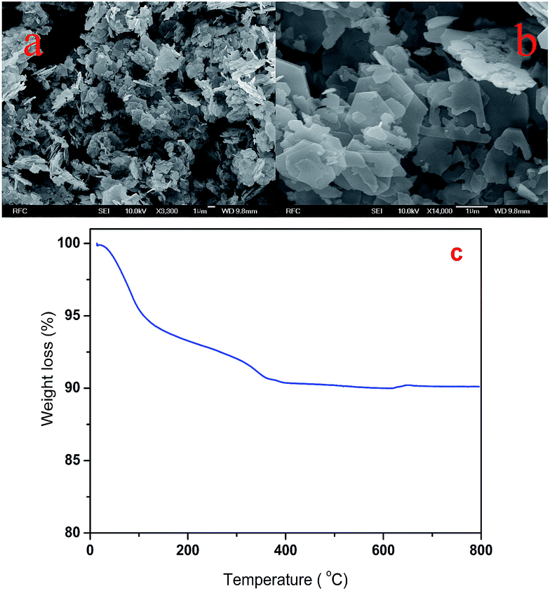 | ||
| Fig. 1 (a and b) FESEM images and (c) TGA curve of precursor sample. | ||
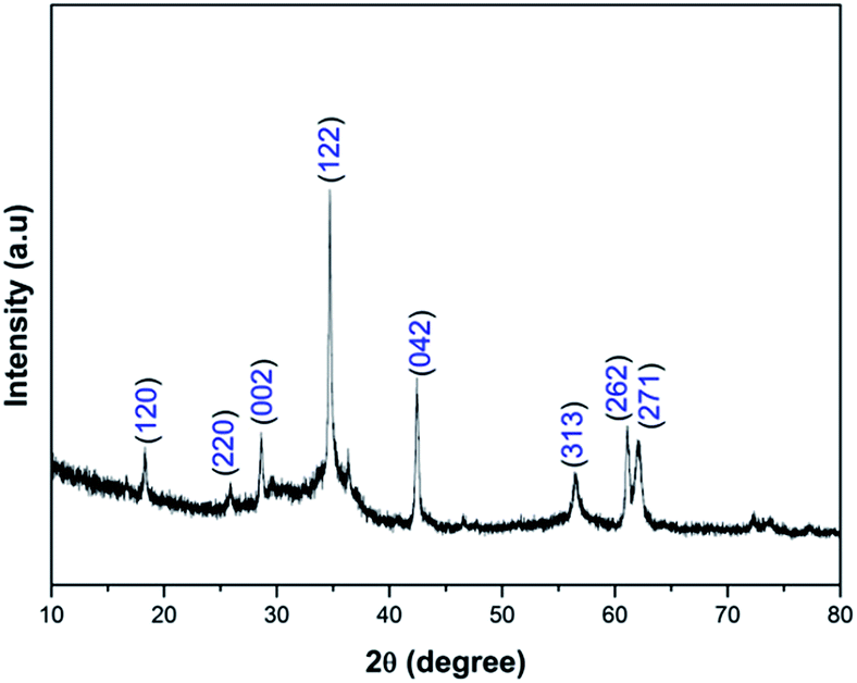 | ||
| Fig. 2 XRD pattern of Zn3V2O8 nanoplatelets. | ||
The purity and oxidation state of Zn3V2O8 nanoplatelets were characterized using X-ray photoelectron spectroscopy (XPS). Fig. 3a shows the complete survey spectrum of Zn3V2O8 nanoplatelets. It shows the presence of V 2p, O 1s, C 1s, and Zn 2p states. Fig. 3b shows the Zn 2p spectrum. The two peaks in the Zn 2p spectrum at 1021.58 and 1044.68 eV correspond to Zn 2p3/2 and Zn 2p1/2, respectively.18 The V 2p spectrum is shown in Fig. 3c. The peak positions at 516.78 (V 2p3/2) and 524.38 eV (V 2p1/2) were attributed to the V5+ state.19Fig. 3d shows the XPS spectrum of O 1s. The O 1s spectrum was deconvoluted into three components (O1, O2, and O3), with binding energies of 529.58, 531.0 and 532.4 eV, respectively. The O1 peak at 529.58 eV was attributed to a metal–oxygen bond, i.e. V–O binding in Zn3V2O8.20 The O2 peak at 531.0 eV was attributed to a large number of defect sites with minimum oxygen coordination and small particle size.21 The O3 component at the higher binding energy of 532.4 eV corresponds to chemically and physically bonded water within the surface.21
 | ||
| Fig. 3 XPS spectra of (a) survey scan, (b) Zn 2p, (c) V 2p, and (d) O 1s for Zn3V2O8 nanoplatelets. | ||
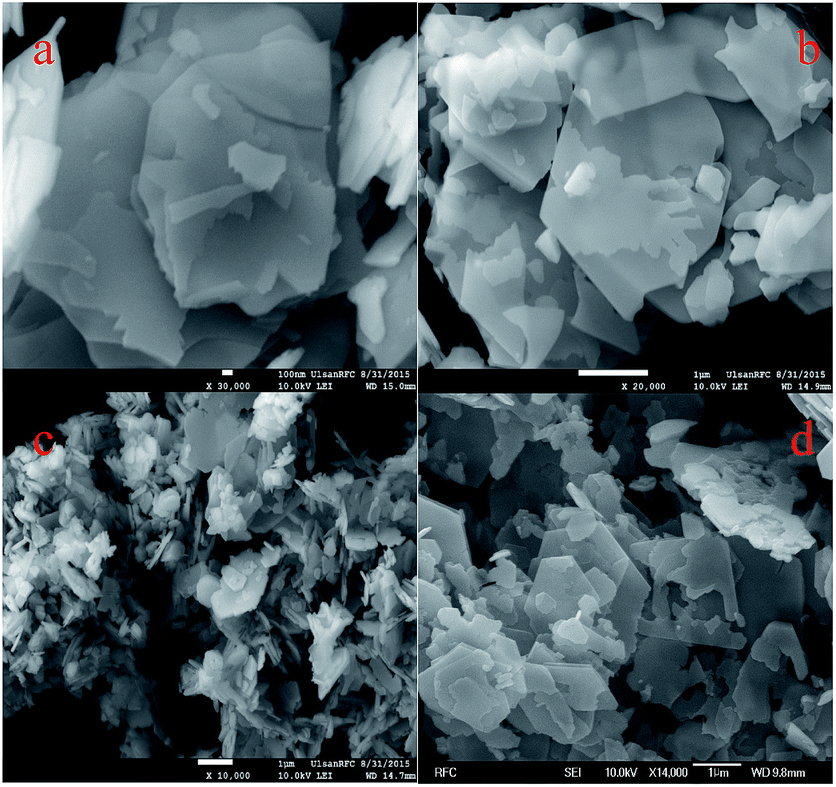
Fig. 4a–c shows FESEM images of Zn3V2O8 nanomaterials at different magnifications. The FESEM images of the prepared Zn3V2O8 nanomaterials show nanoplatelet morphology. Fig. 4a and b clearly show the even distribution of nanoplatelets with a flake-like arrangement. Fig. 4c depicts the nanoplatelet morphology and dimensions. The platelets are arranged with spacing between neighboring nanoplatelets, which offers effective electron and ion transport and enables the improvement of electrochemical performance in batteries and supercapacitors. The length of the nanoplatelets is about 1 μm and their breadth is between 500 nm and 1 μm.
 | ||
| Fig. 4 (a–c) FESEM images of Zn3V2O8 nanoplatelets at different magnifications, (d) AFM image of Zn3V2O8 nanoplatelets, (e–g) HRTEM images of Zn3V2O8 nanoplatelets, and (h) SAED pattern of Zn3V2O8 nanoplatelets. | ||
Fig. 4d shows an AFM image of zinc vanadate nanoplatelets. The thickness of the nanoplatelets measured by AFM is 27.9 nm. The structure of the Zn3V2O8 nanoplatelets was further characterized using HRTEM. Fig. 4e and f shows HRTEM images of Zn3V2O8 nanoplatelets. The TEM images clearly reveal the nanoplatelet arrangement and dimensions. Fig. 4g shows a higher-magnification HRTEM image. The image clearly shows the crystalline structure of the sample. The d-spacing corresponds to the (271) plane of Zn3V2O8. Fig. 4h shows the selected-area electron diffraction (SAED) pattern of Zn3V2O8 nanoplatelets. The SAED pattern can be indexed to Zn3V2O8.
A time-dependent experiment was carried out to explain the formation of nanoplatelets. Fig. 5 shows FESEM images of the precursor sample prepared at different durations of hydrothermal treatment. A mechanism of the formation of the Zn3V2O8 nanoplatelets based on the results of the time-dependent experiment is shown in Fig. 6. The formation of Zn3V2O8 nanoplatelets includes nucleation, growth, splitting and exfoliation processes, as well as self-aggregation. Initially, zinc and vanadium ions reacted to form zinc vanadate nuclei. After nucleation, a growth process started to form a zinc vanadate nanostructure. Herein, the role of SDS was important.
 | ||
| Fig. 5 FESEM images of Zn3V2O8 precursor sample at different durations of hydrothermal treatment: (a) 6 h, (b) 12 h, (c) 18 h, and (d) 24 h. | ||
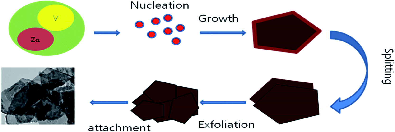 | ||
| Fig. 6 Schematic representation of the formation of Zn3V2O8 nanoplatelets. | ||
SDS acted as a structure-directing agent that controls the surface free energy.22 Therefore, it promoted the growth process. The structure of the final product was dependent upon the hydrothermal temperature, duration of treatment, concentration of the surfactant and pH of the solution. Initially, thick zinc vanadate platelets were formed. As the hydrothermal reaction continues, the platelets split into thin sheets.19
Fig. 5a shows (after 6 h of hydrothermal reaction) thick nanoplates with sheets. These sheets may have formed due to a splitting process. The splitting process continued for up to 12 hours of hydrothermal treatment (Fig. 5b). The sheets were then exfoliated to form thin platelets (Fig. 5c).19 After exfoliation, the sheets were attached together by self-aggregation, forming zinc vanadate nanoplatelets (Fig. 5d).
The Zn3V2O8 nanoplatelets were subjected to BET measurement to determine the details of specific surface area and pore size distribution. Fig. S2† shows the BET isotherm and corresponding pore size distribution curve of Zn3V2O8 nanoplatelets. The BET surface area of the Zn3V2O8 nanoplatelets was 11.53 m2 g−1. The pore size distribution curve for adsorption by the BJH method clearly indicates that most of the pores were distributed in the region of 2–10 nm. The mean pore diameter of the Zn3V2O8 nanoplatelets was 5.9 nm. The large surface area with a porous nature helps to enhance the diffusion of ions, which improves the electrochemical performance in both Li-ion batteries and supercapacitors.23
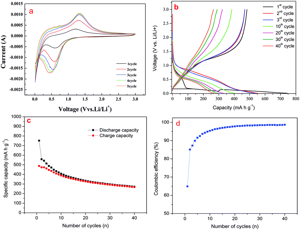
The electrochemical performance of Zn3V2O8 nanoplatelets was studied using cyclic voltammetry and charge–discharge analysis and their potential as the active material for lithium-ion batteries was determined. Fig. 7a shows CV curves of Zn3V2O8 nanoplatelets in the potential range of 0.01 to 3.0 V at a scan rate of 0.1 mV s−1. The cathodic scans of the CV curves show reduction peaks at 0.59 V, which are due to the reduction of zinc (Zn2+ to Zn0) and reduction of vanadium (V5+ to V4+ and further to V3+).16,24 The oxidation peaks at 1.23 V are due to the oxidation of vanadium (V3+ to V5+) and zinc (Zn0 to Zn2+). The charge–discharge measurement of Zn3V2O8 nanoplatelets was carried out using a potential range of 0.01 to 3.0 V at a current density of 100 mA g−1. Fig. 7b shows the charge–discharge profile of Zn3V2O8 nanoplatelets at cycles 1, 2, 3, 10, 20, 30 and 40. The discharge curve of the first cycle is different from those of the subsequent cycles. It shows a plateau at about 1.1 to 0.19 V. The capacity of the first-cycle plateau at less than 0.19 V is due to a slow process of lithium storage into orthorhombic-type Zn3V2O8, and the plateau at higher voltage can be attributed to the faradic nature of the material.16
 | ||
| Fig. 7 (a) CV curves of Zn3V2O8 at a scan rate of 0.1 mV s−1, (b) charge–discharge curve of Zn3V2O8 nanoplatelets, (c) curve of specific capacity against cycle number and (d) curve of coulombic efficiency against number of cycles. | ||
The initial discharge and charge capacities were 752 and 488 mA h g−1, respectively. The irreversible capacity loss in the first cycle was 35.1%. After the first cycle, the electrochemical process was highly reversible. The discharge and charge capacities in the second cycle decreased to 558 and 474 mA h g−1, respectively, and the capacity loss in the second cycle was only 15%. The irreversible capacity loss in the first cycle may be attributed to the loss of lithium due to the formation of a solid electrolyte interphase (SEI) layer.24Fig. 7c and d show the cycling stability curve and coulombic efficiency of Zn3V2O8 nanoplatelets. The specific capacity decreased from the first cycle to the second cycle and further large decreases in specific capacity were observed up to the 10th cycle. Thereafter, the specific capacity decreased linearly. The coulombic efficiency of Zn3V2O8 nanoplatelets increased from the first cycle (64.9%) to the second cycle (85.1%), rising to greater than 95% after the 10th cycle and greater than 99% at the 40th cycle. Zhang et al.15 synthesized Zn3(VO4)2 microspheres as an anode and reported first and second discharge capacities of 1180 and 676 mA h g−1, respectively, at a current density of 20 mA g−1. Although the discharge capacities were high, the current density was very low. Gan et al.16 reported a first discharge capacity of 1522 mA h g−1 and excellent cycling stability for Zn3V2O8 material. However, they used 30% conductive material, which likely resulted in a low specific volumetric capacity.
The electrochemical supercapacitor performance of the Zn3V2O8 nanoplatelets was studied using cyclic voltammetry and galvanostatic charge–discharge studies. The CV study was performed within the potential window of 0–0.5 V at different scan rates. Fig. 8a shows the CV curves of the Zn3V2O8 nanoplatelet electrode at scan rates of 5, 10, 25, and 50 mV s−1. The shapes of the CV curves do not reveal electric double-layer capacitance, which indicates that the capacitance was mainly due to pseudocapacitive behavior.25 The CV curves show a pair of redox peaks at about 0.37 and 0.25 V. These redox peaks may originate from the intercalation and de-intercalation of K+ from the electrolyte into zinc oxide.26 Fig. S3† shows the curve of scan rate versus peak current. It is interesting to note that the variation of peak current with scan rate is linear. This linearity of peak current with scan rate confirms that the electrochemical reaction was a surface redox reaction that led to pseudocapacitive behavior of the electrode.27 The specific capacitance Cc (F g−1) was calculated using the following formula:28
 | (1) |
 | ||
| Fig. 8 (a) CV curve of Zn3V2O8 nanoplatelets, (b) curve of specific capacitance versus scan rate, (c) charge–discharge curve of Zn3V2O8 nanoplatelets, and (d) curve of specific capacitance versus specific current. | ||
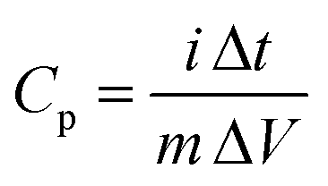
The supercapacitor performance was further evaluated for its specific capacitance and cycling stability using charge–discharge analysis. Fig. 8c shows the discharge curves of the Zn3V2O8 electrode at different specific currents: 2, 4, 6, and 8 A g−1. The discharge profiles reveal nonlinear rather than linear behavior of the electric double-layer capacitance, which suggests that the capacitance was mainly due to pseudocapacitance.30 The specific capacitance Cp (F g−1) was calculated using the following formula:31
 | (2) |
Long-term cycling stability is important for practical applications. The long-term cycling stability of the Zn3V2O8 electrode was tested over 2000 continuous charge–discharge cycles at a specific current of 8 A g−1. Fig. 9 shows the cycling stability curve for 2000 cycles. The inset of Fig. 9 shows the charge–discharge curve of the first and last five cycles (cycles 1–5 and 1996–2000). The specific capacitance increased from 100% to 120% after 15 cycles, decreasing slowly thereafter. After 2000 cycles, about 98% of the initial specific capacitance was retained. The Zn3V2O8 nanoplatelet electrode exhibited good specific capacitance with high cycling stability. This appreciable performance is mainly due to the crystal structure and morphology of the Zn3V2O8 nanoplatelets. The crystal structure of Zn3V2O8 is similar to that of Ni3V2O8. Three types of O atom constitute the tetrahedron and octahedron. V is located in the center of the tetrahedron and Zn is located in the center of the octahedron.16 The gap between the octahedron and the tetrahedron, as well as channels of the supercell, allows more effective electron and ion transport, which leads to an improved electrochemical performance.
 | ||
| Fig. 9 Curve of cycling stability against number of cycles. (Inset shows the initial and final five cycles of charge–discharge.) | ||
Conclusion
In conclusion, Zn3V2O8 nanoplatelets were synthesized by a hydrothermal method using SDS as a surfactant. SDS played an important role in the control of morphology, acting as a structure-directing agent that promoted the growth process to form zinc vanadate platelets. FESEM and HRTEM revealed the morphology of the nanoplatelets. Battery studies revealed a high discharge capacity at the second cycle (558 mA h g−1). The coulombic efficiency was about 99% at the 40th cycle. Based on CV and charge–discharge measurements, it was shown that the Zn3V2O8 nanoplatelets exhibited a maximum specific capacitance of 302 F g−1 (5 mV s−1) and 207 F g−1 (2 A g−1). The high specific capacitance with excellent cycling stability confirms the suitability of Zn3V2O8 nanoplatelets for supercapacitor applications.Acknowledgements
This study was supported by the Priority Research Centers Program through the National Research Foundation of Korea (NRF), funded by the Ministry of Education, Science and Technology (MEST) of the Korean government (2009-0093818).Notes and references
- P. Simon and Y. Gogotsi, Nat. Mater., 2008, 7, 845–854 CrossRef CAS PubMed.
- C. Yuan, H. B. Wu, Y. Xie and X. W. Lou, Angew. Chem., Int. Ed., 2014, 53, 1488–1504 CrossRef CAS PubMed.
- X. Zhao, B. M. Sanchez, P. J. Dobson and P. S. Grant, Nanoscale, 2011, 3, 839–855 RSC.
- D. P. Dubal, P. Gomez-romero, B. R. Sankapal and R. Holze, Nano Energy, 2015, 11, 377–399 CrossRef CAS PubMed.
- S. G. Mohamed, C.-J. Chen, C. K. Chen, S.-F. Hu and R.-S. Liu, ACS Appl. Mater. Interfaces, 2014, 6, 22701–22708 CAS.
- W.-B. Zhang, L.-B. Kong, X.-J. Ma, Y.-C. Luo and L. Kang, RSC Adv., 2014, 4, 41772–41777 RSC.
- Y. Zhang, Y. Liu, J. Chen, Q. Guo, T. Wang and H. Pang, Sci. Rep., 2014, 4, 5687 CAS.
- F. Cheng and J. Chen, J. Mater. Chem., 2011, 21, 9841–9848 RSC.
- S. Zhang, R. Hu, L. Liu and D. Wang, Mater. Lett., 2014, 124, 57–60 CrossRef CAS PubMed.
- L. Xiao, Y. Zhao, J. Yin and L. Zhang, Chem.–Eur. J., 2009, 15, 9442–9450 CrossRef CAS PubMed.
- L. Zeng, F. Xiao, J. Wang, S. Gao, X. Ding and M. Wei, J. Mater. Chem., 2012, 22, 14284–14288 RSC.
- C. Zheng, L. Zeng, M. Wang, H. Zheng and M. Wei, CrystEngComm, 2014, 16, 10309–10313 RSC.
- S. S. Pitale, M. Gohain, I. M. Nagpure, O. M. Ntwaeaborwa, B. C. B. Bezuidenhoudt and H. C. Swart, Phys. B, 2012, 407, 1485–1488 CrossRef CAS PubMed.
- R. Shi, Y. Wang, F. Zhou and Y. Zhu, J. Mater. Chem., 2011, 21, 6313–6320 RSC.
- S. Y. Zhang, X. Xiao, M. Lu and Z. Q. Li, J. Mater. Sci., 2013, 48, 3679–3685 CrossRef CAS.
- L.-H. Gan, D. Deng, Y. Zhang, G. Li, X. Wang, L. Jiang and C.-R. Wang, J. Mater. Chem. A, 2014, 2, 2461–2466 CAS.
- S. Zhang, N. Lei, W. Ma, Z. Zhang, Z. Sun and Y. Wang, Mater. Lett., 2014, 129, 91–94 CrossRef CAS PubMed.
- P. Yang, X. Xiao, Y. Li, Y. Ding, P. Qiang, X. Tan, W. Mai, Z. Lin, W. Wu, T. Li, H. Jin, P. Liu, J. Zhou, C. P. Wong and Z. L. Wang, ACS Nano, 2013, 7, 2617–2626 CrossRef CAS PubMed.
- G. Yang, H. Cui, G. Yang and C. Wang, ACS Nano, 2014, 8, 4474–4487 CrossRef CAS PubMed.
- L. Jiang, Y. Qu, Z. Ren, P. Yu, D. Zhao, W. Zhou, L. Wang and H. Fu, ACS Appl. Mater. Interfaces, 2015, 7, 1595–1601 CAS.
- X.-F. Lu, D.-J. Wu, R.-Z. Li, Q. Li, S.-H. Ye, Y. X. Tong and G.-R. Li, J. Mater. Chem. A, 2014, 2, 4706–4713 CAS.
- L. Z. Pei, N. Lin, T. Wei, H. D. Liu and H. Y. Yu, J. Alloys Compd., 2015, 631, 90–98 CrossRef CAS PubMed.
- F. Wu, C. Yu, W. Liu, T. Wang, J. Feng and S. Xiong, J. Mater. Chem. A, 2015, 3, 16728–16736 CAS.
- S. Zhang, W. An and G. Wu, Chem. Phys. Lett., 2015, 621, 1–4 CrossRef CAS PubMed.
- K. K. Purushothaman, I. M. Babu, B. Sethuraman and G. Muralidharan, ACS Appl. Mater. Interfaces, 2013, 5, 10767–10773 CAS.
- X. Dong, Y. Cao, J. Wang, M. B. Chan-park, L. Wang, W. Huang and P. Chen, RSC Adv., 2012, 2, 4364–4369 RSC.
- S. Vijayakumar, A. K. Ponnalagi, S. Nagamuthu and G. Muralidharan, Electrochim. Acta, 2013, 106, 500–505 CrossRef CAS PubMed.
- J. W. Lee, A. S. Hall, J.-D. Kim and T. E. Mallouk, Chem. Mater., 2012, 24, 1158–1164 CrossRef CAS.
- P. Justin, S. K. Meher and G. R. Rao, J. Phys. Chem. C, 2010, 114, 5203–5210 CAS.
- S. Vijayakumar, S. Nagamuthu and G. Muralidharan, ACS Sustainable Chem. Eng., 2013, 1, 1110–1118 CrossRef CAS.
- S. Nagamuthu, S. Vijayakumar and G. Muralidharan, Energy Fuels, 2013, 27, 3508–3515 CrossRef CAS.
- P. Syedvali, G. Rajeshkhanna, E. Umesh babu, G. U. kiran, G. R. Rao and P. Justin, RSC Adv., 2015, 5, 38407–38416 RSC.
- S. Shi, X. Zhuang, B. Cheng and X. Wang, J. Mater. Chem. A, 2013, 1, 13779–13788 CAS.
- F. K. Butt, M. Tahir, C. Cao, F. Idress, R. Ahmed, W. S. Khan, Z. Ali, N. Mahmood, M. Tanveer, A. Mahmood and I. Aslam, ACS Appl. Mater. Interfaces, 2014, 6, 13635–13641 CAS.
- M. Shahid, J. Liu, Z. Ali, I. Shakir and M. F. Warsi, J. Power Sources, 2013, 230, 277–281 CrossRef CAS PubMed.
Footnote |
| † Electronic supplementary information (ESI) available: XRD pattern of Zn3V2O8 precursor sample, BET isotherm of Zn3V2O8 and scan rate versus peak current curve. See DOI: 10.1039/c5ra13904j |
| This journal is © The Royal Society of Chemistry 2015 |