Enhanced corneal permeation of coumarin-6 using nanoliposomes containing dipotassium glycyrrhizinate: in vitro mechanism and in vivo permeation evaluation

Chuanlong Guo, Fenghua Cui, Mengshuang Li, Fengjie Li and Xianggen Wu*
State Key Laboratory Cultivation Base, Shandong Provincial Key Laboratory of Ophthalmology, Shandong Eye Institute, Shandong Academy of Medical Sciences, Qingdao 266071, China. E-mail: wuxianggen@126.com

Received 14th July 2015 , Accepted 1st September 2015

First published on 1st September 2015


Abstract

The objective of the present study was to investigate the elasticity of the lipid bilayer of nanoliposomes regarding in vitro cellular uptake/mechanics and in vivo corneal permeation through ocular topical routes. Flexible nanoliposomes, using dipotassium glycyrrhizinate as an edge activator, and their physical properties, membrane elasticity, cellular uptake characterizations and mechanisms, as well as in vivo corneal permeation using rabbits and mice as experimental animals, were investigated and compared with the conventional liposomal formulation composed of soybean phosphatidylcholine and cholesterol. Flexible nanoliposomes required less energy to prepare and had elastic lipid membranes. Compared with nanoliposomes, flexible nanoliposomes showed a significantly higher cellular uptake of coumarin-6. Moreover and interestingly, the flexible nanoliposomes showed different cellular uptake mechanisms in cells. Flexible nanoliposomes also showed significantly higher corneal penetrating ability in in vivo testing. Therefore, the fluidity of the liposomal membrane differently affected cellular uptake/internalization and in vivo corneal penetration of the nanoliposomes, and flexible nanoliposomes might be a promising therapeutic tool for the treatment of ocular surface disorders.
Introduction

Topical instillation represents the most convenient route for ocular drug delivery, and the majority (nearly 90%) of ocular diseases are treated with topical application of drug solutions (i.e., eyedrops), particularly to the anterior segment of the eye. However, this route is impeded by poor ocular bioavailability (<5%), mainly attributed to the low corneal permeability of drugs, tear turnover, and drug elimination via the conjunctiva and sclera. Efforts to enhance ocular bioavailability from topical instillations have been accomplished using novel formulations, such as hydrogels, polymeric micelles, nanosuspensions, and lipid-based nanocarriers.1

Among these novel formulations, liposomes, particularly nanoliposomes, have been widely evaluated as ocular drug delivery systems to enhance the absorption of therapeutic drugs, improve bioavailability, reduce systemic side effects, and sustain intraocular drug levels. However, some conflicting results on corneal permeation and efficacy have been reported. For example, Morimoto K et al. reported that liposomes did not increase the corneal permeabilities of 6-carboxyfluorescein, FITC-dextran or rhodamine B,2 and Taniguchi K et al. revealed that the corneal permeability of dexamethasone and dexamethasone valerate was not affected by liposomes.3 To pursue enhanced corneal permeation and efficacy, some novel liposomes, such as cationic liposome,4 N-trimethyl chitosan (TMC)-coated liposomes,5 and in situ thermosensitive liposomal hydrogel,6 have been investigated.

Conventional liposomes are composed mainly of phospholipids and cholesterol. Cholesterol usually acts as a stabilizer for liposomal formulations, and the hardness of liposomes increases with increasing cholesterol content when unsaturated phospholipid was used as a lipid component of liposomes.7 The fluidity of the lipid bilayer of liposomes could affect penetration ability, but reports of effectiveness between cholesterol and the permeation of ocular topical liposomes have been controversial. For example, a recent report from Shafaa MW et al. revealed that the cholesterol molar ratio in prepared liposomal formulations served to decrease the permeability of the lipid bilayer, manifested by a low rate of drug release, an increased percentage of entrapment efficiency and consequently lower bioavailability.8 In contrast, Inokuchi Y et al. reported that liposome rigidity seemed to be necessary to maintain the liposomal structure and entrapment of substances under the ocular surface biological conditions.7 Over the past decade, flexible liposomes, as a new class of liposome, have been developed with the character of more fluidity of the lipid bilayer, compared to that of the conventional liposomes. Flexible liposomes, also known as deformable liposomes, elastic liposomes, or transfersomes, are liposomes that contain edge activators, including bile salts,9 polysorbates, or sorbitan esters. These edge activators destabilize the lipid bilayer of the liposomes and increase the flexibility of the liposomes. Some reports from different medical fields have shown that flexible liposomes were more effective than rigid liposomes, such as conventional liposomes.10–12

Because liposome rigidity might affect the corneal permeation of liposomal formulations, and the effects of flexible liposome elasticity on ocular topical delivery have not been yet studied, the aim of the present study was to investigate the elasticity of the lipid bilayer of liposomes regarding in vitro cellular uptake/mechanics and in vivo corneal permeation via an ocular topical route. For this purpose, coumarin-6 (cou-6), a fluorescence dye used as a hydrophobic model compound, was incorporated into flexible nanoliposomes using soybean phosphatidylcholine (SPC) and dipotassium glycyrrhizinate (DG) (as an edge activator), and the physical properties, membrane elasticity, cellular uptake characterizations and mechanisms, as well as its in vivo corneal permeation using rabbits and mice as experimental animals, of cou-6 were investigated and compared with the conventional liposomal formulation composed of SPC and cholesterol, to elucidate how nanoliposome rigidity affected delivery efficiency and corneal permeation after eyedrop administration.

Materials and methods

Materials and animals

Chemical reagents. DG was kindly donated by Tianshan Pharmaceutical Industrial Co., Ltd (Xinjiang, China), and it was used as received. SPC was purchased from Lipoid GmbH (Ludwigshafen, German). Cholesterol was kindly provided by Shanghai Advanced Vehicle Technology Pharmaceutical Ltd (Shanghai, China). Cou-6 and glucose were purchased from Sigma-Aldrich, Co., (St. Louis, MO, USA). Benzalkonium bromide solution (5%, Jiangxi Jingdong Pharmaceutical Ltd [Jiangxi, China]) was used as the original solution and was diluted to the test concentration with cell culture media in 72 h cytotoxicity testing, and it was diluted to the test concentration with phosphate buffered salines (PBS) in formulation cytotoxicity testing.
Animals. New Zealand white rabbits were obtained from Qingdao Kangda Foodstuffs Co., Ltd, (Qingdao, Shandong, China) (License No. SCXK [Lu] 20070023). BALB/c mice were purchased from Beijing HFK Bioscience Co., Ltd (Beijing, China) (License No. SCXK [Jing] 2014-0004). All of the animals were healthy and free of clinically observable ocular abnormalities. The animal care and procedures were conducted according to the Principles of Laboratory Animal Care. The use of animals in this study adhered to the ARVO Statement for the Use of Animals in Ophthalmic and Vision Research, and the animal study was approved by the Shandong Eye Institute Ethics Committee for Animal Experimentation in Qingdao, Shandong, China.

Preparation of cou-6 nanoliposomes and flexible nanoliposomes

The flexible liposomal formulation was prepared by an improved dry-film dispersing method (Lu et al., 2007). Cou-6 (0.5 mg) and SPC (150 mg) were first dissolved in absolute ethyl alcohol in a 50 mL round-bottom flask. The alcohol was then removed using a rotary evaporator (Huxi RE-52-3 rotatory evaporator, Shanghai, China) under reduced pressure, and the final traces of alcohol was removed under a vacuum overnight. A thin film of SPC formed on the wall of the flask. Ten milliliters of PBS containing 85.6 mg of DG (SPC[thin space (1/6-em)]:[thin space (1/6-em)]DG = 2[thin space (1/6-em)]:[thin space (1/6-em)]1, mole ratio) were added to the lipid film and rotated for 30 min in a water bath at 40 °C to obtain a crude dispersion of liposomes. The flexible liposome dispersion was then sonicated for 5 min by intermittent probe sonication (SCIENTZ-IID, Ningbo Scientz Biotechnology Co., Ltd, Zhejiang, China) with a procedure of 5% amplitude and 2 s/2 s of sonications/interval under cooling in an ice-water bath, to obtain an opalescent dispersion of flexible nanoliposomes. After sterile filtration with a 0.22 μm filter (Merck Millipore Ltd, IRL),13 the flexible nanoliposome dispersion was stored at 4 °C for further analysis. For the preparation of the conventional liposomal formulation, similar procedures were conducted. Briefly, cou-6 (0.5 mg) and SPC (150 mg) were dissolved in absolute ethyl alcohol, and 37.6 mg of cholesterol (SPC[thin space (1/6-em)]:[thin space (1/6-em)]cholesterol = 2[thin space (1/6-em)]:[thin space (1/6-em)]1, mole ratio) were dissolved in methylene dichloride; then, these two solutions were mixed and evaporated to remove the solvent completely and to form a thin-film layer on the wall of the flask. Blank PBS was added to hydrate the thin film and obtain a crude dispersion of liposomes; then, the liposome dispersion was sonicated for 5 min by intermittent probe sonication under cooling in an ice-water bath with a procedure of 5% amplitude and 2 s/2 s of sonications/interval. An additional sonication, at 15 min of 30% amplitude and 2 s/2 s of sonications/interval, was needed to obtain an opalescent dispersion of nanoliposomes with diameters similar to flexible nanoliposomes. The osmotic pressure of both formulations was measured and was adjusted to be in the range of 290–320 mOsmol per kg (STY-1E Osmometer, Tianjin, China) with glucose, and the pH was adjusted to be in the range of 6.5–6.7 (MODEL 828, Orion, USA).

The entrapment efficiency of cou-6-loaded nanoliposomes and flexible nanoliposomes was determined using a high-performance liquid chromatographic (HPLC) method. Briefly, 0.1 mL of cou-6-loaded nanoliposomes or flexible nanoliposomes was dissolved in 10 mL of methanol and was centrifuged at 12[thin space (1/6-em)]000 rpm for 10 min. The cou-6 content in the supernatant was measured by HPLC. The encapsulation efficiency was expressed as the ratio of detected and added cou-6 amount.14 The HPLC system was fitted with a G1321A FLD Detector (detection at excitation-emission wavelengths of 465/502 nm, Agilent, US) and a G1311A Quat Pump (Agilent, US). Reverse-phase Agilent C18 columns (250 mm × 4.60 mm, 5 μm, Agilent, US) were used for sample separation. The elution of cou-6 consisted of 90% methanol and 10% water. The flow rate was kept constant at 1.0 mL min−1. The detection was performed at 60 °C. The retention time of cou-6 was 5.7 min.

Size analysis and the zeta potential

The mean particle size of the liposomal and flexible liposomal formulations was determined by photo-correlation spectroscopy, using an OP-90S nanoparticle sizer (Optek Instrument, Inc. [Zibo, Shandong, China]), and the zeta potential was determined by photo-correlation spectroscopy, using a Zetasizer (Malvern Nano-ZS90, UK), using the original formulation without any dilution.

Morphological characterization

Nanoliposomes and flexible nanoliposomes were observed and photographed with transmission electron microscopy (TEM) (JEM-1200EX, JEOL Ltd, Tokyo, Japan). The samples were stained with an aqueous solution of phosphotungstic acid (1%, w/v) for approximately 2 min. Then, a drop of each sample was dipped onto a carbon-coated copper grid, and the excess solution was absorbed using filter paper. The grid was allowed to air dry thoroughly, and the sample was observed and imaged.

Measurement of elasticity

The elasticity of the prepared vesicle bilayer was measured by the extrusion method as reported earlier, with minor modifications.15 Briefly, the vesicle carriers were extruded for 5 min with approximately 5 kg of pressure through a 20 nm pore size membrane filter (Anotop 25 Plus 0.02 μm, Whatman GmbH, Germany) equipped with a 5 mL syringe (BD Company, Becton Dickinson S.A., Spain). The elasticity of the vesicles was evaluated by particle size, particle polydispersity index, volume filtered per filter, and the percentage of cou-6 in the solution after 20 nm filtering.

Cell culture tests

A human corneal epithelial cell line (HCECs) (ATCC CRL-11135, kindly donated by Prof. Chonn-Ki Joo, the Catholic University of Korea) was used in this study. Briefly, the HCECs were grown at 37 °C and were humidified in a 5% CO2/95% air atmosphere in a culture medium of D-MEM/F-12 Dulbecco's modified eagle medium supplemented with 10% (v/v) fetal bovine serum. The culture medium was replaced every other day. The cells were subcultured after 3–4 days (subculture ratio, 1[thin space (1/6-em)]:[thin space (1/6-em)]3) with 0.25% trypsin containing 0.02% EDTA.
In vitro cytotoxicity testing. The cytotoxicity of the DG was tested on HCECs with standard 3-(4,5-dimethylthiazol-2-yl)-2,5-diphenyltetrazolium bromide (MTT) testing. The concentrations of the DG were 0.313, 0.625, 1.25, 2.5, 5, 10 and 20 mg mL−1, and the incubation time was 24 h or 72 h. Benzalkonium bromide, a preservative widely used in China in ophthalmic solutions was also tested in 72 h incubation as controls with concentrations of 0.01, 0.1, 1, and 10 μg mL−1. The MTT transformed crystals were dissolved in DMSO, and their absorbance at 490 nm was measured using a microplate reader (Molecular Devices, Sunnyvale, CA, USA). Experiments were performed in triplicate on six wells for each measurement.

Regarding the cytotoxicity of liposomal and flexible liposomal formulations, because eyedrops are rapidly cleared from the surface of the eye, it was assumed that a 1 h incubation time would be sufficient to observe any toxic effects.16 The cells were incubated for 1 h, followed by 4 hours of incubation with MTT; then, benzalkonium bromide were also used as controls with concentrations of 10 and 100 μg mL−1.

In vitro HCECs uptake and mechanical characters. Uptake studies were conducted according to standard protocols, with minor modifications.17 Briefly, after the cells were grown to confluence as determined by light microscopy, the medium was aspirated, and the cells were rinsed with Dulbecco's phosphate buffered salines (DPBS) (composition: 130 mM NaCl, 7.5 mM Na2HPO4, 1.5 mM KH2PO4, 0.5 mM MgSO4, 1 mM CaCl2, 0.03 mM KCl, and 5 mM glucose at pH 7.4) at 37 °C and then were equilibrated in 1 mL of DPBS for 30 min at 37 °C for all of the tests. Time-dependent accumulation was determined. Since the contact of topically applied ophthalmic drops with epithelial cells of the ocular surface is limited to a rather short time of 2–5 minutes, the incubation time was restricted.18 Because cou-6 was insoluble in the PBS solution, so it was firstly dissolved in DMSO (the final concentration of DMSO in the incubation solution was 0.1%) and then was diluted with PBS to the test concentration of 50 μg mL−1, and this solution was used as the cou-6 free solution group (control group) in the uptake test. At the end of the experiment, to each time point, the drug solution was removed, and the cells were rinsed three times with 2 mL of ice-cold stop solution (210 mM KCl, 2 mM HEPES at pH 7.4) to terminate uptake. The cells were then solubilized in 1 mL of lysis solution (0.3 M NaOH, 0.1% Triton X-100) overnight at room temperature. The lysate was transferred to a 96-well plate and was assayed using a 96-well fluorescent microplate reader. Cou-6 fluorescence was measured at excitation-emission wavelengths of 465/502 nm. The fluorescence of the cell lysate was corrected for the autofluorescence of untreated cells. The uptake was normalized to the protein content of the cells, which was measured by the Bradford assay method (Beyotime BCA Protein Assay Kit, Beyotime Institute of Biotechnology), with bovine serum albumin used as the standard. The results are reported as the mean fluorescence intensity per microgram of protein.

To investigate the endocytosis pathway, 5 × 10⁵ cells were seeded in 12-well plates and incubated for 24 h prior. Thus, after 24 hours of incubation, the cells were pre-incubated for 30 min with the different inhibitors at the concentrations listed in Table 2. After this pre-incubation, the liposomal or flexible liposomal formulation was added and incubated for an additional 1 h. Negative controls, i.e., cells without the presence of inhibitors and/or the liposomal or flexible liposomal formulation, were also performed. To determine whether the uptake procedure was energy-dependent or not, the uptake was also performed at 4 °C or NaN3. After incubation, the medium was removed, and the cells were rinsed three times with ice-cold stop solution to terminate uptake and to ensure nanoliposome removal from the outer cell membrane. The cells were then digested from the plate and collected to form single cell suspension. Then, the mean intracellular fluorescence intensity was measured by flow cytometry system (FCS) with excitation of 488 nm. The results are reported as the means of the distributions of cell fluorescence intensity obtained by measuring ∼10[thin space (1/6-em)]000 cells, averaged between 3 independent replications of 3 independent experiments. Error bars indicate the standard deviations among these independent experiments.

Table 1 The parameters of the nanoliposomes and flexible nanoliposomes (both of the formulations contained 50 μg mL−1 cou-6 and 15 mg mL−1 SPC, n = 3)
Formulation Entrapment efficacy (%) Particle size (nm) Polydispersity index Zeta potential (mV) Elasticity evaluation
Particle size (nm) after 20 nm filtration Polydispersity index after 20 nm filtration Volume filtered (mL) Percentage of cou-6 in the solution after 20 nm filtration
a Note: the volume was not sufficient for detection.
Nanoliposomes 98.73 ± 0.25 107 ± 4 0.278 ± 0.014 −23.20 ± 4.60 NDa NDa <0.20 44.88 ± 4.52%
Flexible nanoliposomes 98.82 ± 0.23 99 ± 5 0.264 ± 0.007 −34.07 ± 1.71 65 ± 5 0.225 ± 0.080 6.50 ± 0.97 96.16 ± 6.21%


Table 2 Inhibitors and their concentrations used in the mechanism study
  Concentration Effect
Hypertonic sucrose 0.45 M Inhibitor of clathrin-mediated endocytosis by the K+ depletion effect
Chlorpromazine 6 μg mL−1 Specific inhibitor of clathrin-mediated endocytosis
Chloroquine 125 μM Disrupts endosomes and lysosomes, prevents endosome acidification and causes swelling to endosomes and lysosomes
Indomethacin 100 μM Inhibitor of caveolar-mediated endocytosis
NaN3 0.10% General inhibitor of endocytic processes
Nystatin 10 μg mL−1 Inhibitor of lipid raft/caveola-dependent endocytosis by the cholesterol sequestration effect
Methyl-β-cyclodextrin (MβCD) 10 mM Cholesterol depletion agent, effective inhibitor of lipid raft/caveola-dependent endocytosis
Phloridizin 200 μM Nontransportable competitive inhibitor, sodium glucose cotransporter inhibitor
Heparin 100 μg mL−1 Specific inhibitor of heparin sulfate proteoglycans (HSPGs)
Amiloride 10 μM Specific inhibitor of macropinocytosis


To visualize the different intracellular distribution characteristics of cou-6/nanoliposomes or cou-6/flexible nanoliposomes, HCECs were seeded in a sterile glass-bottom dish and were incubated with 1 μM DiI at 37 °C for 1 h to label the lipid membranes. After washing with PBS, the HCECs were incubated with the liposomal or flexible liposomal formulation for 5 min, 30 min and 60 min, respectively. Finally, fluorescence images were obtained using confocal laser scanning microscopy (CLSM), and the excitation wavelength for cou-6/nanoliposome or cou-6/flexible nanoliposome detection was set at 488 nm and adjusted to 561 nm for lipid membranes.

In vivo permeation testing

Corneal penetration was performed both in rabbits and mice. The rabbits were divided into two groups as follows: one group received eyedrops of the flexible liposomal formulation, and the other received eyedrops of the liposomal formulation. Four instillations of 50 μL of the eyedrops were administered to both eyes of the animals at 10 min intervals. At 0.5, 1 and 2 h following the last instillation of the formulation, the rabbits were sacrificed with a sodium pentobarbital overdose (four rabbits for each formulation and time point analyzed). Then, the ocular surface was rinsed with normal saline and dried with filter paper to remove tear fluid. After approximately 100 μL of aqueous humor was aspirated from the anterior chamber using a 29-gauge needle, corneas were excised. The aqueous humor samples were mixed with equal volumes of methanol, followed by centrifugation, and the resulting supernatant was analyzed by HPLC. For each time point, six corneas were weighed, suspended (50 mg of cornea per 1 mL of methanol) in methanol, and homogenized. Samples of corneal tissue were stored at −80 °C. For analysis, 500 μL of each sample were centrifuged, and the supernatant was analyzed by HPLC. Another two corneas were fixed in 4% paraformaldehyde at 4 °C for 3 h and then observed with the CLSM.

In the mouse tests, the test procedure was similar to with the rabbits but with five mice and ten corneas for each formulation and time point analyzed (eight corneas for cou-6 determination and two for CLSM observation), and 5 μL of the eyedrops were administered each time.

In vivo ocular irritation tests

Ocular tolerance was tested using 8.56 mg mL−1 DG in PBS, flexible liposomal formulation and liposomal formulation, with blank PBS as control formulations. Each formulation was studied in 6 rabbits. Each formulation was instilled in the right eye for 30 minutes for a total of 13 times, leaving the left eye untouched as a control. Clinical signs were evaluated before the test and at 1, 6, and 24 h after the last instillation. The degree of eye irritation was scored using the modified Draize test.20–22 Irritation was classified as one of four grades: practically non-irritating, score 0–3; slightly irritating, score 4–8; moderately irritating, score 9–12; and severely irritating (or corrosive), score 13–16.23

Anti-inflammatory effect

The anti-inflammatory efficacy was tested using 8.56 mg mL−1 DG in PBS, flexible liposomal formulation and liposomal formulation, with blank PBS, dexamethasone sodium phosphate (DSP) eye drops (5 mL[thin space (1/6-em)]:[thin space (1/6-em)]1.25 mg, Xinxiang Huaqing Pharmaceutical industry Co., Ltd) and pranoprofen eye drops (5 mL[thin space (1/6-em)]:[thin space (1/6-em)]5 mg, Senju Pharmaceutical Co., Ltd) as control formulations. This test was performed in rabbits by instillation of a single dose of 50 μL of each formulation in the conjunctival sac of the left eye. The contralateral eye was used as untreated control. Each formulation was tested in 5 rabbits. After 30 min, 50 μL of 0.5% (w/v) sodium arachidonate a solution (SAS) in PBS (pH 7.4) was instilled in the left eye.19,20 Inflammation was quantified 30, 60, 90, 120, 180, 240, and 360 min after instillation of SAS. Ocular changes were graded according the scores as described previously.19,20

Data analysis

The data were analyzed using SPSS software, version 11.5. MTT tests were performed using ANOVA with multiple comparisons, and comparisons of cou-6 in corneas between the liposomal and flexible liposomal formulation groups were determined using the independent-samples t test. For all of the evaluations, a P-value less than 0.05 was considered significant.

Results

Formulation and characterization of nanoliposomes and flexible nanoliposomes

Both the liposomal and flexible liposomal formulations were transparent and slightly opalescent with green fluorescence compared to water, and the detail parameters are listed in Table 1. Further, both the nanoliposomes and flexible nanoliposomes had high and consistent encapsulation efficiency. TEM analysis showed that both the nanoliposomes and flexible nanoliposomes were small unilamellar vesicles with a spherical and homogenous appearance, and no aggregates were presented (Fig. 1 shows the appearance of these two formulations). No differences were observed between these two kinds of nanoliposomes in terms of morphology when observed under low magnification (×25K), whereas the flexible nanoliposomes showed shrinkage rims under high magnification (×150K), but this observation was not observed to the nanoliposomes, and their rims were smooth. The elasticity evaluation was consistent with the phenomenon observed by the TEM, and the particle size obtained by photo-correlation spectroscopy agreed with the size visualized by TEM.
image file: c5ra13830b-f1.tif
Fig. 1 Characterization and appearance of the nanoliposomes and flexible nanoliposomes. (A) The appearance of the nanoliposomes and flexible nanoliposomes, (B) transmission electron microscope (TEM) morphology of the flexible nanoliposomes, (C) TEM morphology of the nanoliposomes.

In vitro cellular tests

Cytotoxicity tests. The results of cell survival after treatment with DG are presented in Fig. 2A and B. The cytotoxicity was time and concentration dependent. No obvious cytotoxicity was observed at a concentration ≦10 mg mL−1 (92.13% cell survival to 10 mg mL−1), but there was 56.8% cell survival to 20 mg mL−1 after 24 h of incubation, and the calculated IC50 (24 h) was 73.97 mg mL−1. When the incubation increased to 72 h, no obvious cytotoxicity was observed at a concentration ≦0.625 mg mL−1 (91.42% cell survival to 0.625 mg mL−1), and the calculated IC50 (72 h) was 1.86 mg mL−1. As a control, benzalkonium bromide (commonly used at a concentration of 100 μg mL−1) had significant cytotoxicity to as low as 1 μg mL−1 after 72 h of incubation, and the calculated IC50 (72 h) was 1.434 μg mL−1 (Fig. 2C).
image file: c5ra13830b-f2.tif
Fig. 2 Cytotoxicity evaluation of DG and the liposomal and flexible liposomal formulations (n = 3). (A) DG with 24 h of incubation in HCECs, (B) DG with 72 h of incubation in HCECs, (C) benzalkonium bromide with 72 h of incubation in HCECs, used as a reference, (D) liposomal and flexible liposomal formulations with 1 h of incubation in HCECs and benzalkonium bromide used as a reference (*P < 0.05 when compared to PBS group).

The results of cell survival after treatment with the liposomal or flexible liposomal formulation are presented in Fig. 2D. After 1 h of incubation, no obvious cytotoxicity was detected for either the liposomal or flexible liposomal formulation, while benzalkonium bromide showed significant cytotoxicity at the concentration of 100 μg mL−1, which is the concentration commonly used in marketed ophthalmic solutions.

In vitro HCECs uptake and mechanism evaluation. As shown in Fig. 3, the level of cou-6 uptake in the liposomal or flexible liposomal formulation was significantly greater than that in free cou-6 solution at the indicated time points, except for in the nanoliposome group at the 5 min time point (P > 0.05 compared to the cou-6 solution group). However, when comparing the cellular uptake of the liposomal and flexible liposomal formulations, some different characteristics could be observed. Following brief incubation for 5 min, the average uptake rates were 0.034, 0.146, and 0.278 fluorescence intensity/μg protein in the cou-6 solution, liposomal formulation, and flexible liposomal formulation, respectively. When the incubation time increased to 60 min, the average uptake rates were 0.057, 0.242, and 1.767 fluorescence intensity/μg protein in the cou-6 solution, liposomal formulation, and flexible liposomal formulation, respectively. There was a greater increase in the flexible nanoliposome group than in the nanoliposome group, and it was conceivable that the flexible liposomal formulations improved their cellular uptake significantly. Longer incubation failed to increase cou-6 uptake in the cou-6 solution or liposomal formulation, indicating that the uptake of the liposomal formulation reached equilibration at 5 min, while in the flexible liposomal formulation, longer incubation induced significantly more cou-6 uptake, indicating that the uptake of the flexible liposomal formulation by the HCECs was time dependent from 5 min to 60 min. The results from CLSM observation agreed with these uptake characterizations. In the liposomal formulation, the fluorescence intensity was not greater increased from 5 min of incubation to 60 min of incubation, while in the flexible liposomal formulation, the fluorescence intensity was obviously increased.
image file: c5ra13830b-f3.tif
Fig. 3 Uptake of cou-6 in HCECs (*P < 0.05 compared to the cellular uptake in the cou-6 group, and #P < 0.05 compared to the nanoliposome group at the same time interval, n = 3).

To determine whether the liposomal and flexible liposomal formulations under investigation in this study followed energy-dependent or -independent pathway, the cellular uptake of nanoliposomes and flexible nanoliposomes was evaluated at 4 °C or in the presence of a metabolic inhibitor (sodium azide). The nanoliposomes were efficiently taken up by the cells incubated at 37 °C. However, compared with the controls, the cellular uptake of nanoliposomes at 4 °C in the presence of sodium azide at 37 °C significantly decreased by 20.47% and 23.10%, respectively (Fig. 4B), so it could be regarded as an index of energy dependence and active trafficking of nanoliposomes in HCECs. Different inhibitors of endocytosis were further used to determine the pathways involved in the uptake of nanoliposomes by HCECs, and the concentration of each inhibitor was evaluated with MTT assay and showed that it caused little change in cell viability (data not shown). The cellular uptake of nanoliposomes was inhibited to different extents with exposure to some inhibitors. Compared with the controls, the inhibitory effect of chlorpromazine on the cellular uptake of nanoliposomes was most obvious when compared to the other inhibitors used in this study, reducing the cellular uptake by 34.34%, and the inhibitory efficacy to hypertonic sucrose and MβCD was 26.95% and 32.00%, respectively, while nystatin had the least effect, decreasing by only 20.18%. However, other inhibitors, such as chloroquine, indomethacin, phloridizin, heparin and amiloride, had no effects on cellular uptake.


image file: c5ra13830b-f4.tif
Fig. 4 In vitro HCECs uptake and mechanical characteristics. (A) and (C) are CLSM observations of the uptake in HCECs of liposomal and flexible liposomal formulations, respectively. The green staining in the cells in the CLSM images represent cou-6, and the red staining indicates DiI. (B) and (D) are endocytosis pathway analyses of the liposomal and flexible liposomal formulations, respectively. Cells were pre-incubated for 30 min with the different inhibitors at the concentrations listed in Table 2 or at 4 °C in NaN3. After pre-incubation, the liposomal or flexible liposomal formulation was added and incubated for an additional 1 h. The data are expressed as the fluorescence intensity (%) of negative controls (*P < 0.05 compared with control group; n = 3).

The flexible liposomal formulations were efficiently taken up by the cells incubated at 37 °C; however, compared with the controls, the cellular uptake failed to be observed to decrease significantly when incubated at 4 °C or in the presence of sodium azide at 37 °C (Fig. 4D), so it could be regarded as index of energy independence and inactive trafficking of flexible liposomal formulations in HCECs. Different inhibitors of endocytosis were also used to verify further the uptake mechanisms of flexible nanoliposomes. The cellular uptake of flexible nanoliposomes was not inhibited by exposure to different inhibitors, except for MβCD and hypertonic sucrose. The inhibitory effect of MβCD on the cellular uptake of flexible nanoliposomes was more obvious than that of the other inhibitors used in this study, reducing the cellular uptake by 45.85%, while hypertonic sucrose was less effective, decreasing by only 17.69%.

From CLSM observation, a difference of cou-6 fluorescence distribution could be observed. When HCECs were incubated with nanoliposomes, the results included punctuated fluorescence in their cytoplasm with different times of incubation (Fig. 4A), while in flexible nanoliposomes, uniform and diffuse fluorescence in the cytoplasm of these cells, as well as in the nuclei, was observed (Fig. 4C), suggesting that the mechanism of uptake and intracellular internalization was actually somewhat different between the nanoliposomes and the flexible nanoliposomes, and these CLSM observation results were consistent with the results of the uptake mechanism evaluation with inhibitors.

In vivo corneal permeation

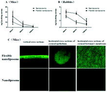
The concentrations of cou-6 in the mouse corneas following topical administration of these two formulations are shown in Fig. 5A. The cou-6 levels of the flexible liposomal formulation were 107.31%, 228.26%, and 136.50% higher than those of the liposomal formulation at the 30, 60 and 120 min time points, respectively. The CLSM observation results also supported the results mentioned above (Fig. 5C). From vertical cross-sectional observation, there was high fluorescence of cou-6 in the corneal epithelium, and the fluorescence became weaker in the deeper tissue of the cornea in both the liposomal and flexible liposomal formulation groups, but the fluorescence intensity was stronger in the flexible nanoliposome group than in the nanoliposome group. The observed results from horizontal observation supported the results of the vertical cross-sectional observation. There was obvious fluorescence in the corneal epithelium and Bowman's membrane (∼30 μm from the surface of the cornea) in the flexible nanoliposome group, while the fluorescence was much weaker in the relevant sites in the nanoliposome group.
image file: c5ra13830b-f5.tif
Fig. 5 In vivo corneal permeation. (A) Cou-6 concentration in mouse corneas after four instillations (5 μL/instillation at 10 min intervals) (*P < 0.05 compared to the liposomal formulation, n = 8). (B) Cou-6 concentration in rabbit corneas after four instillations (50 μL/instillation at 10 min intervals) (*P < 0.05 compared to the liposomal formulation, n = 6). (C) CLSM of horizontal and vertical cross-sections through the cornea at the 30 min time point in the mouse tests.

The results of the concentrations of cou-6 in the rabbit corneas are shown in Fig. 5B, and the results were similar to those of the mouse tests. The cou-6 levels of the flexible liposomal formulation were 39.64%, 172.09%, and 103.27% higher than those of the liposomal formulation at the 30, 60 and 120 min time points, respectively. Regarding the concentrations of cou-6 in the aqueous humor, we detected only 1.90 ± 0.09 ng mL−1 in the flexible nanoliposome group and 1.20 ± 0.32 ng mL−1 in the conventional nanoliposome group at the 30 min time point, and we failed to detect any cou-6 in the 60 min and 120 min time point samples in the aqueous humor of both of these groups.

Ocular tolerance

Values of clinical scores were 0–2 to different timepoints in all four groups. Then, flexible liposomal formulation and liposomal formulation were classified as non-irritating and safe for ophthalmic administration.

Anti-inflammatory efficacy

Topical SAS produced a mild ocular inflammation in rabbit. As seen in Fig. 6, the DG (except for 90 min time point), the nanoliposomes, and the flexible nanoliposomes showed no obvious anti-inflammatory activity. The DSP, a widely used corticosteroid eye drops, showed significant anti-inflammatory efficacy during the whole observation period. While the pranoprofen eye drops, a widely used nonsteroidal anti-inflammatory drug eye drops, just exhibited efficacy at some time points.
image file: c5ra13830b-f6.tif
Fig. 6 Anti-inflammatory efficacy of DG, nanoliposome, and flexible nanoliposome after SAS induced inflammation in the rabbit eye, with the dexamethasone sodium phosphate (DSP) eye drops (5 mL[thin space (1/6-em)]:[thin space (1/6-em)]1.25 mg) and pranoprofen eye drops (5 mL[thin space (1/6-em)]:[thin space (1/6-em)]5 mg) as control formulations (mean ± SD, n = 5, *P < 0.05 compared to the PBS group).

Discussion

In this investigation, cou-6-loaded nanoliposomes or flexible nanoliposomes were prepared using a simple solvent evaporation/film hydration method. The procedure was simple, and the finally obtained nanoliposomes or flexible nanoliposomes were well dispersed in aqueous solution, with a narrow particle size distribution. Because the non-encapsulated cou-6 was insoluble in the aqueous solution, a 0.22 μm filter was used not only to obtain sterility but also to separate non-encapsulated cou-6, although the encapsulation efficacy was high.13,21 One highlight of the preparation procedure for flexible nanoliposomes was that it used much less energy saving than the procedure for conventional nanoliposomes, because a higher amplitude of sonication was needed to obtain an opalescent dispersion of nanoliposomes with diameters similar to those of flexible nanoliposomes. This difference could be explained by DG, as an edge activator, having high affinity to interact with lipid bilayers, and it penetrated into liposomal lipid bilayers and disrupted the vesicular structure, so it saved much energy to create the multilamellar liposomes with small unilamellar vesicles. The other highlight was that it was environmentally friendly, because only ethanol was used during the preparation of the flexible nanoliposomes, while methylene dichloride is usually needed to dissolve cholesterol in conventional liposomal formulations.

In the research field of flexible liposomes, bile salts, including primarily sodium glycocholate, sodium deoxycholate, and sodium taurocholate, have been widely used as edge activators, particularly in those applied to skin. However, bile salts have shown ocular toxicity, and liposomes containing sodium deoxycholate caused toxicity or irritation to both spontaneously derived human corneal epithelial cells and rabbit corneas.22,23 Other edge activators, including surfactants of polysorbates or sorbitan esters, have also shown some extent of toxicity or irritation to the eye. In this investigation, DG was used and showed promising results. DG is a compound obtained by extraction with water from licorice root, and it has been widely used in internal and external drugs, as well as in cosmetics.24 DG is also widely used in ophthalmic solutions, such as potassium aspartate compound, penthenol and dipotassium glycyrrhizate eyedrops (manufacturer: ROHTO-MENTHOLATUM). Moreover, continuous application can be performed almost without side effects. In this study, DG cytotoxicity was determined, and the results were promising. DG showed slight time-dependent and concentration-dependent cytotoxicity, and it only showed some cytotoxicity when the concentration reached 20 mg mL−1 in 24 h incubation testing, as well as showing some cytotoxicity when the concentration reached 1.25 mg mL−1 in 72 h incubation testing. Regarding the toxicity of the liposomal and flexible liposomal formulations, neither of these two formulations showed cytotoxicity after 1 h of incubation. In the in vivo testing, the animals, particularly the rabbits, showed no irritation during the testing, consistent with the results for cytotoxicity. In summary, all of the tests revealed that DG and the formulations containing it should be safe for topical ocular application.

DG is a pharmacological active ingredient which can serve as an antiallergic and/or anti-inflammatory agent. While DG and the flexible nanoliposome exhibited no decrease in the ocular inflammation caused by instillation of SAS in rabbits' eyes in this test. One reason might be that the DG's concentration in our test was not high enough to perform the anti-inflammatory activity, as usually 10 mg mL−1 of DG solution showed improving allergic conjunctivitis,25 and only glycyrrhizin in a 50 mg mL−1 solution showed a comparable anti-inflammatory effect to that of dexamethasone (1 mg mL−1) in the quantitative evaluation of ocular anti-inflammatory measurements in rabbits,26 while the DG's concentration in our test was just 8.56 mg mL−1 to the DG solution and the flexible nanoliposomal formulation. The other reason might be the ocular inflammation animal model used in this test was not sensitive enough to fully show the anti-inflammatory effect, as the pranoprofen eye drops, a widely used nonsteroidal anti-inflammatory drug eye drops, just exhibited efficacy at some time points, though this ocular inflammation animal model used in this test was used to the evaluation of ocular anti-Inflammatory activity elsewhere.19,20 However, we still should give sufficient consideration to the potential therapeutic effect of DG if it is used in drug delivery system such as flexible liposome involved in this text. There are many diseases that are involved in the treatment of ocular inflammation, such as the prevention and curing of corneal immunologic rejection after keratoplasty, and the anti-inflammatory eye drops are one of the most used drugs in Ophthalmology. We could get a synergistic effect if we have a fully consideration about these. The flexible nanoliposomes containing cyclosporine to the prevention and curing of corneal immunologic rejection after keratoplasty is under texting in our group, and we anticipate a synergistic effect could be found in the pharmacodynamics testing.

Elasticity of lipid membranes is an important nanomechanical property to flexible liposome, and there were several methodologies reported to perform this evaluation. Atomic force microscopy based measurements has been turned out to be a valuable imaging technique to assess the evaluation, and some quantified parameters such as Young's modulus could be obtained and evaluated in this methodology.27 Electron spin resonance28–30 and fluorescence anisotropy measurement11 were also reported to be used to assess elasticity. Extrusion measurement was one of the most widely used methodology, and the particle size changing characters was performed to evaluate the elasticity, and the volume was also touched in some reports.31–33 The extrusion measurement was performed in this test. During the elasticity evaluation, almost none of the nanoliposomes could pass through the 20 nm filter smoothly, because the particle size was much larger than the filter size. However, the flexible nanoliposomes could be filtered, and the particle size in the solution after filtration was still much larger than the filter size, although it was somewhat smaller than the original formulation. Moreover and interestingly, the cou-6 concentration in the flexible nanoliposomes was nearly equal to that of the original solution, indicating that the whole solution was filtered, while in the nanoliposomes, only a small proportion of the particles less 20 nm in size were initially filtered at the beginning of filtration, and the filter was blocked completely, so only a low concentration of cou-6 was detected in the filtered solution. In the elasticity evaluation, it could be confirmed that the DG added to the formulation truly increased the elasticity of the lipid bilayer of nanoliposomes.

The mechanisms of interaction of nanoliposomes with cell membranes that result into intracellular drug delivery have been studied extensively, but they are poorly understood. Four mechanisms of intracellular drug delivery by liposomes – adsorption, endocytosis, fusion, and lipid exchange – have been widely accepted.34,35 In this investigation, the internalization of nanoliposomes by HCECs was significantly reduced following incubation at 4 °C in the presence of sodium azide. This result clearly demonstrated that the uptake of nanoliposomes occurred via an energy-dependent process, while the process occurred along the active endocytosis pathway. Further clarification of the mechanism revealed that the internalization of nanoliposomes by HCECs was reduced with two kinds of inhibitors: one was inhibitors of clathrin-mediated endocytosis (hypertonic sucrose and chlorpromazine), and the other was inhibitors of lipid raft/caveola – dependent endocytosis (MβCD and nystatin). This active endocytosis might be mainly mediated via these two pathways: clathrin-mediated endocytosis and lipid raft/caveola-dependent endocytosis. However, it is worthwhile to mention that the uptake of this nanoliposome in 4 °C sodium azide was still obvious, indicating the existence of other energy-independent pathways not included in this test, and no inhibitor used in this test could completely block the uptake of nanoliposomes into the HCECs, also indicating the complicated mechanisms involved in the internalization of the nanoliposomes by HCECs.

In flexible nanoliposomes, the uptake and internalization mechanisms appeared different from those of conventional nanoliposomes. The internalization of flexible nanoliposomes failed to be reduced following incubation at 4 °C or in the presence of sodium azide. This finding indicated that the uptake of the flexible nanoliposomes was mainly an energy-independent process. Further mechanism analysis through inhibitors was somewhat consistent with this result, apart from MβCD and hypertonic sucrose having inhibitory effects. It is still worthwhile to mention that chlorpromazine and nystatin had no inhibitory effects. These findings could be explained by chlorpromazine and hypertonic sucrose having different mechanisms of inhibition than MβCD and nystatin, although the final results were clathrin-mediated endocytosis inhibition with chlorpromazine and hypertonic sucrose and lipid raft/caveola-dependent endocytosis inhibition with MβCD and nystatin. These results provided us with the information that clathrin-mediated endocytosis and lipid raft/caveola-dependent endocytosis might be involved in flexible nanoliposomes, while they were still different from conventional nanoliposomes. The CLSM observations provided further evidence of the differences in the mechanisms. Punctuated fluorescence was found in their cytoplasm with different times of incubation, consistent with active endocytosis being a process mainly mediated by conventional nanoliposomes, while with flexible nanoliposomes, uniform and diffuse fluorescence in the cytoplasm, as well as in the nuclei, was observed, and this phenomenon occurred according to the fusion.18 Considering the results of inhibitory effects, the fusion process might be among the main mechanisms of the flexible nanoliposomes, although some endocytosis processes were still involved, as MβCD and hypertonic sucrose had inhibitory effects. The different mechanisms between the conventional nanoliposomes and the flexible nanoliposomes should be greatly affected by cholesterol and DG. Because the conventional nanoliposomes were composed of phospholipids and cholesterol and had a highly rigid lipid bilayer, it was somewhat difficult to be fused to the cell membrane, so endocytosis was the main process. In contrast, in the flexible nanoliposomes, DG was added to destabilize and increase the flexibility of the lipid bilayer of nanoliposomes, which was somewhat easier to fuse with the cell membrane, compared to the conventional nanoliposomes. Above all, the fluidity of the liposomal membrane differently affected cellular uptake and internalization of the nanoliposomes.

In the animal tests, there were higher concentrations of cou-6 in the corneas of mice and rabbits in the flexible nanoliposome group than in the conventional nanoliposome group, revealing that the flexible nanoliposomes had excellent capacity for corneal penetration. From the CLSM observations, both the flexible nanoliposomes and the conventional nanoliposomes were mainly found in the corneal epithelium, failing to penetrate the deeper tissues of the cornea. The concentration testing in the aqueous humor of rabbits was somewhat consistent with these CLSM results. These results in the conventional nanoliposome group were similar to some reports of nanoliposomes in ocular topical drug delivery,36 and the results in the flexible nanoliposome group were also similar to some reports in dermatological drug delivery, in which the flexible nanoliposomes were not able to penetrate the lower layers of the corneal stratum.24,37 Above all, flexible nanoliposomes still constitute a promising therapeutic tool for the immunomodulatory treatment of ocular surface disorders, such as keratoconjunctivitis sicca, vernal conjunctivitis, and atopical blepharitis, although they were not suitable for achieving therapeutic concentrations in the aqueous humor of intact corneas.

Although some of these results with flexible nanoliposomes were promising, no pharmaceutically active ingredients were tested in this investigation, and the molecular characteristics of the medical reagents encapsulated might exert an influence on the in vitro/in vivo fate of flexible nanoliposomes. Further research is needed to develop medical reagent formulations with these flexible nanoliposomes to promote their use. As an inherent obstacle to liposomal formulation, the stability and shelf life of flexible liposomal formulations were not a concern in this study, although we found that the flexible liposomal formulation was slightly more stable in 4 °C storage; however, instability and leakage of entrapped cou-6 remained obstacles, requiring further investigation into formulation procedures.

Conclusions

The results of this investigation showed that the fluidity of the liposomal membrane differently affected cellular uptake and internalization of nanoliposomes, and flexible nanoliposomes had excellent capability for in vivo corneal penetration, particularly to the corneal epithelium. Therefore, flexible nanoliposomes might be a promising therapeutic tool for the treatment of ocular surface disorders.

Author contributions

X. W. designed the research. C. G., F. C., M. L. and F. L. performed the experiments. C. G. and X. W. analyzed data and participated in the discussion. X. W. wrote and revised the paper. All authors reviewed the manuscript.

Conflict of interest

The authors declare no competing financial interests.

Acknowledgements

This research was supported by the National Natural Science Foundation of China (project nos. 81000369, 81271036 and 81500751), the Natural Science Foundation of Shandong Province, China (project no. ZR2015PH062), and the Science and Technology Foundation of Qingdao, Shandong Province, China (project no. 13-1-4-177-jch).

References

  1. A. Reijerkerk, C. C. Appeldoorn, J. Rip, M. de Boer and P. J. Gaillard, Invest. Ophthalmol. Visual Sci., 2014, 55, 2788–2794 CAS.
  2. K. Morimoto, T. Nakamura, T. Nakai and K. Morisaka, Arch. Int. Pharmacodyn. Ther., 1988, 293, 7–13 CAS.
  3. K. Taniguchi, K. Itakura, K. Morisaki and S. Hayashi, J. Pharmacobio-Dyn., 1988, 11, 685–693 CrossRef CAS.
  4. W. He, X. Guo, M. Feng and N. Mao, Int. J. Pharm., 2013, 458, 10 CrossRef PubMed.
  5. S. Wang, J. Zhang, T. Jiang, L. Zheng, Z. Wang, J. Zhang and P. Yu, Int. J. Pharm., 2011, 403, 219–229 CrossRef CAS PubMed.
  6. K. M. Hosny, AAPS PharmSciTech, 2009, 10, 1336–1342 CrossRef CAS PubMed.
  7. Y. Inokuchi, K. Hironaka, T. Fujisawa, Y. Tozuka, K. Tsuruma, M. Shimazawa, H. Takeuchi and H. Hara, Invest. Ophthalmol. Visual Sci., 2010, 51, 3162–3170 Search PubMed.
  8. M. W. Shafaa, N. M. Sabra and R. A. Fouad, Biopharm. Drug Dispos., 2011, 32, 507–517 CrossRef CAS PubMed.
  9. M. Niu, Y. Lu, L. Hovgaard and W. Wu, Int. J. Nanomed., 2011, 6, 1155–1166 CrossRef CAS PubMed.
  10. H. A. Salama, A. A. Mahmoud, A. O. Kamel, M. Abdel Hady and G. A. Awad, J. Liposome Res., 2012, 22, 336–345 CrossRef CAS PubMed.
  11. Y. Chen, Y. Lu, J. Chen, J. Lai, J. Sun, F. Hu and W. Wu, Int. J. Pharm., 2009, 376, 153–160 CrossRef CAS PubMed.
  12. P. Utreja, S. Jain and A. K. Tiwary, Drug Delivery, 2012, 19, 11–20 CrossRef CAS PubMed.
  13. J. Li, H. Xu, X. Ke and J. Tian, J. Drug Targeting, 2012, 20, 467–473 CrossRef CAS PubMed.
  14. X. G. Wu, G. Li and Y. L. Gao, Chem. Pharm. Bull., 2006, 54, 977–981 CrossRef CAS.
  15. N. Aggarwal and S. Goindi, Int. J. Pharm., 2012, 437, 277–287 CrossRef CAS PubMed.
  16. C. Di Tommaso, A. Torriglia, P. Furrer, F. Behar-Cohen, R. Gurny and M. Moller, Int. J. Pharm., 2011, 416, 515–524 CrossRef CAS PubMed.
  17. S. Dey, J. Patel, B. S. Anand, B. Jain-Vakkalagadda, P. Kaliki, D. Pal, V. Ganapathy and A. K. Mitra, Invest. Ophthalmol. Visual Sci., 2003, 44, 2909–2918 Search PubMed.
  18. U. Pleyer, J. Grammer, P. Kosmidis and D. G. Ruckert, Surv. Ophthalmol., 1995, 39(suppl 1), S3–S16 CrossRef.
  19. H. L. Alvarado, G. Abrego, M. L. Garduno-Ramirez, B. Clares, A. C. Calpena and M. L. Garcia, Nanomedicine, 2015, 11, 521–530 CrossRef CAS PubMed.
  20. C. Bucolo, A. Maltese, G. Puglisi and R. Pignatello, Ophthalmic Res., 2002, 34, 319–323 CrossRef CAS PubMed.
  21. H. Li, Y. Xiao, J. Niu, X. Chen and Q. Ping, J. Nanosci. Nanotechnol., 2011, 11, 8547–8555 CrossRef CAS PubMed.
  22. Y. Dai, R. Zhou, L. Liu, Y. Lu, J. Qi and W. Wu, Int. J. Nanomed., 2013, 8, 1921–1933 Search PubMed.
  23. A. Kumar, K. Pathak and V. Bali, Drug Discovery Today, 2012, 17, 1233–1241 CrossRef CAS PubMed.
  24. M. Trotta, E. Peira, F. Debernardi and M. Gallarate, Int. J. Pharm., 2002, 241, 319–327 CrossRef CAS.
  25. S. Hayasaka, T. Kodama and A. Ohira, Am. J. Chin. Med., 2012, 40, 887–904 CrossRef CAS PubMed.
  26. H. Tanaka, T. Hasegawa, M. Matsushita, H. Miichi and S. Hayashi, Ophthalmic Res., 1987, 19, 213–220 CrossRef CAS PubMed.
  27. F. W. Stetter and T. Hugel, Biophys. J., 2013, 104, 1049–1055 CrossRef CAS PubMed.
  28. A. Gillet, A. Grammenos, P. Compere, B. Evrard and G. Piel, Int. J. Pharm., 2009, 380, 174–180 CrossRef CAS PubMed.
  29. L. Coderch, J. Fonollosa, M. de Pera, J. Estelrich, A. de La Maza and J. L. Parra, J. Controlled Release, 2000, 68, 85–95 CrossRef CAS.
  30. B. A. van den Bergh, P. W. Wertz, H. E. Junginger and J. A. Bouwstra, Int. J. Pharm., 2001, 217, 13–24 CrossRef CAS.
  31. B. Geusens, J. Lambert, S. C. de Smedt, K. Buyens, N. N. Sanders and M. van Gele, J. Controlled Release, 2009, 133, 214–220 CrossRef CAS PubMed.
  32. L. H. Higa, P. Schilrreff, A. P. Perez, M. A. Iriarte, D. I. Roncaglia, M. J. Morilla and E. L. Romero, Nanomedicine, 2012, 8, 1319–1328 CrossRef CAS PubMed.
  33. C. K. Song, P. Balakrishnan, C. K. Shim, S. J. Chung, S. Chong and D. D. Kim, Colloids Surf., B, 2012, 92, 299–304 CrossRef CAS PubMed.
  34. R. Agarwal, I. Iezhitsa, P. Agarwal, N. A. Abdul Nasir, N. Razali, R. Alyautdin and N. M. Ismail, Drug Delivery, 2014, 1–17 CrossRef PubMed.
  35. V. H. Lee, P. T. Urrea, R. E. Smith and D. J. Schanzlin, Surv. Ophthalmol., 1985, 29, 335–348 CrossRef CAS.
  36. G. Buech, E. Bertelmann, U. Pleyer, I. Siebenbrodt and H. H. Borchert, J. Ocul. Pharmacol. Ther., 2007, 23, 292–303 CrossRef CAS PubMed.
  37. G. M. El Maghraby, A. C. Williams and B. W. Barry, J. Pharm. Pharmacol., 2001, 53, 1069–1077 CrossRef CAS.

This journal is © The Royal Society of Chemistry 2015
Click here to see how this site uses Cookies. View our privacy policy here.