DOI:
10.1039/C5RA13684A
(Paper)
RSC Adv., 2015,
5, 90255-90264
Microwave-assisted synthesis of monoclinic–tetragonal BiVO4 heterojunctions with enhanced visible-light-driven photocatalytic degradation of tetracycline†
Received
13th July 2015
, Accepted 8th October 2015
First published on 8th October 2015
Abstract
Recently, the controlled synthesis of semiconductor photocatalysts has received much attention in the field of pollutant degradation. In this study, BiVO4 photocatalysts of different crystalline phases (monoclinic scheelite (M-phase), tetragonal zircon (T-phase) and monoclinic–tetragonal heterojunction (M–T)) have been successfully synthesized via a facile microwave-assisted method. The crystalline phases of the products were controlled by switching the organic additives. The photocatalytic activities of the as-prepared samples were evaluated by the degradation of tetracycline (TC) under visible light irradiation. The obtained results clearly showed that the M–T BiVO4 exhibited the highest photodegradation ratio of the three samples of 80.5% in 1 h, which can be attributed to the heterojunction structure between different phases.
1. Introduction
Tetracycline (TC) is a well known broad-spectrum antibacterial agent widely applied in bacterial infection treatment, which is often found in significant quantities in aquatic environments and thus poses a serious threat to ecosystems and human health by causing proliferation of bacterial drug resistance.1 Therefore, the elimination of TC from aqueous environments has become an important environmental problem. However, traditional solutions are often constrained by their low efficiency and high cost. Recently, photocatalytic oxidation has been established to be a promising technique for environmental remediation2,3 and provides an efficient tool for the degradation of TC.4–8 From a viewpoint of energy conversion, visible light counts for more than 43% of incoming solar energy. Thus, developing high efficiency visible-light-driven materials for the photocatalytic degradation of TC is significant but still a challenge.9
Recent research into heterogeneous photocatalyst systems has tended to focus on the development of new alternatives to conventional UV-driven TiO2 to utilize visible light.10 Bismuth vanadate (BiVO4), as a ternary oxide semiconductor with a narrow band gap, has been found to be a promising photocatalyst with high stability, nontoxicity, and visible-light response for applications both in oxygen evolution from water splitting11–14 and environmental remediation.15–18 As we all know, BiVO4 has three crystalline types: monoclinic scheelite (M-phase), tetragonal zircon (T-phase) and tetragonal scheelite.19 However, previous research indicated that the M-phase BiVO4 exhibited higher photocatalytic activity than the other phases.20 Unfortunately, the efficiency of single-phase BiVO4 is still limited by the fast recombination of photo-induced charge carriers.21
Over the past years, construction of a heterojunction structure has proven to be a very efficient method to overcome this defect22–26 by realizing interfacial charge transfer between phases to inhibit the internal recombination of photogenerated electrons and holes.27–29 For example, researchers have reported the synthesis of a V2O5–BiVO4 heterostructure and found that the coupled semiconductor photocatalyst exhibited a higher photoactivity for the degradation of methylene blue (MB) than pure BiVO4 under visible light illumination.30 Moreover, a BiOCl–BiVO4 heterojunction photocatalyst was also reported31 which showed an increased activity by improving the separation of charge carriers. Very recently, the construction of a heterojunction structure from different types of crystalline phases of BiVO4 has drawn much attention.32 Unlike the heterojunction of BiVO4 with other semiconductors, heterojunctions between phases often possess a higher interfacial or inter-particle charge transfer rate and can be easily obtained by simply adjusting the pH of the substrate or the heating temperature in the crystallization process.33,34 Therefore, it is reasonable to expect a high TC degradation efficiency of M–T BiVO4 heterojunction photocatalysts.
More recently, microwave-assisted methods have been developed as a very facile method for the synthesis of tailor-made inorganic materials.35–38 Compared with other traditional heating methods, microwave irradiation can heat the substrate quickly and uniformly with a plastic (Teflon) reaction container, and thusly obtain a more homogeneous nucleation process and shorter crystallization time, which is very favorable to the synthesis of a high efficiency M–T BiVO4 heterojunction photocatalyst.
Herein, we report efficient visible-light-driven TC degradation on a M–T BiVO4 heterojunction photocatalyst which was synthesized by a microwave-assisted method. It should be pointed out that the crystalline phases of BiVO4 were controlled simply by switching the organic additives employed in the synthesis. In addition, the M–T BiVO4 showed a higher TC degradation activity than the other samples. Furthermore, a tentative mechanism of the enhanced photocatalytic activities is also discussed in this paper.
2. Experimental section
2.1. Chemicals
Bismuth nitrate, ammonium metavanadate and tetracycline were purchased from Aladdin (China). The rest of the reagents used were all purchased from Sinopharm (China). All reagents were of analytical grade and used without further purification. Deionized water was used in all experiments.
2.2. Synthesis
The BiVO4 photocatalyst was obtained by a simple microwave-assisted method. A typical synthesis preparation was as follows: 1 mmol Bi(NO3)3·5H2O was dissolved in 15 mL deionized water to make a hydrolyzed white flocculated suspension. 1 mmol NH4VO3 was dissolved in 20 mL of distilled water and then heated at about 70 °C with stirring for 10 min to produce a clear solution. Then these two solutions were mixed together to form a light yellow solution under vigorous stirring. After stirring for 30 min, 0.2 g EDTA or 0.4 g EDTA-Na2 was added to the light yellow solution which was stirred for another 30 min. The obtained mixture was transferred into a plastic (Teflon) reaction container. A microwave reaction was carried out in a microwave reactor (MDS-6, Xinyi microwave Chemical Company) with an operating power of 1000 W and a working temperature of 100 °C, for 1 h. After the container cooled down to room temperature, the precipitates were collected by centrifugation, washed with deionized water and absolute ethanol three times, and dried at 60 °C for 12 h. The obtained samples were T-phase, M-phase and M–T samples, in which the M–T sample was prepared by 1 h microwave heating with no additive, the M-phase sample was obtained by 1 h heating with 0.2 g EDTA and the T-phase sample was formed by 1 h heating with 0.4 g EDTA-Na2.
2.3. Characterization
The crystalline phases of the as-synthesized samples were determined by X-ray diffraction (XRD) patterns using a D/MAX-2500 diffractometer (Rigaku, Japan) with a Cu Kα radiation source (λ = 1.54056 Å) at a scan rate of 5° min−1. The applied current was 300 mA and the accelerating voltage was 50 kV. The morphologies of the samples were obtained by scanning electron microscopy (SEM), for which the images were collected on a S-4800 field emission SEM (FESEM, Hitachi, Japan). Transmission electron microscopy (TEM) and high-resolution transmission electron microscopy (HRTEM) were performed on an F20 S-TWIN electron microscope (Tecnai G2, FEI Co.) with a 200 kV accelerating voltage. UV-vis absorption spectra were recorded on a UV 2550 (Shimadzu, Japan) UV-vis spectrophotometer. BaSO4 was used as a reflectance standard. Steady state luminescence experiments were performed using a Photon Technology International Model Quantamaster-QM4m spectrofluorimeter equipped with a 75 W lamp and dual excitation monochromators.
2.4. Photocatalytic activity
The photocatalytic activity of the as-prepared samples was determined by the photodegradation of a TC solution, which was carried out at 308 K in a photochemical reactor under visible light irradiation. The as-prepared BiVO4 (0.1 g) was mixed with the TC solution (0.1 L, 10 mg L−1), followed by magnetic stirring in darkness for 30 min to reach an adsorption equilibrium. The photochemical reactor was irradiated with a 150 W xenon lamp which was located at a distance of 8 cm from one side of the reactor containing the solution. UV light with a wavelength of less than 420 nm was removed by a UV-cutoff filter. In 10 min irradiation intervals, a series of aqueous solution samples (6 mL) were collected and separated from the suspended catalyst particles for analysis. The photocatalytic degradation ratio (DR) was calculated via the following formula:| | |
DR = (1 − Ai/A0) × 100%
| (1) |
where A0 is the foremost absorbence of TC at the adsorption equilibrium and Ai is the absorbence after the sampling analysis. The TC concentration of different samples was measured on a UV-vis spectrophotometer by monitoring its characteristic absorption wavelength at 357 nm.
3. Results and discussion
3.1. Morphology and structure
Three BiVO4 materials with different crystalline phases were obtained by developing the microwave-assisted method and different reaction conditions, namely the T-phase sample, the M-phase sample and the M–T sample. Fig. 1 displays the XRD patterns of the different BiVO4 samples. All peaks of the T-phase sample in the XRD pattern can be well indexed to the tetragonal zircon phase (JCPDS no.14-0133), while the peaks of the M-phase sample are indexed to the monoclinic scheelite phase (JCPDS no.14-0688). The M–T sample has an XRD pattern showing both M-phase and T-phase peaks, which demonstrates the binary composition of the M–T BiVO4 sample. The crystalline structures of the different samples were adjusted only by changing the additive reagent during the synthesis process.
 |
| | Fig. 1 XRD patterns of the T-phase sample, the M-phase sample and the M–T sample. | |
Fig. 2 shows the SEM images of the different samples. As shown in Fig. 2a and b, the M–T sample exhibits a rod-like structure with nanoparticles scattered on the surface. The rod-like structures have a length of 3–5 μm while the scattered nanoparticles have diameters of about 100 nm. From Fig. 2c and d, the M-phase sample exhibits a massive 2D sheet structure with an average thickness of 30 nm. Fig. 2e and f show that the T-phase sample exhibits a spherical structure in the micro-scale with a rough surface. The diameter of the T-phase microspheres is about 2.5 μm. SEM studies indicate that changing the additives can also control the morphology of BiVO4 as well as the crystalline structure.
 |
| | Fig. 2 SEM images of the M–T sample: (a) and (b), the M-phase sample: (c) and (d), and the T-phase sample: (e) and (f). | |
Further insight into the morphology and detailed surface nature of the M–T sample was obtained using transmission electron microscopy (TEM) and high-resolution transmission electron microscopy (HRTEM). As shown in Fig. 3a, the rod structure and scattering nanocrystals of the M–T sample can be easily recognized. From the HRTEM image (Fig. 3b) of an individual micro-rod surface, distinct lattice fringes with a spacing of d = 0.365 nm for the rod structure and d = 0.312 nm for the surface nanocrystal can be observed, which coincide with the (200) plane of the T-phase and the (−130) plane of the M-phase of BiVO4, respectively. The above obtained results clearly demonstrate that an inter-facial (or inter-particle) junction between the M- and T- crystalline phases of BiVO4 has been formed.
 |
| | Fig. 3 TEM (a) and HRTEM (b) images of the M–T sample. | |
To figure out the crystallization process of M–T BiVO4, a series of time-resolved experiments were carried out. In these experiments, only the reaction time was changed without altering any other reaction conditions. Fig. 4 shows the SEM images and the corresponding XRD patterns of the obtained samples at different reaction times (from 1 min, 15 min, 30 min and 45 min), from which we envision that both the morphology and crystalline structure changed during the microwave heating. At the early stage of crystallization after only a very short time (1 min) of heating, some M-phase BiVO4 can be recognized in the XRD pattern, even though the T-phase is predominant. With continuous heating, the presence of the M-phase BiVO4 was enhanced (Fig. 4e) with the morphology of the sample changing from amorphous nanoparticles to a micro-rod structure (Fig. 4a–d).
 |
| | Fig. 4 SEM images of the obtained samples after different microwave reaction times of (a) 1 min, (b) 15 min, (c) 30 min, (d) 45 min and (e) XRD patterns of M–T BiVO4. | |
3.2. UV-vis absorption spectra and specific surface area
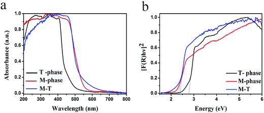
The light harvesting abilities of different BiVO4 samples were measured by UV-vis absorption spectroscopy. From Fig. 5a, all the samples exhibit excellent visible-light absorption ability. Among them, the T-phase sample shows a weaker light absorption ability than the M-phase sample and the M–T sample, with a light absorption edge at 510 nm. The M-phase sample has an absorption edge at 550 nm, while the M–T sample has a light absorption edge at 560 nm. The energy band gaps (Eg) of different samples were also estimated using the following equation:39where α, h, ν, A and Eg are the absorption coefficient, Planck’s constant, the incident light frequency, a constant and the band gap energy, respectively. Among them, n depends on the characteristics of the optical transition of the semiconductor, n = 1 for direct transition semiconductors and n = 4 for indirect transition semiconductors. BiVO4 is a typical kind of direct transition semiconductor and so the value of n is chosen to be 1.40–42 The estimated band gaps from the plots of (αhν)2 versus hν are shown in Fig. 5b, from which, the energy band gaps of the T-phase, M-phase and M–T samples are shown to be 2.75, 2.4 and 2.3 eV, respectively.
 |
| | Fig. 5 (a) UV-vis absorption spectra, (b) calculated band gap of the T-phase sample, M-phase sample and M–T sample. | |
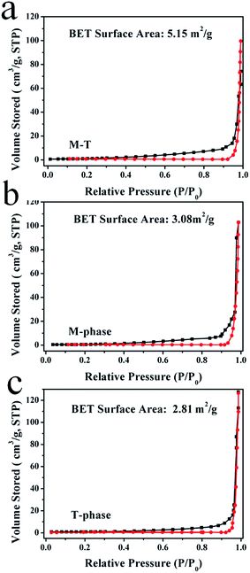
The nitrogen adsorption–desorption isotherm and the BET surface areas of the as-prepared samples are displayed in Fig. 6. From the figure, it can be clearly observed that the BET surface areas of the T-phase sample, M-phase sample and M–T sample are 2.81 m2 g−1, 3.08 m2 g−1 and 5.15 m2 g−1, respectively. Moreover, it is known that because the photocatalytic activity depends highly on specific surface area, the larger surface areas of the BiVO4 samples can facilitate the adsorption of more TC molecules, which is beneficial for improving their visible light activities.
 |
| | Fig. 6 Nitrogen adsorption–desorption isotherms and BET surface areas of the T-phase, M-phase and M–T samples. | |
3.3. Photocatalytic degradation of TC
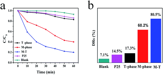
The photocatalytic activities of the different BiVO4 samples and commercial Degussa P25 TiO2 powder were evaluated by the degradation of TC under visible-light irradiation. In order to make a comparison, a blank experiment was also conducted without a catalyst, and little decrease of TC concentration could be observed. As shown in Fig. 7, the degradation ratio over P25 was 14.5%, indicating that its activity with regard to TC degradation is very poor. However, the M–T sample exhibits a high activity with a degradation ratio of 80.5%, which is much higher than for the M-phase sample and the T-phase sample (60.2% and 17.3%, respectively). In addition, time-resolved TC absorption spectra for the M–T sample under visible light irradiation are displayed in Fig. 8, in which the intensities of the TC absorption peaks at 357 nm gradually decrease. Many studies have pointed out that the photocatalytic activity of BiVO4 has a close relationship with the crystal structure.33,34 In this work, we also found that the M–T BiVO4 sample showed a higher photocatalytic activity than both the M-phase and T-phase samples, which can be attributed to the heterojunction structure between the M-phase and T-phase that facilitates the charge transfer between phases, inhibiting the recombination of photo-induced charge carriers. In order to examine the credibility of our results for the photodegradation ratio, dark experiments were carried out to remove the effect of physical adsorption (Fig. 9). Within 20 min, all the samples reached an adsorption equilibrium, suggesting that the effect of the physical adsorption was removed and the activity of the as-prepared samples were correctly evaluated.
 |
| | Fig. 7 Photocatalytic degradation of TC versus visible light irradiation time with the different samples. | |
 |
| | Fig. 8 Time-dependent UV-vis absorption spectra of the TC solution in the presence of the M–T sample. | |
 |
| | Fig. 9 Adsorption equilibrium curves of the different samples in 60 min kept in darkness. | |
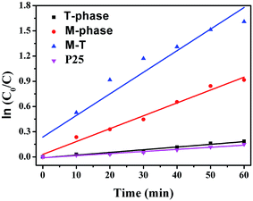
A kinetics study was employed to further illustrate the photocatalytic activities (Fig. 10). The kinetic rate constants were gained by a Langmuir–Hinshelwood model and the decomposition of TC matched the first order kinetic equation:
where
C and
C0 represent the TC concentration remaining in the solution at an irradiation time of
t and the initial concentration at
t = 0, respectively.
kapp expresses the rate constant of degradation. The
kapp values are 0.00321, 0.01532, 0.02569 and 0.00249 for the T-phase, M-phase M–T and P25 samples. Obviously, the
kapp value of the M–T sample (0.02569) is the highest among all the samples, which is consistent with the conclusion that it possesses the highest rate of photocatalytic degradation.
 |
| | Fig. 10 Kinetics of the photocatalytic degradation ratio with the different samples. | |
3.4. Investigation of reactive species
As is well-known, many reactive species such as h+, ˙O2−, and ˙OH play major roles in TC decomposition.43,44 In our work, ˙O2− could not be produced by electrons because of the more positive reduction potential of −0.33 eV/NHE (O2/˙O2−),45 compared to the M-phase (EVB = 0.34 eV) and the T-phase (EVB = 0.16 eV). However, due to the reduction potential of O2/H2O2 being 0.695 eV,43 the electrons can react to generate ˙OH via the O2 and H+ with the equation: O2 + 2e− + 2H+ = H2O2, H2O2 + e− = ˙OH + OH−. In comparison to the potential of ˙OH/OH− (2.38 eV/NHE) and ˙OH/H2O (2.72 eV/NHE), the VB of the M-phase BiVO4 is 2.74 eV, so the ˙OH could be produced via the reaction of h+, OH− and H2O on the M–T BiVO4 surface. The ˙OH can just react with the TC immediately.
A series of active species trapping experiments were conducted to further investigate the photocatalytic oxidation mechanism of TC degradation by the M–T sample. AgNO3, iso-propanol (IPA) and triethanolamine (TEA) were employed as trapping reagents of photogenerated electrons (e−), ˙OH radicals and holes (h+), respectively (Fig. 11). As the TEA (the scavenger of holes) was added to the reaction system, the DR decreased significantly compared to the reaction in the absence of TEA, indicating that the photogenerated holes were the major oxidative species for the oxidation of TC.45–49 When the IPA was added into the reaction system to trap ˙OH, the DR of the TC was slightly decreased to 62.2%, which indicates that the ˙OH took part in the oxidation reaction.50 However, when AgNO3 was used as the scavenger of electrons,45–49 the DR increased rather than decreased, which demonstrated that the scavenger of e− meant there was less opportunity for recombination of electron–hole pairs and more holes were produced to take part in the reaction process.
 |
| | Fig. 11 Photocatalytic degradation ratios of TC using different radical scavengers over the M–T sample. | |
3.5. The intermediates of TC degradation
The intermediates of TC degradation were investigated via LC-MS and the results are exhibited in Fig. 12. As shown in Fig. 12a, an intense prominent ion with m/z = 445 can be clearly observed, which can be attributed to TC. From the analysis of LC-MS, it can be seen that the TC was attacked by the major reactive oxidated species h+. Successive ion fragmentations during collision-induced dissociation occur in the following order: m/z = 445 → m/z = 406 (by loss of OH, H and CH3) → m/z = 362 (by loss of CONH2) → m/z = 318 (by loss of N, CH2 and CH3) → m/z = 274 (then by loss of CH, C, H and OH). On the basis of all the above experimental results and the previous studies,51 the possible processes of the degradation are displayed in Fig. 13. Finally, the intermediate products would be degraded to small inorganic molecular material.
 |
| | Fig. 12 Typical LC-MS chromatogram after irradiation for 1 h. | |
 |
| | Fig. 13 Proposed degradation pathways for the photocatalytic degradation of TC with the M–T photocatalyst. | |
3.6. Possible mechanism of TC degradation by M–T BiVO4
The possible mechanism of TC degradation by the M–T BiVO4 is now discussed in detail. As shown in Fig. 14, T-phase BiVO4 has a larger energy band gap than M-phase BiVO4 with both higher CB bottom and lower VB top positions. Under visible-light irradiation, both the T-phase and M-phase BiVO4 were excited and produced photo-induced electrons and holes at the CB and VB. Due to the heterojunction structure between the T-phase and the M-phase of BiVO4, electrons at the T-phase CB can be easily injected into the CB of the M-phase. On the other hand, holes at the T-phase VB can also be injected into the M-phase VB, which effectively inhibits the recombination of electrons and holes, and thus promotes the photocatalytic activity.52
 |
| | Fig. 14 Photocatalytic mechanism at the M–T BiVO4 heterojunction under visible light irradiation. | |
3.7. Photoluminescence spectra analysis
Photoluminescence (PL) analysis was also conducted to evaluate the separation ability of photogenerated charge carriers.53 The measurement was conducted at an excitation wavelength of 360 nm with a photomultiplier tube voltage of 500 V. From Fig. 15, all the samples have a PL emission peak at 469 nm and the peak intensities of different specific samples were in the order of the T-phase sample, the M-phase sample and the M–T sample, which is the exact reverse order of DR. It is well-acknowledged that the PL emission intensity of a semiconductor is proportional to the opportunity for the recombination of photoinduced electron–hole pairs.54 In other words, a lower PL intensity implies a stronger charge-separation ability. The completely opposite PL and DR data sufficiently demonstrate the validity of our assumption.
 |
| | Fig. 15 Photoluminescence spectra of the T-phase sample, M-phase sample and M–T sample. | |
3.8. Photostability
The stability of the M–T sample was evaluated by repeating the experiments. After each run, the catalysts were collected and washed by a simple filtration followed by ultrasonic cleaning with deionized water. As shown in Fig. 16a, the M–T sample exhibits a high stability, resulting in a high decomposition ratio even after four cycles. Meanwhile, the XRD pattern of the M–T sample is barely changed after four cycling experiments (Fig. 16b). In addition, the SEM images after four cycles remain unchanged, which is displayed in Fig. S1.† These results clearly suggest that the photocatalyst has an excellent photostability.
 |
| | Fig. 16 (a) Four cycling experiments of the M–T sample, (b) XRD patterns of the M–T sample before and after four cycling experiments. | |
4. Conclusions
In summary, different BiVO4 photocatalysts were prepared by a microwave-assisted method. The crystalline phases of the products were controlled by varying the organic additives. Visible-light-driven TC degradation was conducted by the obtained BiVO4 photocatalysts. Among them, the M–T sample showed the highest DR of 80.5% in 1 h. The high activity of the M–T BiVO4 photocatalyst was likely to result from the fast charge transfer between the heterojunction of the different phases of BiVO4.
Acknowledgements
The authors greatly gratefully acknowledge the financial support of the National Natural Science Foundation of China (21276116, 21477050, 21301076 and 21303074), Excellent Youth Foundation of Jiangsu Scientific Committee (BK20140011), Chinese-German Cooperation Research Project (GZ1091), Program for High-Level Innovative and Entrepreneurial Talents in Jiangsu Province, Program for New Century Excellent Talents in University (NCET-13-0835), Henry Fok Education Foundation (141068) and Six Talents Peak Project in Jiangsu Province (XCL-025).
References
- M. R. Hoffmann, S. T. Martin, W. Choi and D. W. Bahnemann, Environmental applications of semiconductor photocatalysis, Chem. Rev., 1995, 95, 69–96 CrossRef CAS
 .
. - C. C. Pei and W. W. Leung, Photocatalytic degradation of Rhodamine B by TiO2/ZnO nanofibers under visible-light irradiation, Sep. Purif. Technol., 2013, 114, 108–116 CrossRef CAS PubMed
 .
. - Y. Jiang and R. Amal, Selective synthesis of TiO2-based nanoparticles with highly active surface sites for gas-phase photocatalytic oxidation, Appl. Catal., B, 2013, 138–139, 260–267 CrossRef CAS PubMed
 .
. - Y. B. Liu, X. J. Gan, B. X. Zhou, B. T. Xiong, J. H. Li, C. P. Dong, J. Bai and W. M. Cai, Photoelectrocatalytic degradation of tetracycline by highly effective TiO2 nanopore arrays electrode, J. Hazard. Mater., 2009, 171, 678–683 CrossRef CAS PubMed
 .
. - R. A. Palominos, M. A. Mondaca, A. Giraldo, G. Penuela, M. Perez-Moya and H. D. Mansilla, Photocatalytic oxidation of the antibiotic tetracycline on TiO2 and ZnO suspensions, Catal. Today, 2009, 144, 100–105 CrossRef CAS PubMed
 .
. - C. Reyes, J. Fernández, J. Freer, M. A. Mondaca, C. Zaror, S. Malato and H. D. Mansilla, Degradation and inactivation of tetracycline by TiO2 photocatalysis, J. Photochem. Photobiol., A, 2006, 184, 141–146 CrossRef CAS PubMed
 .
. - J. Jeong, W. Song, W. J. Cooper, J. Jung and J. Greaves, Degradation of tetracycline antibiotics: mechanisms and kinetic studies for advanced oxidation/reduction processes, Chemosphere, 2010, 78, 533–540 CrossRef CAS PubMed
 .
. - J. Huang, L. Xiao and X. Yang, WO3 nanoplates, hierarchical flower-like assemblies and their photocatalytic properties, Mater. Res. Bull., 2013, 48, 2782–2785 CrossRef CAS PubMed
 .
. - P. W. Huo, Z. Y. Lu, X. L. Liu, X. Gao, J. M. Pan, D. Wu, J. Ying, H. M. Li and Y. S. Yan, Preparation molecular/ions imprinted photocatalysts of La3+@POPD/TiO2/fly-ash cenospheres: preferential photodegradation of TCs antibiotics, Chem. Eng. J., 2012, 198–199, 73–80 CrossRef CAS PubMed
 .
. - A. Kubacka, M. Fernández-García and G. Colón, Nanoarchitectures for solar photocatalytic applications, Chem. Rev., 2012, 112, 1555–1614 CrossRef CAS PubMed
 .
. - S. Tokunaga, H. Kato and A. Kudo, Selective preparation of monoclinic and tetragonal BiVO4 with scheelite structure and their photocatalytic properties, Chem. Mater., 2001, 13, 4624–4628 CrossRef CAS
 .
. - K. Sayama, A. Nomura and T. Arai, Photoelectrochemical decomposition of water into H2 and O2 on porous BiVO4 thin-film electrodes under visible light and significant effect of Ag ion treatment, J. Phys. Chem. B, 2006, 110, 11352–11360 CrossRef CAS PubMed
 .
. - A. Kudo, K. Omori and H. Kato, A novel aqueous process for preparation of crystal form-controlled and highly crystalline BiVO4 powder from layered vanadates at room temperature and its photocatalytic and photophysical properties, J. Am. Chem. Soc., 1999, 121, 11459–11467 CrossRef CAS
 .
. - J. Z. Su, L. J. Guo and S. Yoriya, Aqueous growth of pyramidal-shaped BiVO4 nanowire arrays and structural characterization: application to photoelectrochemical water splitting, Cryst. Growth Des., 2010, 10, 856–861 CAS
 .
. - L. Zhang, D. R. Chen and X. L. Jiao, Monoclinic structured BiVO4 nanosheets: hydrothermal preparation, formation mechanism, and coloristic and photocatalytic properties, J. Phys. Chem. B, 2006, 110, 2668–2673 CrossRef CAS PubMed
 .
. - W. Z. Yin, W. Z. Wang and L. Zhou, CTAB-assisted synthesis of monoclinic BiVO4 photocatalyst and its highly efficient degradation of organic dye under visible-light irradiation, J. Hazard. Mater., 2010, 173, 194–199 CrossRef CAS PubMed
 .
. - H. Q. Jiang, H. Endo and H. Natori, Fabrication and photoactivities of spherical-shaped BiVO4 photocatalysts through solution combustion synthesis method, J. Eur. Ceram. Soc., 2008, 28, 2955–2962 CrossRef CAS PubMed
 .
. - L. Zhou, W. Wang and S. Liu, A sonochemical route to visible-light-driven high-activity BiVO4 photocatalyst, J. Mol. Catal. A: Chem., 2006, 252, 120–124 CrossRef CAS PubMed
 .
. - A. Martínez-de la Cruz, U. M. García-Pérez and S. Sepúlveda-Guzmán, Characterization of the visible-light-driven BiVO4 photocatalyst synthesized via a polymer-assisted hydrothermal method, Res. Chem. Intermed., 2013, 39, 881–894 CrossRef
 .
. - S. J. Hong, S. Lee, J. S. Jang and J. S. Lee, Heterojunction BiVO4/WO3 electrodes for enhanced photoactivity of water oxidation, Energy Environ. Sci., 2011, 4, 1781–1787 CAS
 .
. - W. Yao, H. Iwai and J. Ye, Effects of molybdenum substitution on the photocatalytic behavior of BiVO4, Dalton Trans., 2008, 1426–1430 RSC
 .
. - M. Long, W. Cai and H. Kisch, Visible light induced photoelectrochemical properties of n-BiVO4 and n-BiVO4/p-Co3O4, J. Phys. Chem. C., 2008, 112, 548–554 CAS
 .
. - X. Zhang, Y. Gong, X. Dong, X. Zhang, C. Ma and F. Shi, Fabrication and efficient visible light-induced photocatalytic activity of Bi2WO6/BiVO4 heterojunction, Mater. Chem. Phys., 2012, 136, 72–476 Search PubMed
 .
. - H. T. Li, X. D. He, Z. H. Kang, H. Huang, Y. Liu, J. L. Liu, S. Y. Lian, C. Him, A. Tsang, X. B. Yang and S. T. Lee, Water-Soluble Fluorescent Carbon Quantum Dots and Photocatalyst Design, Angew. Chem., Int. Ed., 2010, 49, 4430–4434 CrossRef CAS PubMed
 .
. - X. H. Gao, H. B. Wu, L. X. Zheng, Y. J. Zhong, Y. Hu and X. W. Lou, Formation of Mesoporous Heterostructured BiVO4/Bi2S3 Hollow Discoids with Enhanced Photoactivity, Angew. Chem., Int. Ed., 2014, 53, 5917–5921 CrossRef CAS PubMed
 .
. - C. Eley, T. Li, F. L. Liao, S. M. Fairclough, J. M. Smith, G. Smith and S. C. Edman Tsang, Nanojunction-Mediated Photocatalytic Enhancement in Heterostructured CdS/ZnO, CdSe/ZnO, and CdTe/ZnO Nanocrystals, Angew. Chem., Int. Ed., 2014, 53, 7838–7842 CrossRef CAS PubMed
 .
. - J. Z. Su, L. J. Guo, N. Z. Bao and C. A. Grimes, Nanostructured WO3/BiVO4 heterojunction films for efficient photoelectrochemical water splitting, Nano Lett., 2011, 11, 1928–1933 CrossRef CAS PubMed
 .
. - P. Ju, P. Wang, B. Li, H. Fan, S. Y. Ai, D. Zhang and Y. Wang, A novel calcined Bi2WO6/BiVO4 heterojunction photocatalyst with highly enhanced photocatalytic activity, Chem. Eng. J., 2014, 236, 430–437 CrossRef CAS PubMed
 .
. - W. R. Zhao, Y. Wang, Y. Yang, J. Tang and Y. A. Yang, Carbon spheres supported visible-light-driven CuO–BiVO4 heterojunction: preparation, characterization, and photocatalytic properties, Appl. Catal., B, 2012, 115–116, 90–99 CrossRef CAS PubMed
 .
. - J. Su, X. X. Zou, G. D. Li, X. Wei, C. Yan, Y. N. Wang, J. Zhao, L. J. Zhou and J. S. Chen, Macroporous V2O5/BiVO4 composites: effect of heterojunction on the behavior of photogenerated charges, J. Phys. Chem. C, 2011, 115, 8064–8071 CAS
 .
. - Z. Q. He, Y. Q. Shi, C. Gao, L. N. Wen, J. M. Chen and S. Song, BiOCl/BiVO4 p–n heterojunction with enhanced photocatalytic activity under visible-light irradiation, J. Phys. Chem. C, 2014, 118, 389–398 CAS
 .
. - S. Usai, S. Obregón, A. I. Becerro and G. Colón, Monoclinic–tetragonal heterostructured BiVO4 by yttrium doping with improved photocatalytic activity, J. Phys. Chem. C, 2013, 117, 24479–24484 CAS
 .
. - H. M. Fan, T. F. Jiang, H. Y. Li, D. J. Wang, L. L. Wang, J. L. Zhai, D. Q. He, P. Wang and T. F. Xie, Effect of BiVO4 crystalline phases on the photoinduced carriers behavior and photocatalytic activity, J. Phys. Chem. C, 2012, 116, 2425–2430 CAS
 .
. - G. Q. Tan, L. L. Zhang, H. J. Ren, S. S. Wei, J. Huang and A. Xia, Effects of pH on the hierarchicalstructures and photocatalytic performance of BiVO4 powders prepared via the microwave hydrothermal method, ACS Appl. Mater. Interfaces, 2013, 5, 5186–5193 CAS
 .
. - W. D. Shi, Y. Yan and X. Yan, Microwave-assisted synthesis of nano-scale BiVO4 photocatalysts and their excellent visible-light-driven photocatalytic activity for the degradation of ciprofloxacin, Chem. Eng. J., 2013, 215–216, 740–746 CrossRef CAS PubMed
 .
. - M. Yan, Y. Yan, C. Wang, W. Lu and W. D. Shi, Ni2+ doped InVO4 nanocrystals: one-pot microwave-assisted synthesis and enhanced photocatalytic O2 production activity under visible-light, Mater. Lett., 2014, 121, 215–218 CrossRef CAS PubMed
 .
. - Y. Yan, S. F. Sun, Y. Song, X. Yan, W. S. Guan, X. L. Liu and W. D. Shi, Microwave-assisted in situ synthesis of reduced graphene oxide–BiVO4 composite photocatalysts and their enhanced photocatalytic performance for the degradation of ciprofloxacin, J. Hazard. Mater., 2013, 250–251, 106–114 CrossRef CAS PubMed
 .
. - Y. Yan, F. P. Cai, Y. Song and W. D. Shi, InVO4 nanocrystal photocatalysts: microwave-assisted synthesis and size-dependent activities of hydrogen production from water splitting under visible light, Chem. Eng. J., 2013, 233, 1–7 CrossRef CAS PubMed
 .
. - M. A. Butler, Photoelectrolysis and physical properties of the semiconducting electrode WO2, J. Appl. Phys., 1977, 48, 1914–1920 CrossRef CAS PubMed
 .
. - M. L. Guan, D. K. Ma, S. W. Hu, Y. J. Chen and S. M. Huang, From hollow olive-shaped BiVO4 to n–p core–shell BiVO4@Bi2O3 microspheres: controlled synthesis and enhanced visible-light-responsive photocatalytic properties, Inorg. Chem., 2011, 50, 800–805 CrossRef CAS PubMed
 .
. - J. Z. Su, L. J. Guo, S. Yoriya and C. A. Grimes, Aqueous growth of pyramidal-shaped BiVO4 nanowire arrays and structural characterization: application to photoelectrochemical water splitting, Cryst. Growth Des., 2010, 10, 856–861 CAS
 .
. - L. Dong, S. Guo, S. Y. Zhu, D. F. Xu, L. L. Zhang, M. X. Huo and X. Yang, Sunlight responsive BiVO4 photocatalyst: effects of pH on L-cysteine-assisted hydrothermal treatment and enhanced degradation of ofloxacin, Catal. Commun., 2011, 16, 250–254 CrossRef CAS PubMed
 .
. - L. S. Zhang, K. H. Wong, H. Y. Yip, C. Hu, J. C. Yu, C. Y. Chan and P. K. Wong, Effective photocatalytic disinfection of E. coli K-12 Using AgBr–Ag–Bi2WO6 nanojunction system irradiated by visible light: the role of diffusing hydroxyl radicals, Environ. Sci. Technol., 2010, 44, 1392–1398 CrossRef CAS PubMed
 .
. - K. Yu, S. Yang, C. Liu, H. Chen, H. Li, C. Sun and S. A. Boyd, Degradation of organic dyes via bismuth silver oxide initiated direct oxidation coupled with sodium bismuthate based visible light photocatalysis, Environ. Sci. Technol., 2012, 46, 7318–7326 CrossRef CAS PubMed
 .
. - S. Q. Liu, N. Zhang, Z. R. Tang and Y. J. Xu, Synthesis of one-dimensional CdS@TiO2 core–shell nanocomposites photocatalyst for selective redox: the dual role of TiO2 shell, ACS Appl. Mater. Interfaces, 2012, 4, 6378–6385 CAS
 .
. - S. C. Yan, Z. S. Li and Z. G. Zou, Photodegradation of rhodamine B and methyl orange over boron-doped g-C3N4 under visible light irradiation, Langmuir, 2010, 26, 3894–3901 CrossRef CAS PubMed
 .
. - X. C. Wang, K. Maeda, X. F. Chen, K. Takanabe, K. Domen, Y. D. Hou, X. Z. Fu and M. Antonietti, Polymer semiconductors for artificial photosynthesis: hydrogen evolution by mesoporous graphitic carbon nitride with visible light, J. Am. Chem. Soc., 2009, 131, 1680–1681 CrossRef CAS PubMed
 .
. - L. Q. Ye, J. Y. Liu, C. Q. Gong, L. H. Tian, T. Y. Peng and L. Zan, Two different roles of metallic Ag on Ag/AgX/BiOX (X = Cl, Br) visible light photocatalysts: surface plasmon resonance and Z-scheme bridge, ACS Catal., 2012, 2, 1677–1683 CrossRef CAS
 .
. - X. Y. Xiao, J. Jiang and L. Z. Zhang, Selective oxidation of benzyl alcohol into benzaldehyde over semiconductors under visible light: the case ofBi12O17Cl2 nanobelts, Appl. Catal., B, 2013, 142–143, 487–493 CrossRef CAS PubMed
 .
. - U. A. Joshi, J. R. Darwent, H. H. P. Yiu and M. J. Rosseinsky, The effect of platinum on the performance of WO3 nanocrystal photocatalysts for the oxidation of methyl orange and iso-propanol, J. Chem. Technol. Biotechnol., 2011, 86, 1018–1023 CrossRef CAS PubMed
 .
. - M. J. Zhou, X. L. Liu, C. C. Ma, H. Q. Wang, Y. F. Tang, P. W. Huo, W. D. Shi, Y. S. Yan and J. H. Yang, Enhanced visible light photocatalytic activity of alkaline earth metal ions-doped CdSe/rGO photocatalysts synthesized by hydrothermal method, Appl. Catal., B, 2015, 172, 174–184 CrossRef PubMed
 .
. - H. P. Li, J. Y. Liu, W. G. Hou, N. Duc, R. J. Zhang and X. T. Tao, Synthesis and characterization of g-C3N4/Bi2MoO6 heterojunctions with enhanced visible light photocatalytic activity, Appl. Catal., B, 2014, 160–161, 89–97 CrossRef CAS PubMed
 .
. - L. S. Zhang, W. Z. Wang, L. Zhou and H. L. Xu, Bi2WO6 nano and microstructures: shape control and associated visible-light-driven photocatalytic activities, Small, 2007, 3, 1618–1625 CrossRef CAS PubMed
 .
. - D. Q. He, L. Q. Wang, H. Y. Li, T. Y. Yan, D. J. Wang and T. F. Xie, Self-assembled 3D hierarchical clew-like Bi2WO6 microspheres: synthesis, photo-induced charges transfer properties, and photocatalytic activities, CrystEngComm, 2011, 13, 4053–4059 RSC
 .
.
Footnote |
| † Electronic supplementary information (ESI) available. See DOI: 10.1039/c5ra13684a |
|
| This journal is © The Royal Society of Chemistry 2015 |
Click here to see how this site uses Cookies. View our privacy policy here. 


.
.
.
.
.
.
.
.
.
.
.
.
.
.
.
.
.
.
.
.
.
.
.
.
.
.
.
.
.
.
.
.
.
.
.
.
.
.
.
.
.
.
.
.
.
.
.
.
.
.
.
.
.
.


