A novel colloidal suspension of TBA+BF4–EG and its applications as a soft solid electrolyte

Anu Renjith and V. Lakshminarayanan*
Raman Research Institute, C.V. Raman Avenue, Sadasivanagar, Bangalore-80, India. E-mail: narayan@rri.res.in

Received 7th July 2015 , Accepted 9th October 2015

First published on 9th October 2015


Abstract

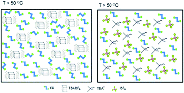
A novel ionically conducting colloidal suspension was prepared from a quaternary ammonium salt and ethylene glycol. The prepared colloidal medium was thoroughly characterized in terms of its ionic conductivity, rheological and thermal properties. The colloid exhibited sharp reversible sol ↔ gel transitions at 50–55 °C. The phase transitions were associated with drastic changes in viscosity (10−2 to 102 Pa s) and ionic conductivity (0.5–12.67 mS cm−1). The sharp transition at ambient temperature helps to preserve the dispersed state of different nanoparticles obtained via sonication in the medium. The soft solid nature of the colloid can be considered to mimick a biological environment and was evaluated as a soft electrolytic medium for monitoring enzyme kinetics.


Introduction

In recent times, designer solvents have been gaining increasing interest in a plethora of applications owing to the fact that these solvents can be custom made for specific applications.1 This has necessitated a good deal of research in preparing novel materials either by synthesizing novel compounds or by exploring novel combinations of commercially available components. The latter is being extensively studied, in view of the ease of preparation and the innumerable possible combinations of existing materials. An excellent example of this class of solvents is the spectrum of eutectic mixtures based on choline chloride ((2-hydroxyethyl) trimethyl ammonium chloride) and its acceptance in almost all possible applications requiring a fluid state.2 Our work aims to develop an ionic conducting soft material from readily available components so as to facilitate its direct use in applications requiring a solid media.

At present, soft solids are prepared by the quasi-solidification of ionic liquids and organic solvents. This is brought about by the addition of polymers,3 inorganic nanoparticles,4 and low molecular weight gelators.5,6 The polymeric analogues of ionic gels are being extensively prepared for various applications. The reversible thermal transitions observed in these polymer based ionic gels are attributed to the reversible cross linking of the polymer with temperature. By appropriate choice of a block polymer, He et al. could prepare a thermally reversible gel by the self assembly of poly (N-isopropyl acrylamide-b-ethylene oxide-b-N-isopropyl acrylamide) in an ionic liquid medium of 1-ethyl-3-methylimidazolium bis (trifluoromethylsulfonyl)-imide ([EMIM][TFSI]).7 Susan et al. prepared polymer based soft solids by in situ polymerization of vinyl monomers in ionic liquids.8 The high cost and moisture sensitivity of the ionic liquid based gels pose limitations for its use in practical applications.

Gels can also be formed by various secondary interactions such as van der Waals forces, hydrogen bonding, π–π interactions occurring within the components. These gels are prepared by the addition of a low concentration of an organogelator to the bulk solvent medium. The organogelator is capable of gelating the bulk solvent medium by promoting the formation of three dimensional network structures based on secondary interactions.9 The organic solvent molecules get trapped within the three dimensional structures causing gelation. These type of gels possess thermal reversibility much better than their polymeric counterpart as the three dimensional network structures are formed by non-covalent interactions unlike that in polymers.6 Smith et al. prepared room temperature gels of tetrafluoroborate based ionic liquid in DMSO solvent by the addition of an organogelator N-carbobenzyloxy-L-isoleucylaminooctadecane.10 Thermo reversible gels have also been prepared from long chain tertiary amines and other related tertiary and quaternary ammonium halide salts (Cn = 12, 18) in organic solvents.11

The soft solid properties of ionic gels together with the inherent ionic conductivity show immense potential in areas of electrochemical capacitors, DSSC, Li ion batteries.12,13 The sol–gel matrices have also been studied as a matrix for protein storage, enzymatic studies, enzyme catalyzed organic reactions and enzyme kinetics.14,15 It has been shown that the structure of the protein in its native state is very well preserved in a gel matrix compared to that in the molecular solvents. The gel matrix when associated with sufficient ionic conductivity are considered ideal medium for electrochemical studies of enzymatic processes. The colloidal suspension reported here, is found to exhibit these physical properties, and hence was explored as a media for the electrochemical monitoring of enzyme kinetics.

The aggregation of nanoparticles in solvent matrices is an important issue that can adversely affect their use in potential applications. The problem can be circumvented using a suitable dispersing medium.16 The nanoparticle dispersions have been prepared earlier in molecular solvents through proper control of surface potentials and surface forces of nanoparticles, yet highly loaded nanoparticle suspensions were found to be difficult to achieve. Through our studies, we demonstrate here that the colloidal suspension can be used for dispersing nanoparticles.

Experimental section

Materials

Tetra butyl ammonium tetraflouroborate (TBABF4), tetra butyl ammonium hexafluorophosphate (TBAPF6) from Aldrich and ethylene glycol, ethanol, acetonitrile, glycerol from Merck were used as received.

Urease from Canavalia ensiformis (Sigma Aldrich) was used as enzyme while urea was used as the substrate.

Pt foils (0.5 mm thickness) purchased from Advent research materials were used as electrodes for the ionic conductivity measurements.

Preparation

The two components shown in Scheme 1 were mixed in a molar ratio of 1[thin space (1/6-em)]:[thin space (1/6-em)]8 and heated at 80 °C under magnetic stirring till a homogeneous colorless solution was obtained. The solution attains a soft solid state as the temperature decreases below 50 °C.
image file: c5ra13303c-s1.tif
Scheme 1 Chemical structure of the components of the colloidal suspension.

Instruments

All the rheological measurements were performed in a commercial modular compact rheometer Anton Paar MCR 501 after loading a fresh sample for every run in concentric cylinder geometry CC17. This geometry has a gap of 0.71 mm, an effective length of 24.99 mm and requires a sample volume of 4.72 ml for every run. The sample temperature was controlled by a water circulation unit Viscotherm VT2. The rheological data was acquired using the Rheoplus software (version 3.40) provided by the manufacturer.

For the cryo-SEM experiments, the samples are transferred to a PP3000T cryo unit (Quorum Technologies) and freeze fractured with a cold knife. The fractured and cut samples are sublimated at −130 °C for 5 minutes and then sputtered with platinum inside the unit. Imaging of the samples were performed using Zeiss Ultra Plus cryo-SEM setup at a temperature of −160 °C. The transition temperature of the material developed was determined by differential scanning calorimetry (DSC) using Perkin-Elmer, Model Pyris-1 at a heating rate of 5 °C min−1. The texture of the colloidal suspension was observed using a polarising optical microscope (POM) (Olympus BX50) equipped with a heating stage (Mettler FP82HT) and a central processor (Mettler FP90).

The electrochemical characterization of the developed medium was performed by electrochemical impedance spectroscopy using Zahner electrochemical workstation in a frequency range of 0.1 to 105 Hz. The kinetics of enzyme–substrate reaction was monitored by a Lock in amplifier (model SR830 DSP, Stanford research systems) using Labview 2012, Stanford research-830 constant frequency.

Electrochemical cell design

A Teflon cell containing ‘H’ shaped grooves as shown in Fig. S1 was designed and prepared in our lab. Two platinum foils (10 × 6 × 0.5 mm) were placed within the vertical segments of the grooves. The Pt foils were placed parallel at a distance of 3 mm. The rear sides of the Pt foils were masked by means of Teflon tapes. The vertical segments of the ‘H’ shaped groove facilitates Pt foils to be placed in a constricted way while the gap formed amidst the Pt foils serves the purpose of a sample well (Fig. S1). The sample well can hold a colloid volume of 110 μL.

Results and discussion

Optimization of the components

The components of the colloidal suspension were chosen such that they possess optimal mutual interactions. This is because of the fact that any excessive mutual interactions among them would lead to the formation of a room temperature liquid which is not desirable.17 In view of this, the cation, anion and the organic solvent were optimized from several preliminary experiments and also based on the literature reports.

The quaternary ammonium cation used in our studies is symmetric with an alkyl chain of Cn = 4. This bulky cation is devoid of any polar functional groups. It is thus least expected for the cation to have any significant secondary interactions with the other components i.e., [BF4] and ethylene glycol (EG). However, recent reports on eutectic solvents prepared from tetra butyl ammonium chloride (TBA+Cl) and EG as well as tetrabutyl ammonium bromide (TBA+Br) and EG show that a strong hydrogen bonding interaction between the Cl/Br anion and EG is sufficient for the formation of a room temperature liquid.18 Thus, together with a symmetric tetrabutyl ammonium cation, a poorly interacting anion also needs to be selected so as to ensure the formation of a soft solid. Abbott et al. studied depressions in freezing point of mixtures containing hydrogen bond donor (HBD) and choline salts with different anions such as Cl, [BF4], [NO3] and F.2,17 The studies showed that the mixtures containing [BF4] were associated with least depression in freezing point and this was correlated with weak hydrogen bonding interactions between the anion and HBD. From our experiments, we have also observed that TBACl and TBABr form corresponding liquids with EG while TBABF4 and TBAPF6 form soft solid with EG. The studies on [C4mim][BF4] assisted gelation of DMSO also reported the crucial role of tetraflouroborate anion in gelating the solvent.10

The effect of solvent on the physical state of the salt on mixing was also studied by the inverted vial experiment. The quaternary ammonium salt [TBA]+[BF4] was fixed and EG (ε – 37) was replaced with other molecular solvents such as acetonitrile (ε − 37.5), glycerol (ε – 42.5) and ethanol (ε – 24). [TBA]+[BF4] dissolved completely in acetonitrile and ethanol while it gelated in glycerol. The results suggest that a structured/associated solvent has greater influence on the physical state of the mixture rather than its dielectric constant.

Based on all these observations, the components were chosen as [TBA]+[BF4] and EG (Scheme 1). A weak hydrogen bonding interaction between hydroxyl protons of EG and BF4 has been earlier confirmed in literature by fluorescence spectroscopy and NMR.19 Thus the symmetric tetrabutyl ammonium cation and the weak hydrogen bonding interactions of BF4 and EG would render a delicate balance of the polar–non polar characteristics to the medium and thus promote the formation of a stable colloidal suspension of [TBA]+[BF4] in EG.

Characterization of the colloidal suspension

As a preliminary test, a vial inversion experiment was carried out on the colloidal suspension. The ability of the suspension to resist flow under its own weight is observed by the vial inversion experiment. A set of vial inversion experiments were conducted with different ratios of TBABF4[thin space (1/6-em)]:[thin space (1/6-em)]EG. A ratio of 1[thin space (1/6-em)]:[thin space (1/6-em)]8 was optimized as the composition exhibited a fine balance in rigidity and fluidity. Photographs of the suspension in an inverted vial are shown in Fig. 1a. The mechanical strength of the composition corresponding to 1[thin space (1/6-em)]:[thin space (1/6-em)]8 was sufficiently high enough that a colloidal volume corresponding to 1.5 ml could hold a Teflon coated magnetic pellet (0.76 g) in an inverted vial as shown in Fig. 1a(ii).
image file: c5ra13303c-f1.tif
Fig. 1 Photographs of the vial containing the colloidal suspension and a Teflon coated magnetic pellet (i) at T > 60 °C and (ii) at RT (a) DSC curve (b) viscosity characteristics with temperature (c) frequency response at a strain amplitude of 0.05% (d) of the colloidal suspension.

The sol–gel transition of the colloidal suspension was monitored by differential scanning calorimetry (DSC) given in Fig. 1b. The gel → sol conversion was observed at 60 °C while the reverse transition corresponding to sol → gel occurred at 51 °C. The gelation and melting processes are accompanied by exothermic and endothermic peaks respectively as usually observed for any freezing or melting process. However, the sharp calorimetric peaks of the colloid observed in DSC reveal a first order transition which indicates that all molecules undergo the transition in unison.20 The driving force for the gelation of the colloid can be attributed to the aggregation of the salt components so as to favor the hydrogen bonding interactions within the solvent EG.

Fig. 1c shows the variation in the viscosity of the colloidal suspension within a temperature range of 25–65 °C. A sharp change in viscosity (almost 104) characteristic of sol–gel transition of the colloid was observed at a temperature close to 55 °C. The temperature associated with the sudden change in the viscosity values corresponds closely with the transition temperature observed in DSC both on heating and cooling.

The mechanical properties of the prepared colloidal suspension were analyzed from the frequency response measurements at room temperature at a strain amplitude of 0.05% (Fig. 1d). The loss modulus and storage modulus shown in figure signifies the fluid and solid phase behavior respectively. The moduli exhibit least dependence to the frequency as is evident from the figure. The loss modulus is lower than that of the storage modulus at all the frequencies studied. These characteristic features observed in the frequency sweep measurements confirm the soft solid behavior of the suspension.21 The elastic and viscous moduli of the suspension at room temperature were measured from the strain amplitude measurements at a constant angular frequency of 0.5 rad s−1 as given in Fig. S2. There is no linearity in the response over a range of strain amplitude between 0.1 to 100%. The elastic modulus lags behind the viscous modulus beyond the critical yield strain of 1% amplitude, indicating a permanent deformation of the structure.

The cryo-SEM image of the colloidal suspension was obtained to examine the sample morphology. Fig. 2a shows the evenly distributed spherical aggregates of ∼50 nm. The spherical features could have formed by the aggregation of TBABF4 in EG. These spherical aggregates of TBABF4 can be stabilized by the weak hydrogen bonding interactions between BF4 and –OH groups of EG. The aggregation behavior of [C4mim][BF4] have been studied by many groups. Bowers et al. observed that these ionic compounds form spherical aggregates in aqueous solution above its critical aggregation concentration.22 Dorbritz et al. observed the aggregation behaviour of [C4mim][BF4] in methanol, 2-propanol, and ethyl acetate by electrospray ionization mass spectrometry.23 The group observed that on decreasing the polarity of the solvent and increasing the concentration of the salt, the size of the aggregates could be increased. However, the three dimensional network structures characteristic of gel structure was not observed in the cryo-SEM images.


image file: c5ra13303c-f2.tif
Fig. 2 (a) Cryo-SEM image of the colloid (b) XRD of the colloid at different temperatures (c) variation in ionic conductivity of the colloidal suspension at different temperatures (d) proposed model of the colloidal suspension (dotted lines around the cube denote the hydrogen bonding interactions).

The birefringence observed in polarizing optical microscope (Fig. S3) at room temperature confirms the crystallinity of the suspension. X-ray diffraction studies were carried out to probe into the nature of the suspension at different temperatures and also with respect to the components. The room temperature XRD patterns of the suspension exhibited additive features of both the components viz., sharp diffraction peaks corresponding to [TBA]+[BF4] throughout the spectra and the broad peak of ethylene glycol in the wide angle region (Fig. 2b). A comparison of the XRD spectra of the colloidal suspension and its components, viz., [TBA]+[BF4] and EG is shown in (Fig. S4). There was no significant shift in peaks or absence of any peak as compared to [TBA]+[BF4] which indicate that the secondary interactions of [BF4] with EG do not hinder the crystallinity of the salt. It can also be noted that beyond the gel → sol transition temperature of the colloid, the entire set of sharp peaks of the suspension disappears due to the melting of salt as observed in Fig. 2b.

The ionic characteristics of the suspension were measured by electrochemical impedance spectroscopy (EIS) using two parallel planar Pt electrodes at the open circuit potential. The Bode phase diagram (Fig. S5a) of the colloid at room temperature shows that the phase angle remains zero at frequencies beyond 6 kHz and hence 10 kHz was optimized for our studies. At this high frequency, the capacitive reactance contribution to impedance is negligible and the system is defined by the solution resistance. The Bode plots at different temperatures from which the ionic conductivities of the suspension were measured are provided in Fig. S5b. Fig. 2c shows the variation in ionic conductivity of the suspension in a temperature range of 25–60 °C.

The ionic conductivity values varied between 0.5 and 12.67 mS cm−1. The values are 2–4 orders of magnitude greater than those reported on polymeric ionic gels (PEG or PEO).24 Sekhon et al. reported an ionic conductivity of the order 10−4 S cm−1 for ionic gels formed from a mixture of ternary solvent, salt and a polymer (PVdF).25 The ionic gels prepared by the gelation of DMSO associated with an ionic conductivity of ∼15 mS cm−1. However the study showed that there was no decrease in ionic conductivity before and after the phase transition. This was attributed to the fact that ions were not part of the gel network.12 The ionic conductivity of the gels formed from ionic liquid compatible cyclic carbonate network varied from 7 μS cm−1 to 3.2 mS cm−1 with increase in ionic liquid composition.26 It is evident from Fig. 2c that the phase transition corresponding to gel → sol at 55 °C, is associated with a seven fold increase in the ionic conductivity. This can be explained by the collapse of the spherical aggregates of quaternary ammonium salt at temperatures above 55 °C, causing the release of a large number of ions. A scheme of the colloid in sol and gel phases are provided in Fig. 2d. Further, it was observed from the rheological characteristics that the viscosity decreases drastically at the phase transition temperature, which can contribute to an increase in ionic mobility.

The colloidal suspension is associated with a fine balance of polar and non-polar components and is expected to have excellent solvent properties. The ambient temperature isotropic phase exhibited by these soft solids helps in solvent free processing of the suspension for experimental studies.

Applications of the TBABF4–EG colloidal suspension

Dispersing medium for nanoparticles. The sharp reversible sol–gel phase transition of the colloid can be utilized as a matrix for dispersing nanomaterials such as CNT, graphene and metal nanoparticles. A few of such dispersions of nanoparticles (CNT, graphene, Fe3O4 NP, AgNP) in the colloid are shown in the Fig. 3a. A vial containing 0.5 mg ml−1 nanoparticles in sol were initially sonicated at a temperature of about 50–60 °C. As soon as a stable dispersion of nanoparticles was achieved, the nanoparticle matrix was plunged into cold water. The sudden drop in temperature brings about the phase transition of sol → gel, thus freezing the well dispersed state of nanoparticles brought about by sonication. The nanoparticles dispersion in the colloid prepared by the method has the potential for use in electrochemical biosensors, electronic and optoelectronic applications.
image file: c5ra13303c-f3.tif
Fig. 3 (a) Inverted vial containing colloid (middle) and the nanoparticle dispersions of CNT, graphene, Fe3O4 NP and AgNP in the colloid (b) variation in the ionic conductance of the colloidal matrix with the progress of urea–urease reaction at different urea concentrations (baseline subtracted).
Electrochemical monitoring of enzyme kinetics in the colloid. The experiments involving enzyme–substrate reaction usually require immobilization of the enzyme. This is done either by the covalent attachment of the enzyme onto an insoluble carrier molecule or entrapment onto a gel matrix. The entrapment onto the gel matrix (usually polyacrylamide or crosslinked gelatin gel) is frequently used owing to the simplicity of the method. To the best of our knowledge, enzyme–substrate reaction has not been previously monitored in a colloidal suspension. To explore this possibility, we have made an attempt to utilize this soft solid as a medium for monitoring enzyme kinetics by using a model reaction of urea–urease. The urea–urease reaction is usually studied in a buffered media by spectrophotometry,27 calorimetry,28 titrimetry or conductivity measurements.29 Osakai et al., developed an amperometric sensor based on direct detection of ammonium ion transferred across a gel–solution interface.30 Recently, changes in the imaginary component of electrochemical impedance spectroscopy were utilized to monitor enzyme kinetics.31

In this work, we show that application of an appropriate high frequency ac potential, as determined from electrochemical impedance spectroscopy and a simultaneous measurement of the ac current response can provide information on the changes, if any, of the ionic content in the bulk medium brought about by the enzyme–substrate reaction. Any transformation that arises due to progress of the enzyme–substrate reaction brings about changes in the bulk ionic characteristics which can very well be monitored by impedance. Since the process is happening in bulk, the accessibility of the active redox centers of the enzyme to the electrode surface is not essential, thereby providing an alternative to the method of complex surface modifications of the electrodes.

In our studies, a small volume (110 μL) of the sol containing a known concentration of urea was injected amidst two parallel Pt electrodes kept at a distance of 3 mm (Fig. S1). The sol solidifies immediately to form a soft solid and the corresponding enzyme urease (1 μL of 1 mg ml−1 aqueous solution) was added. The soft solid nature of the matrix helps to overcome the problems such as solvent seepage and solvent evaporation that are usually associated with molecular solvents when used in microliter volumes. The enzyme–substrate reaction was allowed to take place in the soft solid matrix. The soft matrix would provide an environment that mimics a biological media which is an alternate to an aqueous buffer solution normally used in such studies. The urea–urease reaction is given as

 
(NH2CONH2) + 2H2O → 2NH4+ + CO32− (1)

As is evident from eqn (1), the reaction yields ammonium ions and carbonate ions. The progress of the reaction is expected to increase the ionic conductance of the medium. The real time monitoring of ionic conductance was carried out by impedance spectroscopy at open circuit potential.

To optimize the frequency at which the measurements were to be performed, Bode phase plot of the colloidal suspension containing the substrate was studied over the entire range of frequencies from 100 kHz to 100 mHz at the open circuit potential (Fig. S6). It was observed that at frequencies greater than 1 kHz, the impedance is contributed only by the resistance of the medium. We have optimized 20 kHz for our study as the phase angle was zero at this frequency.

The kinetics of urea–urease reaction was monitored at various concentrations of urea ranging from 0.5–5 mM. The observed changes in the ionic conductance with time were baseline corrected to remove the inherent ionic conductance of the soft solid-urea mixture. An increase in the ionic conductance with time was observed indicating that ions are generated with the progress of the reaction. It can be seen from Fig. 3b that for each concentration, the initial increase in ionic conductance is linear while it tends to plateau with time due to the saturation of the enzyme sites.

The initial rate of the urea–urease reaction was obtained from the slope of the linear segment of t = 0 to t = 80 s of Fig. 3b. Since the progress of the reaction is monitored as change in ionic conductance of the medium with time, the units of rate of reaction are expressed in terms of nS min−1 (Fig. 4). The plot of initial rate of the reaction vs. urea concentration was found to follow Michaelis–Menten eqn (2) with an adj. R2 of 0.96.

 
image file: c5ra13303c-t1.tif(2)
where Vmax represents the maximum velocity achieved by the system, [S] is the substrate concentration and Km is the Michaelis constant.


image file: c5ra13303c-f4.tif
Fig. 4 Michaelis–Menten fitting of the plot of the initial reaction rate vs. concentration of urea for the urease–urea reaction carried out in the TBABF4–EG colloidal matrix.

Km is determined from the concentration of the substrate at which the rate of the reaction is exactly half of Vmax. Thus, a lower Km indicates that the maximum velocity of the reaction Vmax is attained at small substrate concentrations. Km is also a measure of enzyme–substrate binding affinity. It is evident from the eqn (S3) that a low Km signifies a high binding affinity in the medium. The experiment was repeated to check the consistency of measurements and was averaged which gives a Km of 2.1 mM with a standard deviation of 0.1621. The Km values for the urea–urease reaction monitored under different experimental conditions were compared earlier by Qin & Cabral.32 In an unbuffered neutral medium, Km varied between 1.25–3.28 mM whereas the values were in the range of 9.5–20 mM in phosphate buffer and 1.2–3.8 mM in a citrate buffer. The value obtained in our studies is very much close to that obtained (Km = 1.8 mM) for the urea–urease reaction done in an unbuffered gel matrix of polyacrylamide.15

Conclusions

A non-polymeric soft matter was prepared by the appropriate choice of commercially available components viz., [TBA]+[BF4] and EG. The presence of both the polar and non-polar components imparts good solvent properties to the medium. The ambient temperature phase transition of colloid was utilized to freeze the nanoparticles in its dispersed state. The dispersions were stable in the colloidal matrix and were not found to settle with time. The inherent ionic conductivity and the soft solid characteristics of the colloidal suspension have been effectively used in this work for preparing an electrochemical medium for studying biological processes. The kinetics of the enzyme–substrate reaction (urea–urease) studied in the colloidal matrix has been shown to follow the Michaelis–Menten equation with a Km value of 2.1 mM.

Acknowledgements

We thank Mr Rajib Basak for all the rheological studies, Mr Dhason A. for cryo-SEM and Mrs Vasudha K. N. for DSC and XRD characterizations.

References

  1. M. Freemantle, Chem. Eng. News, 1998, 76, 32 Search PubMed.
  2. (a) A. P. Abbott, G. Capper, D. L. Davies, H. L. Munro, R. K. Rasheed and V. Tambyrajah, Chem. Commun., 2001, 2010 RSC; (b) Q. Zhang, K. D. O. Vigier, S. Royer and F. Jerome, Chem. Soc. Rev., 2012, 41, 7108 RSC; (c) A. Hayyan, F. S. Mjalli, I. M. AlNashef, Y. M. Al-Wahaibi, T. Al-Wahaibi and M. Ali Hashim, J. Mol. Liq., 2013, 178, 137 CrossRef CAS PubMed; (d) A. Hayyan, F. S. Mjalli, I. M. AlNashef, Y. M. Al-Wahaibi, T. Al-Wahaibi and M. Ali Hashim, Thermochim. Acta, 2012, 541, 70 CrossRef CAS PubMed.
  3. S. Washiro, M. Yoshizawa, H. Nakajima and H. Ohno, Polymer, 2004, 45, 1577 CrossRef CAS PubMed.
  4. P. Wang, S. M. Zakeeruddin, P. Comte, I. Exnar and M. Gratzel, J. Am. Chem. Soc., 2003, 125, 1166 CrossRef CAS PubMed.
  5. J. C. Ribot, C. G. Sanchez, R. Hoogenboom and U. S. Schubert, J. Mater. Chem., 2010, 20, 8279 RSC.
  6. K. Hanabusa, H. Fukui, M. Suzuki and H. Shirai, Langmuir, 2005, 21, 10383 CrossRef CAS PubMed.
  7. Y. He and T. P. Lodge, Macromolecules, 2008, 41, 167 CrossRef CAS.
  8. M. A. H. Susan, T. Kaneko, A. Noda and M. Watanabe, J. Am. Chem. Soc., 2005, 127, 4976 CrossRef CAS PubMed.
  9. J. Bonnet, G. Suissa, M. Raynal and L. Bouteiller, Soft Matter, 2014, 10, 3154 RSC.
  10. N. W. Smith, J. Knowles, J. G. Albright and S. V. Dzyuba, J. Mol. Liq., 2010, 157, 83 CrossRef CAS PubMed.
  11. L. Lu and R. G. Weiss, Chem. Commun., 1996, 2029 RSC.
  12. (a) G. P. Pandey and S. A. Hashmi, J. Mater. Chem. A, 2013, 1, 3372 RSC; (b) X. Liu and T. Osaka, J. Electrochem. Soc., 1997, 144, 3066 CrossRef CAS PubMed; (c) W. Lu, K. Henry, C. Turchi and J. Pellegrino, J. Electrochem. Soc., 2008, 155, A361 CrossRef CAS PubMed.
  13. (a) W. Zhang, C. Yuan, J. Guo, L. Qiu and F. Yan, ACS Appl. Mater. Interfaces, 2014, 6, 8723 CrossRef CAS PubMed; (b) H. Ye, J. Huang, J. J. Xu, A. Khalfan and S. G. Greenbaum, J. Electrochem. Soc., 2007, 154, A1048 CrossRef CAS PubMed.
  14. (a) H. F. Mullerad and D. Avnir, J. Am. Chem. Soc., 2005, 127, 8077 CrossRef PubMed; (b) M. Yan, J. Ge, Z. Liu and P. Ouyang, J. Am. Chem. Soc., 2006, 128, 11008 CrossRef CAS PubMed.
  15. (a) S. Wu, J. Lin and S. Chan, Appl. Biochem. Biotechnol., 1994, 47, 11 CrossRef CAS; (b) B. Atkinson and I. Rousseau, Biotechnol. Bioeng., 1977, 19, 1065 CrossRef CAS PubMed.
  16. (a) M. E. Mackay, A. Tuteja, P. M. Duxbury, C. J. Hawker, B. V. Horn, Z. Guan, G. Chen and R. S. Krishnan, Science, 2006, 311, 1740 CrossRef CAS PubMed; (b) B. I. Kharisov, H. V. R. Dias, O. V. Kharissova, A. Vázquez, Y. Peña and I. Gómez, RSC Adv., 2014, 4, 45354 RSC.
  17. A. P. Abbott, G. Capper, D. L. Davies, R. K. Rasheed and V. Tambyrajah, Chem. Commun., 2003, 70 RSC.
  18. (a) F. S. Mjalli, J. Naser, B. Jibril, V. Alizadeh and Z. J. Gano, J. Chem. Eng. Data, 2014, 59, 2242 CrossRef CAS; (b) R. Yusof, E. Abdulmalek, K. Sirat and M. B. A. Rahman, Molecules, 2014, 19, 8011 CrossRef PubMed.
  19. T. Singh, K. S. Rao and A. Kumar, ChemPhysChem, 2011, 12, 836 CrossRef CAS PubMed.
  20. Molecular Gels: Materials with Self-Assembled Fibrillar Networks, ed. R. G. Weiss and P. Terech, 2005, pp. 233–244 Search PubMed.
  21. Y. Hea and T. P. Lodge, Chem. Commun., 2007, 2732 RSC.
  22. J. Bowers, C. P. Butts, P. J. Martin, M. C. Vergara-Gutierrez and R. K. Heenan, Langmuir, 2004, 20, 2191 CrossRef CAS.
  23. S. Dorbritz, R. Wolfgang and U. Krag, Adv. Synth. Catal., 2005, 347, 1273 CrossRef CAS PubMed.
  24. S. S. Soni, K. B. Fadadu and A. Gibaud, Langmuir, 2012, 28, 751 CrossRef PubMed.
  25. S. S. Sekhon and H. P. Singh, Solid State Ionics, 2002, 152–153, 169 CrossRef CAS.
  26. S. Jana, A. Parthiban and C. L. L. Chai, Chem. Commun., 2010, 46, 1488 RSC.
  27. F. Lespinas, G. Dupuy, F. Revol and C. Aubry, Clin. Chem., 1989, 35, 654 CAS.
  28. M. J. Todd and J. Gomez, Anal. Biochem., 2001, 296, 179 CrossRef CAS PubMed.
  29. D. D. van Slyke and R. M. Archibald, J. Biol. Chem., 1944, 154, 623 CAS.
  30. T. Osakai, T. Kakutani and M. Senda, Anal. Sci., 1988, 4, 529 CrossRef CAS.
  31. S. Shrikrishnan, K. Sankaran and V. Lakshminarayanan, J. Phys. Chem. C, 2012, 116, 16030 CAS.
  32. Y. Qin and J. M. S. Cabral, Appl. Biochem. Biotechnol., 1994, 49, 217 CrossRef CAS.

Footnote

Electronic supplementary information (ESI) available: Additional details on the characterization of the colloidal suspension. The basics of the electrochemical concepts used for the ionic conductivity studies and enzyme kinetics are also provided. Bode plots of the colloid used for the electrochemical studies are also provided. See DOI: 10.1039/c5ra13303c

This journal is © The Royal Society of Chemistry 2015
Click here to see how this site uses Cookies. View our privacy policy here.