Congli Wang,
Bo Li,
Weifen Niu,
Shasha Hong,
Bassam Saif,
Songbai Wang,
Chuan Dong and
Shaomin Shuang*
Department of Chemistry and Chemical Engineering and Institute of Environmental Science, Shanxi University, Taiyuan 030006, P. R. China. E-mail: smshuang@sxu.edu.cn; Fax: +86-531-7018842; Tel: +86-531-7011688
First published on 13th October 2015
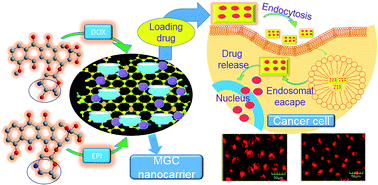
β-Cyclodextrin modified graphene oxide–magnetic (MGC) nanocomposite as an innovative drug carrier was the first to be developed via an effective layer-by-layer-assembly method. Doxorubicin hydrochloride (DOX) and epirubicin hydrochloride (EPI) as model drugs were loaded onto the MGC via π–π stacking, hydrogen bonding and hydrophobic interactions. The MGC exhibits remarkably higher loading capacity for DOX (909.09 mg g−1) and EPI (781.25 mg g−1) than magnetic graphene oxide (MG). The release profiles of the drugs are pH-sensitive which would control the release in the acidic cytoplasm of cancer cells. Furthermore, cellular uptake using fluorescein isothiocyanate (FITC) labeled MGC proves successful internalization of MGC into the cytoplasm of MCF-7 cells. The fluorescence images demonstrate that MGC/DOX, to a certain extent, displays a more excellent delivery and superior release than MGC/EPI, due to the chiral selective function of β-cyclodextrin (β-CD). The pure MGC shows no obvious cytotoxicity while drug loaded MGC reveals significantly high potency for killing MCF-7 breast cancer cells, suggesting that multi-functionalized MGC is an efficient nanoplatform for targeted delivery and controlled release of stereoisomeric anticancer drugs for biomedical applications.
To reduce side effects and increase therapeutic efficacy, intensive research is ongoing to focus on the development of effective delivery systems to facilitate cellular uptake of drug and realize intelligent controlled release. A widely prescribed strategy to achieve efficient external targeting is to introduce various types of magnetic nanoparticles on the large surface of GO, which can drive the drug carriers to specific tumor tissues by a guided magnetic field.14 A variety of magnetic nanoparticles including Fe3O4,10 Co3O4,15 Cu/Ni alloy,16 Ni,17 etc. have been successfully deposited on the basal plane of GO by physical or chemical methods. However, it's relatively hard to control the size distribution, deposition density and morphology of the as-prepared magnetic nanoparticles. Magnetic Fe3O4 has a great deal of advantages over other magnetic nanoparticles such as strong superparamagnetism, low toxicity, excellent biocompatibility and easy preparation process.18 The immobilization of Fe3O4 on the platform of GO can not only avoid aggregating of Fe3O4 nanoparticles but also protect them from oxidation. Unfortunately, the hydrophilic groups of GO are easy to lost by heating during the process of in situ deposition of metallic salts, affecting further dispersion and loading amount of the nanocomposites.19 The further functionalization of magnetic graphene oxide (MG) is essential for applicable purpose.
Various efforts have been made to explore potential applications of MG in drug delivery systems. Yang's group firstly developed superparamagnetic graphene oxide–Fe3O4 nanoparticles and conjugated the MG with folic acid for loading doxorubicin hydrochloride (DOX).10,20 In order to improve the biocompatibility, a series of polymers, such as polyethylene glycol (PEG),21 chitosan (CHI),22 polyethylenimine (PEI),23 poly[2-(dimethylamino) ethyl methacrylate] (PDMAEMA),24 and so on, were employed to modify MG for loading and release of DOX and levofloxacin. However, relative low loading capacity and complicated manipulated process have confined further use. The drug delivery systems are facing a great challenge toward high effective loading. Recently, Qiu et al. firstly introduced β-cyclodextrin (β-CD) to graphene oxide–magnetic nanohybrid as a novel stationary phase for efficient enantioseparation of tryptophan.25 Yan and Liu et al. fabricated β-CD functionalized magnetic graphene nanocomposite as an adsorbent for the removal of organic dyes26 and heavy metal ions.27 These researches inspire us and motivate us to try to expand such a nanocomposite for drug delivery.
DOX and epirubicin hydrochloride (EPI) (illustration in Scheme 1) as stereoisomers of anthracycline-based broad-spectrum antitumor drugs, play an important role in restraining the composition of nucleic acids of cancer cells.28 Only when the drugs are sufficiently migrated inside the cancer cells will it operates. However, most of the drug vehicles could neither be transported into the cell due to the large size nor show adequate release of the drug in cytoplasm,29 resulting in a very low internalization of drug and poor effect of chemotherapy. The combination of β-CD and MG as the drug vessel possesses not only the unique inclusion properties of β-CD but also loading and magnetic targeting capacities of MG, which provides an admirable opportunity for targeted drug delivery.
![]() | ||
Scheme 1 The preparation of β-CD-functionalized magnetic graphene oxide (MGC) and representation of loaded with DOX and EPI. |
Herein, a targeted delivery carrier of β-cyclodextrin modified graphene oxide–magnetic nanohybrid was successfully fabricated. β-CD was assembled onto the surface of MG by hydrophobic, van der Waals and hydrogen bonding interactions. DOX and EPI were used as model drugs to assess the drug-loading and releasing properties and the effect of pH was examined. Cell cytotoxicity and cellular imaging were carried out toward further application. It will be expected that this nanocarrier has a great potential in targeted delivery, controlled release and biomedical applications.
MG sample | mGO![]() ![]() |
MGC sample | mMG![]() ![]() |
β-CD wt% |
---|---|---|---|---|
MG1 | 1![]() ![]() |
MGC1 | 1![]() ![]() |
9.12 |
MGC2 | 1![]() ![]() |
9.69 | ||
MGC3 | 1![]() ![]() |
9.24 | ||
MG2 | 1![]() ![]() |
MGC4 | 1![]() ![]() |
14.08 |
MGC5 | 1![]() ![]() |
21.18 | ||
MGC6 | 1![]() ![]() |
15.25 | ||
MG3 | 1![]() ![]() |
MGC7 | 1![]() ![]() |
8.73 |
MGC8 | 1![]() ![]() |
9.81 | ||
MGC9 | 1![]() ![]() |
9.2 |
![]() | (1) |
![]() | (2) |
To demonstrate the morphology and size of as-prepared nanohybrid, representative TEM was used to characterize the surface morphologies. As shown in Fig. 1, it is obvious that graphite powder has been exfoliated into thin flakes of GO after oxidation (Fig. 1b). The pure Fe3O4 nanoparticles are seriously gathered for clusters and the particle diameter significantly increases, which is disadvantageous for drug delivery (Fig. 1a). After Fe3O4 is immobilized on the surface of GO, it is found that a large amount of uniform Fe3O4 nanoparticles with average size of about 13 ± 1.2 nm are distributed on the surfaces and edges of the GO nanosheet, which is necessary to keep a powerful magnetism (Fig. 1c). Furthermore, no great aggregation of Fe3O4 or large blank on GO is observed, and almost no unbonded Fe3O4 is viewed outside of GO sheets. It is noted that the morphology of MG shows no significant difference from GO, unambiguously indicating that this moderate method would not damage the layered structure during the functionalized process. After the modification of β-CD, it can be clearly observed that GO is the matrix of Fe3O4 nanoparticles and the β-CD is covering on the surface, like a film,32 which may be important to improve the water solubility of the carrier. The TEM images could provide direct evidence for the successful fabrication of MGC nanocomposites. In AFM images (Fig. S1, ESI†), GO reveals a height of 1.0 nm, suggesting a single layer graphene oxide sheet.33 After functionalization of Fe3O4 and β-CD, an obvious increase of thickness is observed, mainly due to the attachment of Fe3O4 and β-CD on both planes of GO.
The loading behaviors of DOX or EPI on the nanocarriers were determined by UV-Vis spectrophotometer. As displayed in Fig. 2A and B, the specific UV-Vis peaks at 252, 290 and around 485 nm are attributed to absorption of DOX (Fig. 2A(a)). The broad peaks at 253 and 298 nm are ascribed to the characteristic absorption of GO (Fig. 2A(b)). After MG and MGC are loaded with DOX, the characteristic absorbance peak at 485 nm presents and the peak around 252 nm shifts to 255 nm, which are corresponded to the peak of free DOX, demonstrating that DOX molecules have been successfully loaded onto MG and MGC (Fig. 2A(c) and (d)). DOX and EPI have stereoisomeric structures with the same absorption peak, so the similar characteristic absorption peaks have been observed in both EPI, MG/EPI and MGC/EPI as shown in Fig. 2B(a), (c) and (d), indicating that EPI can also be loaded on the nanocarrier.
To further characterize the formation of GO, MG and MGC, corresponding FTIR spectra were shown in Fig. 2C. As expected, the spectrum of GO is quite fit in with previous work.34 GO with the layered structure has a lot of oxygen-containing functional groups located on its basal planes and around the sheet edges, including hydroxyl, epoxy and carboxylic acid groups.35 The broad and intense band at 3435 cm−1 is attributed to the stretching of O–H (Fig. 2C(a)). The characteristic band at 1741 cm−1 is ascribed to the CO of carboxylic groups which is shifted to 1610 cm−1, indicating the formation of the –COO– group after modifying with Fe3O4 (Fig. 2C(c)). The peak at 1633 cm−1 is corresponding to the stretching of C
C. The bands at 1267 cm−1 and 1033 cm−1 are associated with the stretching vibration of C–OH and the deformation vibration of C–O, respectively (Fig. 2C(a)). For MG, the FTIR spectrum is different from GO as evidenced by the appearance of an intense peak at 579 cm−1, which belongs to the stretching vibration of Fe–O bond. Moreover, the Fe–O stretching peak has shifted to higher wavenumbers compared to that of 569 cm−1 in bulk Fe3O4 (Fig. 2C(b)), demonstrating that GO has been successfully functionalized with Fe3O4 nanoparticles.36 After MG nanohybrid is further modified with β-CD, it could be observed that the FTIR spectrum of the MGC exhibits characteristic absorbing peaks of β-CD. The band at 1161 cm−1 is assigned to the coupled C–O–C stretching/O–H bending vibrations. The featured peaks at 1010 cm−1 and 1076 cm−1 are attributed to the coupled C–O/C–C stretching/O–H bending vibrations, and the C–H/O–H bending vibration is at 1429 cm−1 (Fig. 2C(d) and (e)). The above results demonstrate that β-CD has been anchored on the surface of MG successfully.
To prove the deposition of Fe3O4 on the surface of GO and the crystal structures of MG and MGC, the XRD patterns of MG and MGC nanocomposite are presented in Fig. 2D. The diffraction peaks of the as-prepared MG nanoparticles at 2θ = 30.31°, 35.58°, 43.19°, 53.58°, 57.18° and 62.87° are corresponding to the (220), (311), (400), (422), (511) and (440) planes of magnetite respectively, which is consistent with the cubic spinel crystal Fe3O4 according to the Joint Committee On Powder Diffraction Standards (JCPDS) Card no. 89-3854 (Fig. 2D(a)).37,38 As compared with MG nanocarrier, the MGC nanocomposites display all diffraction peaks of Fe3O4 (Fig. 2D(b)), and no obvious shifts can be observed, indicating that Fe3O4 nanoparticles have been successfully connected with GO and have no effect on the layered structure during all the experimental processes. What's more, the average particle size of Fe3O4 could be estimated to be 14 nm by the formula of Scherrer, which is in accordance with the data measured from the TEM images (Fig. 1C).
The magnetic properties of MGC nanohybrids were investigated at room temperature on a VSM. As shown in Fig. 2E, the magnetic hysteresis loops of all the samples display S-like curves. The values of specific saturation magnetization (Ms) of Fe3O4 (Fig. 2E(a)), MG (Fig. 2E(b)) and MGC (Fig. 2E(c)) are 65.79, 57.25 and 55.64 emu g−1, respectively. It is evident that the MG possesses good magnetic property while the Ms of MG is weaker than Fe3O4, implying that Fe3O4 has been successfully in situ deposited on the surface of the GO. The Ms of MGC has a slight decrease for MG, which is likely ascribed to the absorption of β-CD leading to a decrease of the relative content of Fe3O4 in the nanohybrid. Furthermore, the magnetization curves have no coercivity and remanence, indicating that there is no remaining magnetization. The insets of Fig. 2E demonstrate that once an external magnetic field is applied, the uniform and rufous suspension of MG and MGC could be attracted quickly toward the magnet, giving rise to the solution clear and transparent. These results reveal that both MG and MGC exhibit excellent superparamagnetic behavior and could be used for targeting drug delivery.
TGA was further used to investigate the amount of β-CD conjugating on the surface of MG. A series of MG and MGC nanocomposites were prepared by the chemical deposition and “grafting to” way. As shown in Table 1, MG1-3 were synthesized with different feed amounts of FeCl3 and FeCl2 by the same of GO, and MGC1-3 were prepared by the same MG1 with different feed quantities of β-CD. Thus, products MG1-3 have the same GO but different contents of Fe3O4, and the generated MGC1–3 or MGC4–6 or MGC7–9 should have the same MG but different contents of β-CD. Fig. 3 displays the thermal behaviors of MG and MGC nanohybrids. As shown in Fig. 3, a slow weight loss at low temperature (<100 °C) is observed, which can be assigned to the loss of residual solvent and water. Another stage of weight reduction occurs below 220 °C, indicating the removal of the labile oxygen-containing functional groups on GO and Fe3O4.39 It is found that the weight reduction region at about 280–600 °C could be attributed to the decomposition of β-CD, and the significant weight loss in the high-temperature region (about 600–800 °C) belongs to the breakdown of GO skeleton.31 Thus, the amount of β-CD molecules could be determined as exhibited in Table 1, demonstrating that large amount of β-CD has been adsorbed on the surface of MG. In Table 1, feed amount means added amount and grafting content means modified content. We notice that the feed amount of β-CD on MGC3, MGC6 and MGC9 with the ratio 1:
30 is greater than that on MGC2, MGC5 and MGC8 with 1
:
20. While modified content for MGC3, MGC6 and MGC9 is lower than that on MGC2, MGC5 and MGC8. The phenomenon may be interpreted as that the modified content of β-CD is mainly related to the efficient surface area of MG. When the feed amount of β-CD is saturated, the grafting amount is optimal. When excessive β-CD is added, the grafting content shows a slight decline due to the existence of space steric hindrance and site competition between molecules in the dynamic process, which is the opposite of rise theoretically. So the MGC5 shows the highest modified amount of β-CD. This is a fascinating result that MG nanohybrid could graft many β-CD molecules, which may provide a good opportunity to load more drugs (the following data will reveal this point). As a comparison, the TGA of MG3 has also been investigated, as shown in Fig. 3C. It is clear that MGC exhibits the much higher thermal stability than MG below 200 °C, which offers a better protection for Fe3O4.
![]() | ||
Fig. 3 TGA curves of MG and MGC hybrid composites, (A) MGC1 (a), MGC2 (b) and MGC3 (c). (B) MGC4 (a), MGC5 (b) and MGC6 (c). (C) MG3 (a), MGC7 (b), MGC8 (c) and MGC9 (d). |
To investigate the effect of pH on the drug loading of MGC5, the same concentrations of MGC5 and DOX or EPI were incubated in different PBS (0.1 M, pH = 2.0–7.0) and the unbound drugs were isolated with a filter membrane. The difference of absorption between pure drug and supernatant fluid is the amount of drug loaded by the carrier. As shown in Fig. 4C, it can be observed that the loading capacity of MGC5 for DOX and EPI rises gradually from pH 2.0 to 6.0 and increases dramatically at pH 7.0, whereas, the loading capacity decreases greatly when the pH is greater than 7.0. The loading experiment is a dynamic equilibrium process.41 In an acidic or basic environment, the lower or higher pH could cause high degree of protonation of DOX and EPI, resulting in the partial dissociation of hydrogen bonding42,43 and the loading capacity is not behaving well. At pH 7.0, multiple strong hydrogen-bonding interactions between –OH, –COOH in MGC and –OH, –NH2 in drugs are conducive to the incorporation of MGC and drugs, leading to a rapid increase of loading amount.42,44 As a result, the subsequent experiments are operated at pH 7.0. With a large specific surface and hydrophobic cavity, MGC is supposed to have excellent loading behavior. To determine the saturated level of DOX and EPI loaded on MGC5, different amounts of DOX or EPI were added into the carrier solution of fixed concentration at pH 7.0. After removal of free DOX, as shown in Fig. 4D(a), it is found that the loading capacity of MGC5 for DOX increases with the increasing initial DOX concentration, and it reaches 720 μg mg−1 when the initial DOX concentration is 360 μg mL−1, which is still unsaturated. For EPI, similar tend can be observed in Fig. 4D(b), and the loading amounts of EPI increase from 92 to 509 μg mg−1 which is less than DOX. Through curve fitting of Langmuir isothermal adsorption, it is found that the maximum adsorption capacities of MGC5 for DOX and EPI are 909.09 and 781.25 mg g−1 respectively (Table 2), considerably higher than the previously reported values.45,46
Models | Langmuir isotherm | Freundlich isotherm | ||||
---|---|---|---|---|---|---|
Drug | DOX | EPI | DOX | EPI | ||
a Note: the unit of qm, KL and Kf is mg g−1, mL mg−1 and mL g−1, respectively. | ||||||
Adsorption parameters | qm | 909.09 | 781.25 | n | 2.1354 | 1.6929 |
KL | 0.01123 | 0.0150 | Kf | 49.472 | 18.733 | |
R2 | 0.9062 | 0.9236 | R2 | 0.9648 | 0.9843 |
In order to study the influence of the content of β-CD on drug loading, the loading amounts of MG1-3 and MGC1-9 for DOX and EPI under the same conditions are also presented for comparison in Fig. 4E. It states clearly that the carriers of MG1-3 possess a low ability about 600 and 500 μg mg−1 for DOX and EPI loading, while MGC1-9 have a loading capacity of almost 850 and 700 μg mg−1 for DOX and EPI, nearly 1.5 times that of MG, indicating that β-CD is responsible for much more drug loading. As shown in Fig. 4E and Table 1, the variation tendency of drug loading is in a good agreement with the content of β-CD. In addition, the loading amount of MGC5 is the largest among the above nanocarrier due to the greatest content of β-CD. These results reveal that the MGC nanocarrier could load of DOX and EPI with exceptionally high loading amounts. Thereby, this nanocarrier could be a potential vehicle for highly efficient load and delivery of DOX or EPI, and MGC5 is chosen for the following experiments.
In addition, to further obtain the distribution of adsorption molecules with more reliability when the adsorption process reaches to an equilibrium state, the adsorption isotherms are built and compared with the common isotherm models of Langmuir (eqn (3)) and Freundlich (eqn (4)), as follows:
![]() | (3) |
![]() | (4) |
It is well known that the Langmuir model is basically used for monolayer adsorption on a surface with a homogeneous system, while the Freundlich model is suitable for multilayer adsorption of a heterogeneous system.47 In order to assess different isotherm models, the fitted plots from the two isotherms are shown in Fig. 5 with the experimental data for adsorption of DOX or EPI onto the nanocarrier of MGC, and the values of parameters for the two models are revealed in Table 2. As shown in Table 2, by comparison of linear correlation coefficients (R2), it is found that the Freundlich model is a better fit for the experimental results of drug loading than the Langmuir model, indicating a multilayer adsorption.
It is reported that this type of nanocarrier could be taken up by tumor cells via the endocytosis at the physiological pH of 7.4, while it declines to an acidic pH value nearly 5.3 in endosomes and lysosomes.48 For purpose of enhancing the efficient release of the anti-cancer drug inside the tumor tissue or cells and avoiding undesired release during the delivery process in blood circulation, it's significant for the release of drug from the carrier to be pH-responsive. Thus, the drug-release behaviors of MGC/DOX and MGC/EPI were performed at pH 5.3 and 7.4 at 37 °C. At various time points, quantitative solutions were taken out and the supernatants were determined by fluorescence measurement. As shown in Fig. 6, at neutral PBS (pH 7.4) to simulate normal physiological condition, the DOX and EPI are released from MGC nanohybrids in a very slow fashion and only below 18% (DOX) and 15% (EPI) of the total bound drugs are released for 50 h, which can be interpreted as that the stronger hydrogen bonding interactions results in a slower and inefficient release. However, in acidic conditions, the release of DOX and EPI from MGC rises dramatically within the first 10 hours and then levels off gradually from 10 to 50 h. The maximum cumulative release of DOX and EPI are about 60% and 35% at pH 5.3 within 50 h, due to the weak hydrogen-bonding interactions between the –OH and –COOH groups on MG and –OH and –NH2 groups in DOX or EPI. The release includes two parts, one is from the desorption of the surface-bound drug molecules on MGC surface,49 another is from the gradual release of drug molecules which have been entrapped in β-CD cavities.50 The complete release of drugs couldn't be achieved at either pH, owing to the diffusion equilibrium between MGC/drug inclusion complexes and the released drug which would inhibit the complete drug release.51 Furthermore, for the steric hindrance of EPI is much higher than DOX, under the same condition, more EPI will remain entrapped in the cavity of β-CD and less EPI than DOX would be released from the MGC carrier. In addition, drug release behaviors are in complete agreement with the loading amounts and the content of β-CD, suggesting that the conjugation of β-CD is conducive to the release of drug. It is well known that the microenvironments in extracellular tissues of tumors and intracellular lysosomes and endosomes are acidic, so pH-responsive drug release from MGC is advantageous to the clinical therapy of cancer.52 As an ideal delivery vehicle, the DOX and EPI can be transported to the targeted site by the water-dispersive MGC, then the drug payload could be internalized to cancer cells through endocytosis. In the acidic microenvironments (pH is about 5.3), protonation of –NH2 groups in DOX or EPI molecules can lead to a positive charge to weaken the part of the hydrogen bond interactions between drugs and the nanocarrier MGC, triggering more desired drug release. Considering the different pH-sensitivity release behaviors of DOX and EPI from MGC, the multi-functionalized nanohybrid MGC could be served as an excellent candidate for intelligent drug release to satisfy different needs.
![]() | ||
Fig. 6 Cumulative release of DOX (A) and EPI (B) from MGC/DOX and MGC/EPI nanoparticles at pH 5.3 and 7.4. |
The following work confirms the anti-tumor efficacy of MGC/DOX and MGC/EPI, for this purpose, MCF-7 cells were incubated with MGC/DOX and MGC/EPI in concentrations of 1 to 20 μg mL−1 for 24 h. The cell viability was determined by standard MTT assay, as shown in Fig. 7C(c) and (d), the dose-dependent cell viability for both MGC/DOX and MGC/EPI decreases with increasing carrier concentrations. Furthermore, at each concentration, the cytotoxicity of MGC/DOX is remarkably higher than that of MGC/EPI, likely due to the different release behaviors of the carrier.53 The results reveal that the MGC loaded of DOX or EPI has the potential for killing MCF-7 cells. As a control, the pure MG and MGC were carried out with the protocol described above, suggesting that the cell viability of pure MG decreases gradually with the increment of the concentration of MG (Fig. 7C(b)). Whereas, it is worthy of note that no obvious toxicity is found at all tested concentrations of MGC without drug loading (Fig. 7C(a)), since the cell growth rates with MGC are the same as that of the medium control, proving that the conjugation of β-CD has greatly reduce cytotoxicity and enhance the biocompatibility of the nanocarrier. It can draw a conclusion that MGC is biocompatible and efficient to provide a probable secure vehicle for drug delivery and cancer therapy.
Footnote |
† Electronic supplementary information (ESI) available. See DOI: 10.1039/c5ra13082d |
This journal is © The Royal Society of Chemistry 2015 |