DOI:
10.1039/C5RA12849H
(Paper)
RSC Adv., 2015,
5, 75348-75353
A sensitive immobilization-free electrochemical assay for T4PNK activity based on exonuclease III-assisted recycling
Received
10th July 2015
, Accepted 25th August 2015
First published on 28th August 2015
Abstract
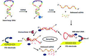
Coupling the properties of T4 polynucleotide kinase (T4PNK) catalyzing the transfer of ATP γ-phosphate residue to 5′-hydroxyl termini of nucleic acids, with lambda exonuclease (λ-exo), a highly processive 5′–3′ exonuclease, digesting the 5′-phosphorylated strand of a double DNA to produce single-strand DNA and mononucleotides, this work develops a highly sensitive immobilization-free electrochemical method for the detection of T4PNK activity based on λ-exo and exonuclease III-assisted signal amplification. Upon the reaction of T4PNK and λ-exo on substrate-DNA, single-stranded DNA segments (released-ssDNA) are dissociated to hybridize with a methylene blue-labeled hairpin probe (MB-DNA) and then the digestion of MB-DNA from the blunt 3 terminus by exonuclease III is activated, resulting in the release of MB-labeled mononucleotides and the complementary DNA segment, followed by the latter hybridizing with another MB-DNA to initiate the cycling process. With a smaller size and less negative charge, the MB-labeled mononucleotide thus diffuses facilely to the negatively charged indium tin oxide (ITO) electrode, actuating an amplified electrochemical signal, and the detection limit of the proposed assay can reach as low as 0.005 U mL−1. Additionally, this assay can avoid the sophisticated probe immobilization processes. Therefore, this strategy exhibits the merits of high sensitivity, simplicity, and being immobilization-free for electrochemical assay of T4PNK activity, which is consequently believed to bear considerable potential as a detection platform for related researches.
Introduction
Originally discovered in protein extracts of Escherichia coli bacteria infected with T-even phage1 and acknowledged to be one of the most frequently used enzymes in molecular biology, T4 polynucleotide kinase (PNK) functions as an indispensable epigenetic event that manages cell function by altering gene expression.2 Briefly, it is capable of phosphorylating DNA at 5′-OH termini through catalyzing the transfer of the γ-phosphate residue of ATP to nucleic acids and oligonucleotides, and also acting as a DNA 3′-phosphatase, consequently playing a vital role in assay of DNA adducts, oligonucleotides, nucleic acid metabolism and repair of DNA lesions.3 Furthermore, it is demonstrated by some reports that aberrant PNK activity is closely related to critical human disorders such as Bloom syndrome, Werner syndrome, and Rothmund–Thomson syndrome.4 Additionally, PNK inhibition is very likely to enhance the sensitivity of human tumors to γ-radiation, also representing a prosperous means of improving the efficacy of existing cancer treatments.5 In this case, it is of tremendous value to develop accordingly a sensitive and simple assay for PNK activity and its potential inhibitor as well.
Conventionally, several approaches were described for the detection of phosphorylation and the activity assay of DNA kinase, including radical isotope 32P-labeling, polyacylamide gel electrophoresis (PAGE), autoradiography and fluorescence.6 Nevertheless, they were somewhat time-consuming, laborious, complex, or involved radio labeling. Recently, to conquer these drawbacks, a great variety of strategies for sensitive detection of PNK activity assays have been developed, including fluorescence assays,7 colorimetric assays8 and electrochemical assays.9 For instance, Song et al. combined β-cyclodextrin polymer-based fluorescence enhancement of pyrene with the exonuclease reaction for sensitive T4PNK activity detection.10 Hou et al. developed a fluorescence assay for the detection of PNK activity based on the coupled λ-exo cleavage reaction and catalytic assembly of bimolecular beacons.11 Lian et al. realized the detection of PNK activity by means of a DNA phosphorylation-accelerated λ-exo cleavage reaction coupled with cationic conjugated polymer-mediated fluorescence resonance energy transfer.12 In spite of remarkable advances toward DNA phosphorylation detection made by these previous protocols, further promotion of the analytical performances, involving convenience, fast response and high sensitivity in particular, has not yet satisfied the increasing needs for biological research, clinic diagnostics, and drug discovery.
Compared with methods such as radioactive, fluorescence and PAGE systems, electrochemical biosensors have otherwise attracted considerable interest in phosphorylation assay because of their simplicity, rapid response, cost-effectiveness, sensitivity and potential ability for real-time and on-site analysis.13 In the past decade, a surging number of biosensors accordingly have been designed for the detection of kinase activity by investigating the current and charge responses of redox probes conjugated during the phosphorylation processes. Besides, to further improve the sensitivity of the biosensors, several methods have been developed through exploiting signal amplification strategies,14 among which target recycling is of high competitiveness, allowing a single target molecule to interact with multiple nucleic acid-based signaling probes and thus offering an intriguing alternative to achieve signal amplification. In the as-proposed approach, after triggering the selective displacement or digestion of the signaling probe, the target probe hybridization sequently releases the intact DNA target to actuate the displacement or digestion of other signaling probes, thus leading to multiple signaling events and a gain in signal amplification. Previously and successfully, these signal amplification strategies have been developed based on fluorescence and electrochemical measurements, issuing an impressive detection sensitivity.15 Worthy of mention, in contrast to traditional immobilization protocols, is the immobilization-free process, which has the merits of giving reproducible measurements performed by a simple electrode washing step. Moreover, it is superior in efficiency to the hybridization of the target to the electrode-bound probe since it can avoid effectively steric hindrance. Hence combining target recycling with the immobilization-free electrochemical detection, this method is very likely to be a robust and promising strategy, enjoying the advantage of improved sensitivity and no longer requiring extra operation processes and instrumentation.
In this study, we demonstrated an immobilization-free electrochemical methylene blue-labeled hairpin probe (MB-DNA)-based method for detection of enzyme activity with signal amplification by exonuclease III-assisted recycling which is believed to be reported for the first time using an electrochemical assay method to detect T4PNK activity. The principle is on the basis of differential diffusivity between electroactive species-labeled oligonucleotides and electroactive species-labeled mononucleotides on a negatively charged electrode surface due to the enzymatic cleavage activity of exonuclease III. In this way, not only does this strategy provide a novel, immobilization-free, simple and sensitive platform for DNA polynucleotide kinase-concerned biological process evolution, but it also holds tremendous potential for application in assays for other essential enzymes and biomolecular diagnosis.
Experimental
Materials and chemicals
T4 polynucleotide kinase (T4PNK), lambda exonuclease (λ-exo), adenosine diphosphate (ADP) and adenosine triphosphate (ATP) were purchased from Invitrogen Technology (Shanghai, China). We designed the substrate-DNA and methylene blue-labeled DNA (MB-DNA) and they were synthesized by Sangon Biotech Co., Ltd (Shanghai, China) and the sequences are listed in Table 1. All other chemicals used were of analytical reagent grade and used without further purification. All aqueous solutions were prepared and diluted using ultrapure water (specific resistance >18.2 MΩ cm) from the Millipore Milli-Q water system (Millipore, Bedford, MA).
Table 1 Sequences of oligonucleotides used in the experiments
Oligonucleotides |
Sequences |
Substrate-DNA |
5′-![[G with combining low line]](https://www.rsc.org/images/entities/b_char_0047_0332.gif) ![[A with combining low line]](https://www.rsc.org/images/entities/b_char_0041_0332.gif) ![[T with combining low line]](https://www.rsc.org/images/entities/b_char_0054_0332.gif) ![[T with combining low line]](https://www.rsc.org/images/entities/b_char_0054_0332.gif) ![[T with combining low line]](https://www.rsc.org/images/entities/b_char_0054_0332.gif) ![[T with combining low line]](https://www.rsc.org/images/entities/b_char_0054_0332.gif) ![[T with combining low line]](https://www.rsc.org/images/entities/b_char_0054_0332.gif) ![[C with combining low line]](https://www.rsc.org/images/entities/b_char_0043_0332.gif) ![[C with combining low line]](https://www.rsc.org/images/entities/b_char_0043_0332.gif) ![[T with combining low line]](https://www.rsc.org/images/entities/b_char_0054_0332.gif) ![[T with combining low line]](https://www.rsc.org/images/entities/b_char_0054_0332.gif) ![[C with combining low line]](https://www.rsc.org/images/entities/b_char_0043_0332.gif) ![[T with combining low line]](https://www.rsc.org/images/entities/b_char_0054_0332.gif) ![[C with combining low line]](https://www.rsc.org/images/entities/b_char_0043_0332.gif) GAACAAAAGGAA GATACGT A![[C with combining low line]](https://www.rsc.org/images/entities/b_char_0043_0332.gif) ![[G with combining low line]](https://www.rsc.org/images/entities/b_char_0047_0332.gif) ![[A with combining low line]](https://www.rsc.org/images/entities/b_char_0041_0332.gif) ![[G with combining low line]](https://www.rsc.org/images/entities/b_char_0047_0332.gif) ![[A with combining low line]](https://www.rsc.org/images/entities/b_char_0041_0332.gif) ![[A with combining low line]](https://www.rsc.org/images/entities/b_char_0041_0332.gif) ![[G with combining low line]](https://www.rsc.org/images/entities/b_char_0047_0332.gif) ![[G with combining low line]](https://www.rsc.org/images/entities/b_char_0047_0332.gif) ![[A with combining low line]](https://www.rsc.org/images/entities/b_char_0041_0332.gif) ![[A with combining low line]](https://www.rsc.org/images/entities/b_char_0041_0332.gif) ![[A with combining low line]](https://www.rsc.org/images/entities/b_char_0041_0332.gif) ![[A with combining low line]](https://www.rsc.org/images/entities/b_char_0041_0332.gif) ![[A with combining low line]](https://www.rsc.org/images/entities/b_char_0041_0332.gif) ![[T with combining low line]](https://www.rsc.org/images/entities/b_char_0054_0332.gif) -3′ |
MB-DNA |
5′-![[A with combining low line]](https://www.rsc.org/images/entities/b_char_0041_0332.gif) ![[G with combining low line]](https://www.rsc.org/images/entities/b_char_0047_0332.gif) ![[G with combining low line]](https://www.rsc.org/images/entities/b_char_0047_0332.gif) ![[A with combining low line]](https://www.rsc.org/images/entities/b_char_0041_0332.gif) ![[A with combining low line]](https://www.rsc.org/images/entities/b_char_0041_0332.gif) ![[G with combining low line]](https://www.rsc.org/images/entities/b_char_0047_0332.gif) CGTACGTA![[T with combining low line]](https://www.rsc.org/images/entities/b_char_0054_0332.gif) ![[C with combining low line]](https://www.rsc.org/images/entities/b_char_0043_0332.gif) ![[T with combining low line]](https://www.rsc.org/images/entities/b_char_0054_0332.gif) ![[T with combining low line]](https://www.rsc.org/images/entities/b_char_0054_0332.gif) ![[C with combining low line]](https://www.rsc.org/images/entities/b_char_0043_0332.gif) ![[C with combining low line]](https://www.rsc.org/images/entities/b_char_0043_0332.gif) TTTGT T-MB-C-3′ |
Instruments
A CHI660E electrochemical workstation (Beijing Chinese Science Days Technology Co., Ltd, Beijing, China) was chosen for differential pulse voltammetry (DPV). The electrochemical workstations had a three-electrode mode comprising an indium tin oxide (ITO) electrode, a platinum counter electrode and an Ag/AgCl reference electrode. All the potentials in this work were with respect to the Ag/AgCl electrode. Before each electrochemical measurement, the ITO electrodes were sequentially sonicated in an Alconox cleaning detergent, acetone, ethanol and water lasting for 15 min each. After these cleaning procedures, a negatively charged surface could be obtained.15e
T4PNK-catalyzed phosphorylation
All DNA samples were prepared in 10 mM Tris–HCl buffer (pH 7.4, 10 mM MgCl2). The concentration of the substrate-DNA stock solution was 10 μM, and the concentration of the MB-DNA stock solutions was 10 μM. Prior to the T4PNK enzyme reaction, all DNA samples were pretreated with the following procedure: heat to 90 °C and incubate for 5 min, and then cool slowly to 37 °C and incubate for 1 h. The obtained DNA solutions were stored at 4 °C for further use.
In a typical phosphorylation and cleavage assay, consisting of 10 mM Tris–HCl, 100 nM substrate-DNA, 10 mM MgCl2, 1.0 mM ATP, 10 units λ-exo and a certain amount of T4PNK, the 200 μL enzyme reaction buffer could introduce phosphorylation sites at the 5′-hydroxyl termini and cause the formation of a 5′-phosphoryl termini product that could be recognized by λ-exo. The resulting solution was kept at 37 °C for a certain period of time before the electrochemical measurements.
Assay optimization
For the purpose of fulfilling the best sensing effect, several important conditions were optimized, including the λ-exo concentration, the ATP concentration, the MB-DNA concentration and the reaction time. The λ-exo concentration range was 1.0–15.0 units, the ATP concentration range was 0.1–5.0 mM, the MB-DNA concentration was 0.1–10 μM, and the reaction time range was 0 to 60 min. Equal volumes of T4PNK storage buffer were added into each enzyme reaction mixture respectively.
Exonuclease III-assisted T4PNK activity detection
A volume of 200 μL Tris–HCl buffer containing 5 μM MB-DNA, 10 units of exonuclease III and the released-ssDNA produced by T4PNK-catalyzed phosphorylation was incubated at 37 °C for a certain period of time. Then, for each DPV scann, 20 μL of the mixture solution was pipetted onto the ITO working electrodes. Before the experiments, the electrolyte buffer was washed by ultrapure water and then thoroughly purged with nitrogen gas. And DPV measurements were conducted in 10 mM Tris–HCl (pH 7.4) containing 50 mM KCl by scanning the potential from −0.2 to 0.5 V, with a modulation amplitude of 25 mV and a scan rate of 100 mV s−1. All the measurements were repeated at least five times.
Influence of inhibitors on T4PNK activity
In order to further evaluate the inhibitor screening ability of the proposed assay, two known inhibitors of T4PNK, namely ADP and (NH4)2SO4 with different concentrations were added in the reaction buffer, respectively. Then 1.0 mM ATP, 10 units of λ-exo, and 10 units of T4PNK were added before the reaction solution was kept at 37 °C for 30 min. The detection processes were recorded using the aforementioned procedure.
Results and discussion
Design strategy of T4PNK activity detection
As shown in Fig. 1, the principle for the strategy was that T4PNK could catalyze the transfer of the γ-phosphate residue of ATP to the 5′-hydroxyl termini of nucleic acids, and λ-exo was a highly processive 5′–3′ exonuclease capable of digesting the 5′-phosphorylated strand of a double DNA to generate single-strand DNA and mononucleotides. In the absence of T4PNK, substrate-DNA could maintain the stem-loop structure due to the binding of the complementary sequences at the ends, in which case the released-ssDNA could not be produced and because of the electrostatic repulsion, MB-DNA could not yet get close to the negative ITO electrode. Therefore, the electrode exhibited a negligible electrochemical signal. Whereas in the presence of T4PNK and λ-exo, T4PNK catalyzed the phosphorylation reaction toward the hydroxyl group at the 5′-termini of the substrate-DNA to yield the 5′-phosphorylated substrate-DNA, which could be immediately cleaved by λ-exo, resulting in the released-ssDNA. Furthermore, the amount of released-ssDNA was positively related to the activity of T4PNK. Then the released-ssDNA hybridized with MB-DNA whose stem-loop structure was opened, forming double-stranded DNA with a receded 3′ terminus, which subsequently initiated the digestion by exonuclease III to degrade MB-DNA from the 3′ terminus and liberate the electroactive species. In this way, an electrochemical signal could be observed. It should be noted that exonuclease III could recognize and cleave from the 3′ end of a duplex DNA with a blunt or receded 3′ terminus, and a duplex DNA with a protruded 3′-terminus was not a substrate for exonuclease III. Since the 3′ terminus of the released-ssDNA in the double-stranded DNA was not fully hybridized to its complementary stand, the released-ssDNA would not be digested by exonuclease III. So the released-ssDNA then could hybridize with MB-DNA and enter a new cycle, therefore amplifying the signal and dramatically increasing the detection sensitivity with an extremely low amount of released-ssDNA. Thus, sensitive electrochemical detection of T4PNK based on exonuclease III-aided signal amplification was fulfilled.
 |
| Fig. 1 Schematic illustration for T4PNK activity detection based on the λ-exo cleavage reaction and exonuclease III-assisted signal amplification. | |
Feasibility of detection of T4PNK activity
To verify the feasibility, agarose gel electrophoresis experiments were employed. The DNA phosphorylation-induced digestion of substrate-DNA by λ-exo was primary in this strategy, which thus was examined by gel electrophoresis. As shown in Fig. 2A, in the absence of T4PNK or λ-exo, an obvious band was observed, indicating no substrate-DNA digestion. When T4PNK and λ-exo were both present, no obvious band was observed, indicating a high efficiency of phosphorylation-induced digestion of substrate-DNA by λ-exo in the presence of T4PNK. Additionally, after the MB-DNA and released-DNA incubated with exonuclease III, the band disappeared. The results were identical to those expected.
 |
| Fig. 2 (A) Agarose (2%) gel electrophoresis image. Lane 1: marker; lane 2: substrate-DNA; lane 3: substrate-DNA + T4PNK; lane 4: substrate-DNA + λ-exo; lane 5: substrate-DNA + T4PNK + λ-exo; lane 6: released-DNA + MB-DNA; lane 7: released-DNA + MB-DNA + exonuclease III. (B) DPV scans of reaction mixtures in response to different controls. (a) T4PNK + λ-exo + exonuclease III, with all three enzymes existing in the reaction mixture; (b) in the absence of T4PNK in the reaction mixture; (c) in the absence of λ-exo in the reaction mixture; (d) in the absence of exonuclease III in the reaction mixture. | |
DPV measurements were further applied to prove the proposed strategy. As shown in Fig. 2B, a significant detectable current signal of methylene blue was observed when all three enzymes of T4PNK, λ-exo, and exonuclease III were present (curve a). Whereas in the absence of T4PNK (curve b), λ-exo (curve c), or exonuclease III (curve d), no obvious current response is shown owing to the electrostatic repulsion between the negatively charged ITO electrode and the negative MB-DNA backbones. In this way, a single released-ssDNA was able to trigger the digestion of multiple MB-DNA into the more diffusive electrochemical indicator (MB), which thereby generated an amplified electrochemical signal. Therefore, this result demonstrated the feasibility of this method for the assay of T4PNK activity.
Optimization of assay conditions
The time of phosphorylation was a vital parameter in optimizing the signal gain of the electrochemical strategy. It was found that the current response of the electrochemical assay was strongly affected by the T4PNK incubation time. As shown in Fig. 3A, with the increase of the phosphorylation time, the current response increased and tended to the relative maximum current signal at 30 min. Nevertheless, with a further increase in the phosphorylation time, the current response otherwise did not increase accordingly, which suggested that the phosphorylation reaction was done in not more than 30 min. Thus, 30 min in the following experiments was chosen as the appropriate time.
 |
| Fig. 3 (A) The curve of the different incubation times for phosphorylation at 37 °C (0–60 min); (B) the curve of different ATP concentrations (0.1–5 mM); (C) the curve of different λ-exo concentrations (0–15 U); (D) the signal-to-noise ratio of different MB-DNA concentrations (0.1–10 μM). | |
ATP played an important role in the phosphorylation process, whose influence of concentration thus was examined, and the results are shown in Fig. 3B. It could be seen that with the ATP concentration increasing, higher current responses of the DNA phosphorylation were observed. While the concentration of ATP was >1.0 mM, a slight decrease is shown. Caused by higher concentrations of ATP during the phosphorylation process, this slight inhibition effect was possibly attributed to a competitive binding reaction between the DNA and ATP. As a result, when the concentration of ATP was relatively high, the binding site of T4PNK was partially blocked. Therefore, 1.0 mM was employed as the optimal ATP concentration to obtain a high sensitivity.
The released-ssDNA from the hairpin substrate DNA could affect the hybridization with the MB-DNA probe, eventually determining the detection sensitivity. In this point of view, different λ-exo concentrations were evaluated that could liberate released-ssDNA upon phosphorylation by T4PNK and digestion by λ-exo. The current intensity was found to increase with the amount of λ-exo and almost reached the saturation value at an amount of 10 units of λ-exo (Fig. 3C). Thus, 10 U λ-exo was selected for the following experiments.
Identically, the initial amount of MB-DNA also served as an indispensable substrate in the performance of the assay. Provided that the initial concentration of MB-DNA was too high, most MB-DNA would overcome the electrostatic repulsion and diffuse to the electrode surface, issuing in a strong background signal. From the other respect, if the initial concentration of MB-DNA was too low, a small electrochemical signal would still be observed though when all the MB was digested by exonuclease III, making it difficult to distinguish the signal from noise. Thus, a concentration of MB-DNA ranging from 0.1 to 10 μM was studied and the condition of 5 μM MB-DNA was fixed due to its best signal-to-noise ratio (Fig. 3D). Therefore, the concentration of MB-DNA was optimized at 5 μM in all the following experiments.
Electrochemical assay of T4PNK activity
Using this fabricated electrochemical platform, a series of different concentrations of T4PNK (from 0.005 to 20 U mL−1) were assayed to demonstrate the ability of the detection to sensitively quantify T4PNK activity under the optimum experimental conditions. As shown in Fig. 4A, it is demonstrated that with an increase in the concentration of T4PNK varying from 0.005 to 20 U mL−1, the DPV signal increased gradually. The corresponding results are shown in Fig. 4B which displays the relationship between the current signal and the T4PNK concentration, and in the Fig. 4B inset, a relatively good linearity is in the range from 0.005 U mL−1 to 1.0 U mL−1. The correlation equation could be described as I = 5.28 × 10⁻⁷X + 1.65 × 10⁻⁸ with the correlation coefficient R2 = 0.9618, where I was current intensity, and X was the T4PNK concentration. The direct detection limit of the method was 0.005 U mL−1. It was demonstrated through the result that the exonuclease III-aided signal amplification strategy could remarkably upgrade the detection sensitivity and it was facile to observe the signal even when the T4PNK concentration was as low as 0.005 U mL−1. The developed method achieved comparable or even better sensitivity against some other signal amplification strategies listed in Table 2. In previous methods employing electrode-bound MB-DNA, compromised by the hindrance effect of the electrode surface and the limited conformational freedom of the bound MB-DNA, the high selectivity with a hairpin structure was often restrained. Different from the traditional MB-DNA-based electrochemical biosensors in which the MB should be immobilized on the electrode surface with the reaction of sequence recognition and hybridization, the immobilization-free process in this method bore the merit of reproducible measurements performed by a simple electrode washing step. Moreover, it was more efficient than hybridization of ssDNA to the electrode-bound probe because it could avoid steric hindrance to a great extent.
 |
| Fig. 4 (A) DPV curve response to different concentrations of T4PNK. The different concentrations of T4PNK are 0.005, 0.01, 0.03, 0.05, 0.08, 0.10, 0.20, 0.50, 1.0, 5.0, 10.0 and 20.0 U mL−1 respectively. (B) Concentration curve for T4PNK. Linear relationship between the current and the concentration of T4PNK from 0.005 to 0.1 U mL−1 is shown in the inset. | |
Table 2 Comparison of analytical performance for T4PNK activity detection by different reported methods
Strategy |
Technique |
LOD (U mL−1) |
Linear range (U mL−1) |
Reference |
β-Cyclodextrin polymer enhancement |
Fluorescence |
0.02 |
0–0.25 |
10 |
Coupled λ-exo and catalytic assembly of bimolecular beacons |
Fluorescence |
0.001 |
0.001–0.1 |
11 |
λ-exo cleavage-induced DNAzyme releasing strategy |
Fluorescence |
0.005 |
0.005–0.2 |
14a |
Coupled λ-exo and exonuclease III recycling |
Fluorescence |
0.01 |
0.01–1 |
16a |
TiO2 nanotubes and Au nanoparticles |
Electrochemical |
0.003 |
0.01–10 |
9a |
Ferrocene-functionalized SWCNTs |
Electrochemical |
0.01 |
0.01–0.1 |
9b |
Dual-signaling amplification coupled with exonuclease reaction |
Electrochemical |
0.02 |
0.05–10 |
16b |
Streptavidin-gold nanoparticles and enzymatic amplification |
Electrochemical |
0.01 |
0.01–5 |
16c |
Coupled exonuclease and graphene oxide platform |
Electrochemical |
0.05 |
0.05–10 |
14d |
Based on exonuclease III-assisted recycling |
Electrochemical |
0.005 |
0.005–1.0 |
This work |
To evaluate the feasibility of the proposed assay, we have further investigated its analytical performance for T4PNK detection in biological samples. The samples were prepared by adding T4PNK in a 50% cell lysates contained reaction buffer, which thus were employed to simulate the intracellular environment during the test procedure. As shown in Fig. 5, in a reaction system containing cell lysates, a gradual increase in current signal was observed as the concentration of T4PNK increases. In this case, the result was in good agreement with those in pure reaction buffer without cell lysates, suggesting that this method could be applicable for T4PNK detection in biological samples.
 |
| Fig. 5 DPV curve response of this system for various activity of T4PNK in 50% cell lysate. | |
Evaluation of T4PNK activity inhibition
In order to exploit the possible application in the inhibition assay, the validity of the developed method to evaluate the inhibitors of T4PNK was proven using ADP and ammonium sulfate as model inhibitors. Both ADP and ammonium sulfate, as shown in Fig. 6, remarkably inhibited the phosphorylation process in a dose-dependent manner. Then, the half-maximal inhibitory concentration values were determined for the T4PNK inhibitors to further demonstrate the utility of this assay used in a quantitative inhibition assay. The addition of 15 mM (NH4)2SO4 or 1.5 mM ADP could inhibit phosphorylation by approximately 50%. The results manifested that this approach could exert various effects on phosphorylation quickly and simply.
 |
| Fig. 6 The effect of different concentrations of T4PNK inhibitors: (A) different concentrations of ADP (0–5 mM); (B) different concentrations of (NH4)2SO4 (0–50 mM). The assay was carried out in a reaction buffer solution containing 10 U T4PNK, 1.0 mM ATP and 10 U λ-exo. | |
Conclusions
In this paper, we described an electrochemical-sensing platform for immobilization-free, signal-on and highly sensitive detection of the activity and inhibition of T4PNK based on an exonuclease III-assisted recycling strategy. Since the step of DNA immobilization was eliminated, the target detection would not demand any labour or time consumption. Taking advantage of the unique strategy of signal amplification, the detection limit could reach as extremely low as 0.005 U mL−1. Besides, this assay could be utilized in screening inhibitors of T4PNK. In terms of its appealing analytical characteristics, this sensing strategy possibly was endorsed with major applications in biomedical research. Provided that it was further developed to an electrochemical microarray assay, it would enjoy an infinitely promising employment in researching the interactions between proteins and nucleic acids.
Acknowledgements
This work was supported in part by the National Natural Science Foundation of China (21305164), State Key Laboratory of Chemo/Biosensing and Chemometrics, Hunan University (2013019).
Notes and references
- C. C. Richards, Proc. Natl. Acad. Sci. U. S. A., 1965, 54, 158 CrossRef.
- C. J. Whitehouse, R. M. Taylor, A. Thistlethwaite, H. Zhang, F. Karimi-Busheri, D. D. Lasko, M. Weinfeld and K. W. Caldecott, Cell, 2001, 104, 107 CrossRef CAS.
-
(a) D. H. Phillips and V. M. Arlt, Nat. Protoc., 2007, 2, 2772 CrossRef CAS PubMed;
(b) A. Rasouli-Nia, F. Karimi-Busheri and M. Weinfeld, Proc. Natl. Acad. Sci. U. S. A., 2004, 101, 6905 CrossRef CAS PubMed;
(c) M. EI-Atifi, I. Dupre, B. Rostaing, A. L. Benabid and F. Berger, BioTechniques, 2003, 35, 262 Search PubMed;
(d) C. Ma, X. Yang, K. Wang, Z. Tang, W. Li, W. Tan and X. Lv, Anal. Biochem., 2008, 372, 131 CrossRef CAS PubMed;
(e) C. Frauendorf, F. Hausch, I. Rohl, A. Lichte, S. Vonhoff and S. Klussmann, Nucleic Acids Res., 2003, 31, e34 CrossRef PubMed;
(f) F. Karimi-Busheri, A. Rasouli-Nia, J. Allalunis-Turner and M. Weinfeld, Cancer Res., 2007, 67, 6619 CrossRef CAS PubMed.
- S. Sharma, K. M. Doherty and R. M. Brosh, J. Biochem., 2006, 398, 319 CrossRef CAS PubMed.
-
(a) G. K. Freschauf, F. Karimi-Busheri, A. Ulaczyk-Lesanko, T. R. Mereniuk, A. Ahrens, J. M. Koshy, A. Rasouli-Nia, P. Pasarj, C. F. Holmes, F. Rininsland, D. G. Hall and M. Weinfeld, Cancer Res., 2009, 69, 7739 CrossRef CAS PubMed;
(b) S. L. Allinson, Future Oncol., 2010, 6, 1031 CrossRef CAS PubMed.
-
(a) L. K. Wang and S. Shuman, J. Biol. Chem., 2001, 276, 26868 CrossRef CAS PubMed;
(b) C. Chappell, L. A. Hanakahi, F. Karimi-Busheri, M. Weinfeld and S. C. West, EMBO J., 2002, 21, 2827 CrossRef CAS PubMed;
(c) M. Meijer, F. Karimi-Busher, T. Y. Huang, M. Weinfeld and D. Young, J. Biol. Chem., 2002, 277, 4050 CrossRef CAS PubMed.
-
(a) C. Ma, H. Fang, K. Wang, K. Xia, H. Chen and W. Zeng, Anal. Biochem., 2013, 443, 166 CrossRef CAS PubMed;
(b) F. Zhou, G. Wang, D. Shi, Y. Sun, L. Sha, Y. Qiu and X. Zhang, Analyst, 2015, 140, 5650 RSC;
(c) M. Tao, Z. Shi, R. Cheng, J. Zhang, B. Li and Y. Jin, Anal. Biochem., 2015, 485, 18 CrossRef CAS PubMed.
-
(a) H.-X. Jiang, D.-M. Kong and H.-X. Shen, Biosens. Bioelectron., 2014, 55, 133 CrossRef CAS PubMed;
(b) C. Jiang, C. Y. Yan, J. H. Jiang and R. Q. Yu, Anal. Chim. Acta, 2013, 766, 88 CrossRef CAS PubMed;
(c) Z. Wang, R. Lévy, D. G. Fernig and M. Brust, J. Am. Chem. Soc., 2006, 128, 2214 CrossRef CAS PubMed.
-
(a) G. Wang, X. He, G. Xu, L. Chen, Y. Zhu, X. Zhang and L. Wang, Biosens. Bioelectron., 2013, 43, 125 CrossRef CAS PubMed;
(b) Y. Wang, X. He, K. Wang, X. Ni, J. Su and Z. Chen, Biosens. Bioelectron., 2012, 32, 213 CrossRef CAS PubMed;
(c) S. Liu, T. Liu and L. Wang, Chem. Commun., 2015, 51, 176 RSC.
- C. Song, X. Yang, K. Wang, Q. Wang, J. Liu, J. Huang, L. He, P. Liu, Z. Qing and W. Liu, Chem. Commun., 2015, 51, 1815 RSC.
- T. Hou, X. Wang, X. Liu, T. Lu, S. Liu and F. Li, Anal. Chem., 2014, 86, 884 CrossRef CAS PubMed.
- S. Lian, C. Liu, X. Zhang, H. Wang and Z. Li, Biosens. Bioelectron., 2015, 66, 316 CrossRef CAS PubMed.
- D. Grieshaber, R. MacKenzie, J. Vörös and E. Reimhult, Sensors, 2008, 8, 1400 CrossRef CAS PubMed.
-
(a) S. F. Liu, J. J. Ming, Y. Lin, C. F. Wang, T. Liu, C. B. Cheng and F. Li, Sens. Actuators, B, 2014, 192, 157 CrossRef CAS PubMed;
(b) L. Zhang, T. Hou, H. Li and F. Li, Analyst, 2015, 140, 4030 RSC;
(c) Y. Wang, Y. Li, H. Ma, X. Ren, W. Cao, T. Yan and Q. Wei, RSC Adv., 2015, 5, 31262 RSC;
(d) L. Lin, Y. Liu, X. Zhao and J. Li, Anal. Chem., 2011, 83, 8396 CrossRef CAS PubMed.
-
(a) X. Wu, Y. Chai, P. Zhang and R. Yuan, ACS Appl. Mater. Interfaces, 2015, 7, 713 CrossRef CAS PubMed;
(b) S. Liu, Y. Wang, C. Zhang, Y. Lin and F. Li, Chem. Commun., 2013, 49, 2335 RSC;
(c) J. Su, H. Zhang, B. Jiang, H. Zheng, Y. Chai, R. Yuan and Y. Xiang, Biosens. Bioelectron., 2011, 29, 184 CrossRef CAS PubMed;
(d) Q. Xu, A. Cao, L. Zhang and C. Zhang, Anal. Chem., 2012, 84, 10845 CrossRef CAS PubMed;
(e) F. Xuan, X. Luo and I.-M. Hsing, Anal. Chem., 2012, 84, 5216 CrossRef CAS PubMed.
-
(a) Z.-M. Zhu, R.-Q. Yu and X. Chu, Anal. Methods, 2014, 6, 6009 RSC;
(b) Y. Peng, J. Jiang and R. Yu, RSC Adv., 2013, 3, 18128 RSC;
(c) T. Hou, X. Wang, X. Liu, C. Pan and F. Li, Sens. Actuators, B, 2014, 202, 588 CrossRef CAS PubMed.
|
This journal is © The Royal Society of Chemistry 2015 |
Click here to see how this site uses Cookies. View our privacy policy here.