
Supporting of mixed ZnS–NiS semiconductors onto clinoptilolite nano-particles to improve its activity in photodegradation of 2-nitrotoluene
Parisa Mohammadyariab and
Alireza Nezamzadeh-Ejhieh*abc
aDepartment of Chemistry, Shahreza Branch, Islamic Azad University, P. O. Box 311-86145, Shahreza, Isfahan, Iran. E-mail: arnezamzadeh@iaush.ac.ir; Fax: +98 31 53291018; Tel: +98 31 53292515
bYoung Researchers and Elite Club, Shahreza Branch, Islamic Azad University, Shahreza, Iran
cRazi Chemistry Research Center (RCRC), Shahreza Branch, Islamic Azad University, Isfahan, Iran. Tel: +98 31 53292500
First published on 1st September 2015
Abstract
Photodegradation of a 2-nitrotoluene (2-NT) aqueous solution was studied using ZnS–NiS supported onto clinoptilolite nano-particles (ZnS–NiS–NCP) and a Hg lamp as the radiation source. A planetary ball-mill was used for preparation of nano-particles of clinoptilolite (NCP). The photocatalyst was prepared via sulfiding of the Zn–Ni exchanged NCP and characterized by Fourier transformation infra red spectroscopy (FT-IR), X-ray diffraction (XRD), UV-Vis diffuse reflectance spectroscopy (DRS) and transmission electron microscopy (TEM). UV-Vis spectrophotometric measurements were performed for the determination of degradation and mineralization of the pollutant. Degradation of the pollutant was also confirmed by chemical oxygen demand (COD) and high performance liquid chromatography (HPLC). The optimal operation parameters were found to be: 0.75 g L−1 of the ZnS9.7%–NiS3.0%–NCP catalyst and 55 ppm of 2-NT at pH 11. Unsupported ZnS and NiS showed smaller degradation activities than the supported one. The degradation process obeyed first-order kinetics.
1. Introduction
Nitro derivatives of toluene such as 2-nitrotoluene (C7H7NO2) cause contamination of groundwater and soil at numerous sites located in packing, explosive manufacturing plants, storage and military-related facilities. They are very toxic, mutagenic and carcinogenic.1 For example, 2-nitrotoluene causes flushing of the face, dizziness, dyspnea (difficult breathing), cyanosis, nausea, vomiting, muscular weakness, increased pulse and respiratory rate, irritability and convulsions [2-nitrotoluene MSDS]. In general, industrial effluents containing toxic, volatile and refractory organic pollutants cause severe environmental problems and thus their removal or degradation is required.2 In this regard, different methods have been developed for the removal of various volatile and non-volatile pollutants from water and wastewater. Common biological treatments are slow or ineffective, while the traditional physicochemical treatments such as adsorption on activated carbon and nano-filtration have inherent limitations in applicability, effectiveness and cost.3 They also transfer the pollutants from one phase to another, leaving a problem of disposal of the transferred materials.4In recent two decades, advanced oxidation processes (AOPs) such as photo-Fenton, photo-oxidation, photo-reduction and photocatalytic technologies have been widely used for the removal of different organic pollutants from water.5 Although common homogeneous Fenton systems offer a cost-effective source of hydroxyl radicals, they have following drawbacks which can limit its industrial applications: (i) the tight working pH range, (ii) the need for recovering the precipitated catalyst after treatment and (iii) deactivation by some ion-complexing agents like phosphate anions. The resulting sludge may also contain organic substances as well as heavy metals and must be treated further, thus increasing the overall costs. Heterogeneous photocatalysis has proven to be the most suitable for the widespread environmental applications due to its chemical inertness, strong oxidizing power, cost effectiveness and long-term stability.6,7 In this method, when a semiconductor catalyst is exposed to photons with at least as much energy as its band gap energy, excitation of an electron from the valence band to conduction band produces electron–hole pairs (e−/h+).8,9 The produced holes can oxidize H2O or OH− (in aqueous solutions) to produce the most oxidative ˙OH radicals, while the conduction band electrons will trap at surface sites and finally absorb by dissolved oxygen to produce superoxide radical anions (O2˙−).10 These radicals (especially hydroxyl one) are strong and nonselective oxidant species that react with the majority of organic pollutants to degrade them into smaller fragments and finally into water and carbon dioxide.11,12
Among various semiconductors, ZnS13 and NiS14 with band gap energies about 3.7 and 0.5 eV, respectively, have potential applications as photocatalyst for the degradation of different organic pollutants.15 NiS is a potential transformation toughening agent for semiconductor materials, while nickel disulfide adopts the pyrite structure and exhibits semiconducting properties. Thus, the synthesis of the 3d transition metal sulfides has attracted great interests for several decades.16
Since the reactions mostly take place on the surface, supported semiconductor on a suitable adsorbent significantly increases the degradation efficiency, because suitable supports can concentrate molecules of pollutants near semiconductor particles and adsorb the generated intermediates.17 Among the different supports, zeolites are important candidates due to their special properties.18 Clinoptilolite, the most abundant zeolite in nature, is iso-structural with heulandites with Si/Al > 4 and monoclinic framework consisting of a ten-membered ring (7.5 × 3.1 Å) and two eight member rings (4.6 × 3.6 Å, and 4.7 × 2.8 Å).19 Due to its abundant in Iran, it was used in this work for decreasing the cost of the proposed method. Also, using of natural zeolite instead of the synthetic one is environmentally friendly and in accordance with the goal of green chemistry.
In this work, ZnS–NiS supported onto clinoptilolite nano-particles was prepared via a precipitation process followed by an ion exchange process. The obtained composite was used as a photocatalyst in the photodegradation of 2-nitrotoluene. Effects of different experimental parameters including: amount of the catalyst, initial concentration of 2-NT, initial solution pH and concentration of ion exchange solution were studied on the degradation extent of 2-NT.
2. Experimental
2.1. Materials
Natural clinoptilolite (CP) tuffs, belong to the Semnan region in the north-east of Iran, purchased from Afrand Tuska Company (Isfahan, Iran). 2-Nitrotoluene (C7H7NO2), zinc nitrate, nickel nitrate, sodium sulfide and other used reagents with analytical grade purity obtained from Merck. pH of the solutions was appropriately adjusted by hydrochloric acid or sodium hydroxide solution (not buffered). Distilled water was used throughout the experiments.2.2. Preparation of nano-particles and catalysts
Natural clinoptilolite was mechanically pretreated by crushing in an agate mortar and sieving in analytical sieves for separating of the particles with mesh size of −352/+400 (44 to 37 μm). The obtained powder was used for preparation of nano-particles using a planetary ball-mill. In order to remove any water soluble and magnetic impurities, the obtained powder was heated at 70 °C in distilled water for 8 h on a magnetic stirrer (n = 3). In order to prepare a sample with fixed water content, after centrifuging, washing and drying, the pretreated powder was stored in a closed bottle containing saturated sodium chloride solution for 2 weeks.For preparation of Ni(II)–Zn(II)-exchanged clinoptilolite nanoparticles (Ni–Zn–NCP), 10 g of NCP powder was added to 100 mL aqueous solution containing 0.1 M Ni2+ and Zn2+ cations (as nitrate salt) and the suspension was shaken for 48 h at room temperature. The procedure was repeated again to complete the ion exchange extent. The suspension was centrifuged and the solid material air dried. Finally, sulfiding of the ion-exchanged sample was carried out in a 0.1 M Na2S solution. For this goal, 1 g Ni–Zn–NCP was added to 10 mL 0.1 M Na2S solution and stirred for 2 h. The suspension was centrifuged and washed with copious amounts of water until the filtrate became free from the sulfide ions. Similar method was used for supporting NiS and ZnS onto micronized clinoptilolite particles. To investigate the effect of the extent of loaded zinc and nickel sulfides on degradation process, different catalysts were prepared by ion exchanging of the zeolite with different concentrations of Zn2+ and Ni2+ covering the rang from 0.05 to 0.3 M.
2.3. Characterization and instruments
All samples were analyzed by X-ray diffraction pattern (XRD) by using a Bruker diffractometer (D8 Advance) with a Ni-filtered copper radiation (Kα=1.5406 Å). Diffuse reflectance UV-Vis spectra (DRS) were recorded by using a Shimadzu UV-3101PC spectrometer equipped with an integrating sphere in the wavelength range of 190–900 nm. FT-IR spectra of the samples, on KBr pellets were recorded with a Nicolet single beam FT-IR (Impact 400D) spectrometer. An UV-Vis spectrophotometer (Carry 100 Scan) was used for recording the absorption spectra of samples. HPLC analysis of samples was performed by an Agilent Technologies 1200 Series instrument with absorbed electron detector, column XDB-C18 (L = 15 cm, id = 4.6 mm and particle size = 5 mm) and UV detector. TEM images of samples were recorded using Transmission Electron Microscope S3500N with Absorbed Electron Detector S-6542 (Hitachi Science System Ltd). Atomic Absorption Spectrometer Perkin Elmer Analyst 400 (air–C2H2, λ = 232.0 nm for nickel and 213.9 nm for zinc) was used for measuring of zinc and nickel in solutions. The pH of point of zero charge of the catalyst, pHpzc, was determined by the method reported in literature.202.4. Photocatalytic degradation experiments
Before performing photocatalytic experiments, the surface adsorption extent of the pollutant on the surface of the ZnS–NiS–NCP photocatalyst was measured at dark condition. For this goal, an appropriate amount of the photocatalyst was added to 20 mL 55 ppm 2-NT aqueous solution to obtain a suspension containing the desired amount of the catalyst (for example: 0.75 g L−1 of the catalyst as the optimized amount, see Section 3.2.3). For performing the photodegradation experiments, the same suspensions were added to a cylindrical Pyrex-glass cell (5 cm inside diameter and 10 cm height) as a reactor. The suspensions were irradiated with a medium pressure Hg lamp (55 W, Philips, located 30 cm above the reactor), under continuous magnetic stirring. The blank solutions had the same condition of analyte solution without the catalyst. During the irradiation, the suspension was sampled out at regular time intervals and the withdrawn suspension was centrifuged. Then absorbance of the supernatants was recorded at λmax = 265 nm (using Carry 100 Scan UV-Vis spectrophotometer) and used to calculate the degradation extent by the following equation:14,15
 | (1) |
 | (2) |
To confirm the mineralization of the mixture, remained COD of the photodegraded solutions was measured at regular time intervals using closed reflux titrimetric method.21 The degradation of the pollutant was also confirmed by HPLC22 (ZORBAX EDB C18 column, L = 15 cm, id = 4.6 mm, particle size = 5 μm, injection volume = 20 μL, flow rate = 0.7 mL min−1, 1![[thin space (1/6-em)]](https://www.rsc.org/images/entities/char_2009.gif) :1 water/methanol as mobile phase and UV-Vis detector at λ = 254 nm).
:1 water/methanol as mobile phase and UV-Vis detector at λ = 254 nm).
3. Results and discussion
3.1. Characterization
| Abbreviation of catalysts | CM2+ (ion-exchange solution (M)) | meq cation/g of the catalysts | ZnS% | NiS% | ||
|---|---|---|---|---|---|---|
| Zn2+ | Ni2+ | Zn2+ | Ni2+ | |||
| C1: ZnS4.1%–NCP | 0.1 | — | 0.85 | — | 4.12 ± 0.06 | — |
| C2: NiS2.9%–NCP | — | 0.1 | — | 0.65 | — | 2.88 ± 0.05 |
| C3: ZnS2.5%–NiS3.0%–NCP | 0.05 | 0.1 | 0.51 | 0.67 | 2.53 ± 0.04 | 3.02 ± 0.05 |
| C4: ZnS4.1%–NiS1.2%–NCP | 0.1 | 0.05 | 0.84 | 0.27 | 4.07 ± 0.10 | 1.22 ± 0.03 |
| C5: ZnS4.5%–NiS2.9%–NCP | 0.1 | 0.1 | 0.93 | 0.64 | 4.46 ± 0.08 | 2.92 ± 0.06 |
| C6: ZnS4.1%–NiS7.3%–NCP | 0.1 | 0.2 | 0.85 | 1.63 | 4.08 ± 0.09 | 7.27 ± 0.11 |
| C7: ZnS4.0%–NiS8.3%–NCP | 0.1 | 0.3 | 0.81 | 1.83 | 4.03 ± 0.07 | 8.33 ± 0.13 |
| C8: ZnS9.7%–NiS3.0%–NCP | 0.2 | 0.1 | 1.98 | 0.67 | 9.72 ± 0.12 | 3.04 ± 0.08 |
| C9: ZnS10.8%–NiS2.8%–NCP | 0.3 | 0.1 | 2.21 | 0.61 | 10.83 ± 0.18 | 2.78 ± 0.06 |
 | ||
| Fig. 1 (A) XRD patterns of NCP (a), ZnS4.1%–NCP (C1) (b), NiS2.9%–NCP (C2) (c) and ZnS9.7%–NiS3.0%–NCP (C8) (d). (B) FT-IR spectra of NCP (a), ZnS4.1%–NCP (C1) (b), NiS2.9%–NCP (C2) (c) and ZnS9.7%–NiS3.0%–NCP (C8) (d). | ||
The average particles size of samples was estimated by the following Scherer's equation using the XRD line broadening method:
|
D = kλ/βcosθ
| (3) |
Comparison of the spectra of NCP and loaded NCP samples shows that all spectra are very similar, confirming that the NCP structure remained un-change during the ion exchange and precipitation processes which in turn show high stability of the NCP structure. Breck30,31 showed that by entering transition metal cations no significant changes occurred in IR spectra of the loaded zeolite and only slight shifts occurred in the peak position for the located peaks below 1000 cm−1. Our results have good agreement with Breck's results confirming relative loading of ZnS and NiS on NCP particles. Because of small amount of loaded NiS and ZnS in the NCP no significant peaks were observed in the IR spectra of loaded samples and this only caused to slight shifts in the peak position for loaded samples.
 | ||
| Fig. 2 (A) Diffuse reflectance spectra of NCP (a), ZnS4.1%–NCP (C1) (b), NiS2.9%–NCP (C2) (c) and ZnS9.7%–NiS3.0%–NCP (C8) (d). (B) TEM images of NCP. (C) TEM images of ZnS9.7%–NiS3.0%–NCP (C8). | ||
3.2. Photodegradation studies
 | ||
| Fig. 3 Photodegradation, photolysis and surface adsorption efficiency of 2-NT at the irradiation time of 20 min, [2-NT]0 = 50 ppm, catalyst = 0.1 g L−1, pH = 6.3. | ||
In the second step, the effect of loading of NiS and ZnS onto micronized clinoptilolite (MCP) was studied on the degradation of 2-NT and the corresponding results are summarized in Fig. 3. Typical plots of ln(C/Co) versus time (inset of Fig. 3) were used for calculation of the rate constants based on the slopes of linear segment of these curves (linear curves were drowning till 160 min irradiation time). Based on the rate constants in Table 2 the following trend was observed for the degradation activity of the used catalysts: NiS–MCP > ZnS–NiS–MCP > ZnS–MCP. The same trend for supported nano-particles obtained as: NiS–NCP > ZnS–NiS–NCP > ZnS–NCP (see Fig. 5A).
| Investigated factor | Value | k × 1000 (min−1) (n = 3) | |
|---|---|---|---|
| Semiconductors | NiS–MCP | 3.197 ± 0.097 | |
| ZnS–NiS–MCP | 2.763 ± 0.032 | ||
| ZnS–MCP | 2.600 ± 0.014 | ||
| Dose of semiconductors | ZnS% | NiS% | |
| 9.7 | 3.0 | 5.633 ± 0.023 | |
| 4.1 | 7.3 | 4.693 ± 0.105 | |
| 4.5 | 2.9 | 4.003 ± 0.127 | |
| 4.1 | 1.2 | 3.870 ± 0.010 | |
| 4.0 | 8.3 | 3.673 ± 0.146 | |
| 2.5 | 3.0 | 3.460 ± 0.080 | |
| 10.8 | 2.8 | 3.243 ± 0.160 | |
| Catalyst mass (g L−1) | 0.75 | 6.483 ± 0.140 | |
| 1.00 | 5.723 ± 0.213 | ||
| 0.50 | 4.975 ± 0.258 | ||
| 0.25 | 3.977 ± 0.221 | ||
| 1.25 | 3.617 ± 0.250 | ||
| C2-NT (ppm) | 55 | 12.120 ± 0.390 | |
| 60 | 9.990 ± 0.323 | ||
| 65 | 9.243 ± 0.252 | ||
| 50 | 7.503 ± 0.285 | ||
| 45 | 4.137 ± 0.284 | ||
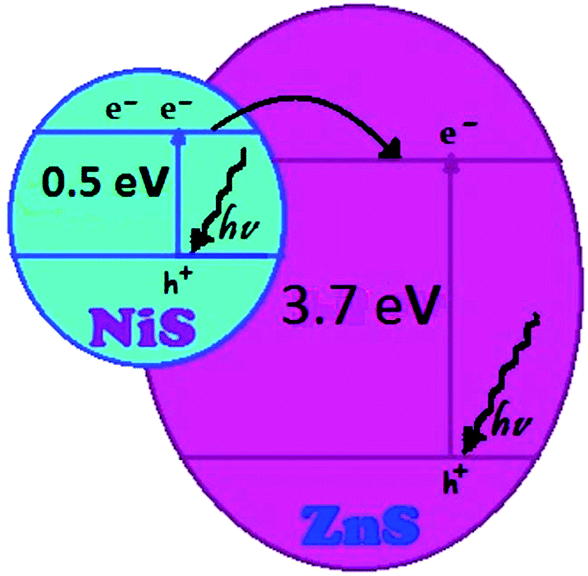 | ||
| Fig. 4 Schematic diagram of energy levels of the semiconductors. | ||
 | ||
| Fig. 5 (A) Effect of role of support on the photodegradation efficiency of 2-NT at the same conditions with Fig. 3, (B) effect of dose of semiconductors on the photodegradation efficiency of 2-NT, [2-NT]0 = 50 ppm, catalyst = 0.1 g L−1, pH = 6.3. | ||
Comparison of the results showed that the activity of NiS–MCP is more than ZnS–MCP because of lower band gap energy of NiS (0.5 eV)37 than ZnS (3.7 eV).38 The conduction and valence bands potentials of these semiconductors are larger that the corresponding redox potentials of H+/H2 and H2O/O2 and the photogenerated electrons and holes can be separated efficiently. In fact, valence band electrons of NiS can be excited more easily than ZnS which contributes the production of more electron–hole pairs. A typical schematic diagram of energy levels of the semiconductors is presented in Fig. 4.
It would be expected that hybridized ZnS–NiS–MCP system to be more effective than ZnS–MCP and NiS–MCP systems because combining of two semiconductors creates a new energy gap which lays between the band gap energies of single semiconductors.39 But in our experiments, NiS–MCP was more active than ZnS–NiS–MCP. In our idea, lower activity of ZnS–NiS–MCP can be related to un-correct mass ratio of NiS/ZnS. Hence, the effect of mass ratio of NiS/ZnS was studied to improve activity of the hybridized photocatalysts which of results will investigate in Section 3.2.2.
In the next step, the effects of raw zeolite, bulk and supported semiconductors and clinoptilolite nano particles (NCP) on the removal of 2-NT were studied and the corresponding results are summarized in Fig. 5A. Results show that the raw MCP and NCP have no significant photocatalytic effect. Bulk zinc and nickel sulfides have also a little photocatalytic effect with respect to the supported one (Fig. 5A). Bulk ZnS–NiS has slight enhancement in activity with respect to the bulk semiconductors alone which confirm the positive role of hybridation. Aggregation of semiconductors particles in the bulk samples decreases the effective surface area of particles available for absorbing photons, results a decrease in the generation of hydroxyl radicals. But, in the supported samples, zeolite prevents to aggregate ZnS, NiS or ZnS–NiS particles due to loading of them on definite ion exchange sites of zeolite, hence the degradation efficiency was increased. These observations are in agreement with literature.40–42 Pure ZnS, NiS or NiS–ZnS have only a simple bond (Ni)Zn–S, but when it forms in the zeolite structure the new bonds of (Ni)Zn–O–Al will form. In this condition, ZnS–NiS is being a part of aluminosilicate framework which has a high thermal and chemical stability. Fitting of ZnS–NiS in the zeolite channels causes to migration of produced photoexcited electrons throughout the zeolite structure and hence prevent from recombination of electron–hole pairs which increases the degradation efficiency. In addition, due to high adsorption capacity of the zeolite, more 2-NT molecules can reach near the catalyst surface where hydroxyl radicals produced. This in turn enhances the chance of OH radicals to attack the 2-NT molecules.
As shown, supported ZnS–NiS onto the nano-particles of clinoptilolite (ZnS4.5%–NiS2.9%–NCP) has higher efficiency with respect to micronized one due to increase in the effective surface area of nano-particles.
As shown, changing the dose of semiconductors led to change in the degradation efficiencies of the hybridized catalysts, so the catalyst containing 9.7% wt of ZnS and 3.0% wt NiS showed the best efficiency in the degradation of 2-NT.
By increasing in the concentration of Ni(II) and Zn(II) in solutions from 0.05 to 0.3 M, entered cations into NCP was increased. Correspondingly, the degradation activities of the prepared catalysts in 0.05 to 0.2 M solutions were increased and thereafter decreased. We believe that the electron–hole pairs were produced in NiS particles (due to the ability to attract a wider range of electromagnetic waves because of its lower energy gap) and then the produced conduction band electrons migrate to ZnS energy levels. This prevents the electron–hole recombination and hence increasing in the NiS dose till to 7.3% wt (in 0.2 M Ni(II) and 0.1 M Zn(II)) caused to increase in the degradation efficiency to 57% (at 160 min). On the other hand, in this trend more NiS particles are available for photons and hence more e–h pairs have been generated. While, when the concentration of Ni(II) in solution was constant, the efficiencies of the obtained catalysts increased with increasing in the Zn(II) concentration to 0.2 M, so the catalyst containing 9.7% ZnS showed 63% degradation efficiency (at 160 min). In this trend, due to the presence of more ZnS particles they play important role to prevent the electron–hole recombination. Hence, ZnS9.7%–NiS3.0%–NCP catalyst showed better activity (63%) than NiS–NCP (49% at 160 min).
To have an estimation for the produced e–h and finally hydroxyl radicals depending to ZnS/NiS ratio in the catalysts, the activities of ZnS9.7%–NiS3.0%–NCP (C8: as the most efficient hybridized catalyst) and ZnS10.8%–NiS2.8%–NCP (C9: as the hybridized catalyst with the lowest efficiency) were compared in the presence of iso-propanol (i-PrOH). In general, because of very weak adsorption power of short aliphatic alcohols on surface of catalysts in aqueous media, direct oxidation of them by photogenerated holes is negligible.44 So alcohols are usually used as a diagnostic tools of ˙OH radicals mediated mechanism. i-PrOH, a good scavenger like methanol, is more easily oxidized by ˙OH radicals.44 Hence, the activities of the catalysts C1 and C2 were studied in the presence of 0.01 and 0.1 M i-PrOH and the photodegradation activities of these catalysts were respectively decreased from their initial values of 61% and 36% to (40% and 17%) and (20% and 6%) in the presence of 0.01 and 0.1 M i-PrOH after 160 min ([2-NT]0 = 50 ppm). As shown by increasing in the concentration of i-PrOH from 0.01 to 0.5 M a sever decrease in the degradation extent of the pollutant was observed. This confirms significant role of hydroxyl radicals to degrade 2-NT. As shown, more sever decrease in the degradation extent was also observed for catalyst C2 in the presence of i-PrOH. This confirms lesser hydroxyl radicals generated by this catalyst. On the other hand, the production of e–h and finally hydroxyl radicals is ZnS/NiS ratio dependent in the hybridized catalysts.
The decrease in the efficiency of the process at higher ZnS and NiS loadings can be considered as the fact that when the concentration of the ZnS and NiS rises, the solid particles increasingly block the penetration of the photons due to aggregation of ZnS and NiS particles. Hence, less ZnS–NiS particles are available, due to decrease in the effective surface area, for receiving photons, so fewer OH radicals were produced. In addition, at higher concentrations of semiconductors, activated molecules collide with ground state molecules and hence deactivated molecules cannot generate electron–hole pairs and finally active hydroxyl radicals.45
| H2O2 ↔ HO2− + H+, pKa = 11.6 | (4) |
| HO2 ↔ O2− + H+, pKa = 4.8 | (5) |
| OH− ↔ O− + H+, pKa = 11.9 | (6) |
 | ||
| Fig. 6 (A) Effect of pH on the photodegradation efficiency of 2-NT at the irradiation time of 20 min, [2-NT]0 = 50 ppm, ZnS9.7%–NiS3.0%–NCP = 0.75 g L−1. (B) Effect of initial 2-NT concentration on its photodegradation efficiency at pH = 11 and the same conditions with (A). (C) Typical diagram of lnC/Co versus time as a function of pollutant concentration. (D) Degradation results in terms of mmol of degraded 2-NT pollutant as a function of its initial concentration. | ||
C/Co versus time as a function of the pollutant concentration. Based on the slopes of the curves rate constants were estimated and collected in Table 2. Due to very short lifetime of hydroxyl radicals (only a few nanosecond), at lower pollutant concentration (here 45 mg L−1) less 2-NT molecules can reach near the catalyst surface, where hydroxyl radicals were generated and hence the produced radicals will deactivate before reaction with molecules of the pollutant, resulting a decrease in the degradation efficiency.52 Hence it would be expected that increasing the pollutant concentration overcomes to this problem and hence the efficiency of the process was increased. But, at high 2-NT concentrations (beyond 55 mg L−1), more quantity of 2-NT molecules were adsorbed on the surface of the ZnS9.7%–NiS3.0%–NCP catalyst. In this case, lesser photons can absorb by the covered semiconductors and more photons can also be absorbed by high present pollutant molecules before they can reach to the catalyst surface. Hence the generation of hydroxyl radicals was reduced.52 In our previous work,31 we suggested that if the photodegradation results state in terms of the mmol of the degraded pollutants instead of degradation%, the better and more real results will be obtained. Hence, the mmol of 2-NT molecules were calculated and the results are shown in Fig. 6C. Based on the results, the best degradation efficiencies were obtained for 55 and 65 mg L−1 of 2-NT when the degradation efficiencies were respectively calculated as percentage and mmol of degraded molecules. The best rate constant was observed for 55 mg L−1 of 2-NT and thus this concentration was chosen for the next experiments.To have an idea about applicability of the proposed method for real samples, a solution containing 55 mg L−1 of 2-NT was prepared in tap water and it was subjected to photodegradation experiment at the optimized conditions. Satisfactory results with respect to distilled water (Fig. 7A) were obtained which confirm that the proposed method can be used as an effective method for the photodegradation of 2-NT in real samples containing common calcium, potassium, sodium, chloride, nitrate and sulfate ions.
 | ||
| Fig. 7 (A) Effect of real sample on the photodegradation efficiency of 2-NT at the irradiation time of 180 min, [2-NT]0 = 55 ppm, ZnS9.7%–NiS3.0%–NCP = 0.75 g L−1, pH 11. (B) Reusability of the ZnS9.7%–NiS3.0%–NCP catalyst in photodegradation of 2-NT at the same conditions with (A). | ||
 | ||
| Fig. 8 HPLC chromatogram of 2-NT solution before and during 85 and 160 min photodegradation of the sample at the same conditions with Fig. 7A. | ||
The initial and final COD values for the system were determined because the chemical oxygen demand is a measure the organic strength of wastewater.14,47 The corresponding results are shown in Fig. 9A. The changing in COD of the 2-NT solution during 0, 60, 120 and 160 min irradiation times of the sample were respectively about 719, 479, 342 and 161 mg L−1 corresponding to degradation percents of 0, 33.4, 52.0 and 78.0, respectively. The COD removal during the photodegradation process indicates the mineralization of 2-NT molecules.
 | ||
| Fig. 9 (A) Change of 2-NT concentrations was attained by COD method plotted versus irradiation time; (B) decrease in absorbance of 2-NT solution during 180 min photodegradation. | ||
Fig. 9B shows the decrease in UV-Vis absorption spectra of 2-NT during the photodegradation experiments (55 ppm 2-NT, 0.75 g L−1 of the catalyst, pH 11). No additional peaks appeared in the UV-Vis spectra during the irradiation process which confirm degradation of 2-NT molecules to smaller fragments. Thus the decrease in the samples' absorbance due to the decrease in the 2-NT concentration was recorded for measurement of the degradation extent in the all above investigated parameters.
In general, decrease in the remaining COD during the photodegradation process is in accordance with the UV-Vis spectrophotometric and HPLC results, all confirming the degradation of 2-NT molecules to smaller fragments.
4. Conclusion
The results confirm that hybridation of the ZnS–NiS increases its photocatalytic activity with respect to non hybridized ZnS and NiS semiconductors. The ratio of ZnS/NiS significantly affects the photocatalytic activity of the prepared catalysts, so the ZnS9.7%–NiS3.0%–NCP was the most active photocatalyst for the degradation of 2-NT. Also, supporting the hybridized system onto clinoptilolite nanoparticles significantly increased its photocatalytic activity with respect to unsupported one. This confirms the ability of the zeolite support to prevent the recombination of the produced electron–pair holes by migration of the conductance band electrons to zeolite structure. In addition, using zeolite nanoparticles improved the activity of the supported ZnS–NiS with respect to the micronized one because of the increased surface area of the nanoparticles.Acknowledgements
The authors thank M. Alizadeh and M. H. Kazemzadeh for performing instrumental analysis of the samples, as experts in laboratory analysis in Shahreza Branch, Islamic Azad University. The authors also thank from Dr A. Sharifzadeh as the university president for supporting of this work.References
- D. Tomova, V. Iliev, S. Rakovsky, M. Anachkov, A. Eliyas and G. L. Puma, J. Photochem. Photobiol., A, 2012, 231, 1–8 CrossRef CAS PubMed.
- B. P. Nenavathu, A. V. R. Krishna Rao, A. Goyal, A. Kapoor and R. K. Dutt, Appl. Catal., A, 2013, 459, 106–113 CrossRef CAS PubMed.
- Z. Qiang, C. Liu, F. Tian and T. Zhang, Chemosphere, 2009, 76, 609–615 CrossRef PubMed.
- U. Bali, E. C. Catalkaya and F. Sengul, J. Environ. Sci. Health, Part A: Toxic/Hazard. Subst. Environ. Eng., 2003, 10, 2259–2275 CrossRef PubMed.
- Z. Lu, P. Huo, Y. Luo, X. Liu, D. Wu, X. Gao, C. Li and Y. Yan, J. Mol. Catal. A: Chem., 2013, 378, 91–98 CrossRef CAS PubMed.
- A. Nezamzadeh-Ejhieh and Z. Salimi, Appl. Catal., A, 2010, 390, 110–118 CrossRef CAS PubMed.
- Y. Zhou, H. Lin, Q. Gu, J. Long and X. Wang, RSC Adv., 2012, 2, 12624–12627 RSC.
- A. Nezamzadeh-Ejhieh and Z. Salimi, Desalination, 2011, 280, 281–287 CrossRef CAS PubMed.
- G. L. Jincai Zhao, New J. Chem., 2000, 24, 411–417 RSC.
- X. Li, G. Liu and J. Zhao, New J. Chem., 1999, 23, 1193–1196 RSC.
- F. Chen, J. He, J. Zhao and J. C. Yu, New J. Chem., 2002, 26, 336–341 RSC.
- A. Nezamzadeh-Ejhieh and M. Karimi-Shamsabadi, Appl. Catal., A, 2014, 477, 83–92 CrossRef CAS PubMed.
- O. P. Yadav, A. Eyasu and R. K. Bachheti, Int. J. ChemTech Res., 2013, 5, 1452–1461 Search PubMed.
- A. Nezamzadeh-Ejhieh and S. Moeinirad, Desalination, 2011, 273, 248–257 CrossRef CAS PubMed.
- A. Nezamzadeh-Ejhieh and H. Zabihi-Mobarakeh, J. Ind. Eng. Chem., 2014, 20, 1421–1431 CrossRef CAS PubMed.
- D. Q. Wang, D. R. Chen and X. L. Jiao, Chin. Chem. Lett., 2004, 15, 79–82 CAS.
- T. Kou, C. Jin, C. Zhang, J. Sun and Z. Zhang, RSC Adv., 2012, 2, 12636–12643 RSC.
- R. M. Mohamed and M. M. Mohamed, Appl. Catal., A, 2008, 340, 16–24 CrossRef CAS PubMed.
- A. Nezamzadeh-Ejhieh and K. Shirvani, J. Chem., 2013, 541736 Search PubMed.
- P. C. C. Faria, J. J. M. Órfão and M. F. R. Pereira, Water Res., 2004, 38, 2043–2052 CrossRef CAS PubMed.
- C. Pulgarín, N. Dela Cruz, J. Giménez, S. Esplugas, D. Grandjean and L. F. de Alencastro, Water Res., 2012, 46, 1947–1957 CrossRef PubMed.
- G. Ledoigt, J. Wiszniowski, A. Ter Halle, C. Richard and A. Hitmi, Chemosphere, 2009, 74, 1224–1230 CrossRef PubMed.
- A. Olad and B. Naseri, Prog. Org. Coat., 2010, 67, 233–238 CrossRef CAS PubMed.
- M. Banerjee, L. Chongad and A. Sharma, Res. J. Recent Sci., 2013, 2, 326–329 Search PubMed.
- A. Nezamzadeh-Ejhieh and M. Shahanshahi, J. Ind. Eng. Chem., 2013, 19, 2026–2033 CrossRef CAS PubMed.
- H. Faghihian, M. Talebi and M. Pirouzi, J. Iran. Chem. Soc., 2008, 5, 394–399 CrossRef CAS.
- M. Brazlauskas and S. Kitrys, Chin. J. Catal., 2008, 29, 25–30 CrossRef CAS.
- A. Nezamzadeh-Ejhieh and S. Khorsandi, J. Ind. Eng. Chem., 2014, 20, 937–946 CrossRef CAS PubMed.
- A. Nezamzadeh-Ejhieh and S. Hushmandrad, Appl. Catal., A, 2010, 388, 149–159 CrossRef CAS PubMed.
- D. W. Breck, Zeolite Molecular Sieves: Structure, Chemistry and Uses, John Wiley & Sons, New York, 1974 Search PubMed.
- A. Nezamzadeh-Ejhieh and M. Karimi-Shamsabadi, Chem. Eng. J., 2013, 228, 631–641 CrossRef CAS PubMed.
- Y. G. Basabe, I. R. Iznaga, L. C. Menorval, P. Liellyn, G. Maurin, D. W. Lewis, R. Binions, M. Autic and A. R. R. Salvador, Microporous Mesoporous Mater., 2010, 135, 187–196 CrossRef PubMed.
- D. Wu, J. Xie, C. Li and L. Chi, Fuel, 2013, 103, 480–485 CrossRef PubMed.
- R. John and S. Florence, Chalcogenide Lett., 2010, 4, 269–273 Search PubMed.
- A. Nezamzadeh-Ejhieh and E. Shahriari, Int. J. Photoenergy, 2011, 518153 Search PubMed.
- S. Pérez, L. Tong, Y. Wang, P. Eichhorn and D. Barceló, Chemosphere, 2011, 83, 340–348 CrossRef PubMed.
- H. Guo, K. Lin, W. Weng, Y. Ke, R. Shen, D. Wang and J. Chen, J. Nanopart. Res., 2013, 15, 1475–1487 CrossRef PubMed.
- R. C. Khandelwal, V. Sharma, N. Gandhi and A. Khant, Int. J. Chem. Sci., 2010, 8, 1965–1972 Search PubMed.
- A. Kar, S. Kundu and A. Patra, RSC Adv., 2012, 2, 10222–10230 RSC.
- A. Nezamzadeh-Ejhieh and F. Khodabakhshi-Chermahini, J. Ind. Eng. Chem., 2014, 20, 695–704 CrossRef CAS PubMed.
- M. Subrahmanyam, P. A. K. Reddy, B. Srinivas and V. Durgakumari, Toxicol. Environ. Chem., 2012, 94, 512–524 CrossRef PubMed.
- J. V. Tolia, M. Chakraborty and Z. V. P. Murthy, Pol. J. Chem. Technol., 2012, 14, 16–21 Search PubMed.
- A. Kar, S. Kundu and A. Patra, RSC Adv., 2012, 2, 10222–10230 RSC.
- Y. Chen, S. Yang, K. wang and L. Lou, J. Photochem. Photobiol., A, 2005, 172, 47–54 CrossRef CAS PubMed.
- M. R. Hoffmann, S. T. Martin, W. Choi and D. W. Bahnemannt, Chem. Rev., 1995, 95, 69–96 CrossRef CAS.
- A. Nezamzadeh-Ejhieh and M. Khorsandi, J. Hazard. Mater., 2010, 176, 629–637 CrossRef PubMed.
- C. Fotiadis, N. P. Xekoukoulotakis and D. Mantzavinos, Catal. Today, 2007, 124, 247–253 CrossRef CAS PubMed.
- A. Nezamzadeh-Ejhieh and M. Amiri, Powder Technol., 2013, 235, 279–288 CrossRef CAS PubMed.
- C. Shifu and L. Yunzhang, Chemosphere, 2007, 67, 1010–1017 CrossRef PubMed.
- A. Nezamzadeh-Ejhieh and N. Moazzeni, J. Ind. Eng. Chem., 2013, 19, 1433–1442 CrossRef CAS PubMed.
- A. Aleboyeh, Y. Moussa and H. Aleboyeh, Dyes Pigm., 2005, 66, 129–134 CrossRef CAS PubMed.
- B. Krishnakumar, R. Velmurugan, B. Subash and M. Swaminathan, Indian J. Chem., Sect. A: Inorg., Bio-inorg., Phys., Theor. Anal. Chem., 2012, 51, 580–585 Search PubMed.
- A. Okte and O. Yilmaz, Appl. Catal., A, 2009, 354, 132–142 CrossRef CAS PubMed.
- H. Bagheri, J. Slobodonik and U. A. T. Brinkman, J. Sci., Islamic Repub. Iran, 2000, 11, 289–299 Search PubMed.
- S. Horikoshi, N. Watanabe, M. Mukae, H. Hidaka and N. Serpone, New J. Chem., 2001, 25, 999–1005 RSC.
| This journal is © The Royal Society of Chemistry 2015 |