DOI:
10.1039/C5RA12527H
(Paper)
RSC Adv., 2015,
5, 61906-61912
Magnetic chemiluminescent immunoassay for human C-reactive protein on the centrifugal microfluidics platform†
Received
29th June 2015
, Accepted 10th July 2015
First published on 13th July 2015
Abstract
Human C-reactive protein (CRP) has been reported as an inflammatory biomarker with the highest reference for use in clinical practice. However, the existing analytical techniques are lacking automation and simplicity, as desired for a prospective immunoassay format for point-of-care (PoC) analysis. We have developed an automated magnetic chemiluminescent immunoassay (MCIA) on a mobile analyser for rapid PoC determination of CRP. The MCIA is fully automated after the initial loading of sample and immunoreagents at the inlet ports. The automated protocol involves the transportation of magnetic capture microparticles between adjacent reaction compartments using a set of stationary magnets, a microfluidic polymer disposable and a specific centrifugal protocol. The developed MCIA has a sample-to-answer time of 25 min and hands-on time of approximately 5 min. It detects the entire pathophysiological range of CRP, as desired for clinically-relevant high sensitivity CRP immunoassay format, i.e. 3–81 ng mL−1 in diluted human serum with a limit of detection (LOD) and limit of quantification (LOQ) of 1.5 ng mL−1 and 1.8 ng mL−1, respectively.
Introduction
Human C-reactive protein (CRP) is an acute-phase serum protein1 produced by hepatocytes in response to inflammatory cytokines that are released during infection or inflammation. It is a pentameric protein of 118 kDa molecular weight comprising of five identical subunits with each subunit composed of 206 amino acids. It has a major role in the human host defense2 by binding to phosphocholine sites or similar molecular configurations that are exposed on the surface of microorganisms.3 In particular, it is of major importance in the nonspecific acute-phase response to inflammation,4 infection, and tissue damage5 or neonatal sepsis6 and is thus associated with clinically useful information.7 The American Heart Association/Center for Disease Control has assigned CRP as the inflammatory biomarker with the highest reference for use in clinical practice.8 Normal CRP levels in serum are usually below 10 μg mL−1 (ref. 9) but can rise to 1000-fold or more3 in case of inflammation. Furthermore, CRP has been identified as a prognostic marker of cardiovascular events,10,11 development of diabetes,12,13 metabolic syndrome,14 depression15 and post-traumatic stress.16 For neonatal sepsis, the relevant clinical cut-off value of CRP is 5 μg mL−1, which is well below the concentrations of CRP in adult sepsis patients as neonates are unable to produce sufficient amounts of acute-phase proteins.17 Therefore, a highly-sensitive immunoassay (IA) with lower detection limits is required for determining the desired high-sensitivity CRP concentration range17 (term used in clinical terminology). Additionally, clinical samples usually have to be diluted prior to analysis, as per the standard guidelines for clinical IAs18 in order to eliminate the higher interference of non-specific substances in undiluted patient samples. Therefore, the desired IA should have the detection range in ng mL−1 taking into account the fact that the patient samples have to be diluted by at least a hundred-fold.
The “gold standard” for the analysis of CRP has always been enzyme-linked immunosorbent assays (ELISAs), although several other assay formats have also been published such as those based on lateral flow,19 surface-plasmon resonance,20 magnetic permeability detection,21 microfluidics,22,23 microchip24 or quartz crystal microbalance.25 However, most of these formats are not fully automated, which impedes the realization of easy-to-use, reliable and quantitative CRP IAs that can be used in the clinical laboratories or at point-of-care (PoC) settings. In contrast, complex and automated workstations (e.g. the Olympus AU640 analyser) that are used in clinics are usually limited to central laboratories, and are associated with high investment, maintenance and operational costs. Moreover, they are bulky and require large laboratory space. On the other hand, lateral flow assays that could be employed for PoC analysis are only qualitative or semi-quantitative26 and lack the desired high analytical sensitivity.
We have employed the centrifugal microfluidics27–33 platform on a portable LabDisk player (28 cm × 18 cm × 15 cm; 2 kg) for the automation of a magnetic chemiluminescent immunoassay (MCIA) that provides a highly prospective format for the PoC detection of CRP. The developed system consists of a plastic disposable disk i.e. “LabDisk”, comprising of reaction cavities and microchannels, which operates automatically via a centrifugation protocol. The process involves the liquid propulsion in the LabDisk by centrifugal forces, thereby providing contact-less and contamination-free actuation. The LabDisk-based MCIA enables rapid determination of CRP levels in human serum with sample-to-answer time of 25 min, as desired for PoC testing. It employs a rapid ELISA procedure with minimum process steps, comprising of a 1-step incubation of all immunoreagents34 for 15 min that results in the formation of a sandwich immuno-complex on capture anti-CRP antibody (Ab)-bound dynabeads®; two wash steps (applying magnetic separation35); and an enzymatic reaction (Fig. 1).
|
| Fig. 1 Schematic of the developed hCRP MCIA procedure. Anti-hCRP capture Ab-bound dynabeads® are incubated with hCRP and biotinylated anti-hCRP detection Ab preconjugated to streptavidin (SA)–HRP for 15 min. The resulting sandwich immuno-complex is then washed twice with washing buffer. Finally, the hCRP concentration is determined by measuring the chemiluminescent signal after enzyme–substrate reaction. | |
Materials & method
Materials
The CRP capture-Ab (Cat.# MAB17071), recombinant CRP (Cat.# 1707-CR-200) and biotinylated CRP detection Ab (Cat.# BAM17072) were procured from R&D Systems, USA, while superparamagnetic dynabeads® (M-280 tosylactivated) were purchased from Life Technologies AS, Norway. The SuperSignal ELISA Femto Maximum Sensitivity Substrate, containing the enzymatic substrate of luminol/H2O2, was from Thermo Fisher Scientific Inc. The bovine serum albumin (BSA), Tween 20, phosphate buffered saline (PBS), streptavidin–bound horseradish peroxidase (SA–HRP) and ultrapure water were bought from Sigma-Aldrich, Carl Roth GmbH, Invitrogen, BioLegend and Gibco, respectively. The recombinant CRP was reconstituted in sterile 20 mM Tris(hydroxymethyl)-aminomethane–hydrochloride (Tris–HCl), pH 8.0, containing 0.1% BSA, while the washing buffer was prepared in 0.05% (v/v) Tween 20 in 0.1 M PBS, pH 7.4. The assay buffer employed was PBS, pH 7.4, containing 0.1% BSA.
Fabrication of LabDisk
The LabDisk with a diameter of 130 mm (depicted in Fig. 2) was fabricated using our previously reported micro-thermoforming procedure.36 Briefly, a micro-milled polymethyl methacrylate (PMMA) structure was casted with polydimethylsiloxane (PDMS) (Elastosil RT607, Wacker, Germany) in order to obtain a negative mold insert for blow molding. This mold insert was replicated using 188 μm thick cyclic olefin polymer (COP) foils (COP ZF 14, Zeon Chemicals, USA) by blow molding. After blow molding, the inlet and venting holes were applied via laser cutting (PLS 3.60, Universal Laser Systems). Prior to use, the LabDisk structure was partially blocked with 0.1 M PBS containing 5.0% BSA (w/v) to prevent non-specific adsorption of proteins37 and coated with 0.5% (w/v) Teflon AF solution (DuPont, USA) diluted in fluorinated liquid (FC-770, Fluorinert FC 770, 3M), as depicted in ESI Fig. S1.† The Teflon AF coating was applied to facilitate transport of the dynabeads® on the COP surface. Finally, the LabDisk was sealed with an adhesive foil (polyolefin foil, Cat. no. 900320, HJ Bioanalytik, Germany).
|
| Fig. 2 LabDisk player setup. (a) The LabDisk including three separate MCIA structures is mounted on the rotational axis of the LabDisk player. (b) A magnet holder is then placed above the LabDisk. The first magnetic stack (magnet 1) is used to transport the dynabeads® into the air-gap towards the receiving chamber, while the second magnetic stack (magnet 2) is used for pulling the microparticles from the air gap into the receiving chamber liquid. A third magnet (mixing magnet) is attached to the holder that is used for homogenization of beads in the detection chamber prior to the detection of chemiluminescent signal. | |
LabDisk player and setup
All processing steps were performed in the mobile LabDisk player prototype (Fig. 2a) manufactured by Qiagen Lake Constance GmbH, Germany. The device can conduct programmed centrifugal microfluidics-based centrifugation protocols and is equipped with an integrated chemiluminescence detection unit (Fluo Sens. QIAGEN Lake Constance, Stockach, Germany). It is further equipped with a PMMA magnetic holder, which contains a set of two permanent magnet stack (each having three neodymium rod-shaped magnets, Cat. no. W-07-N and S 03 01-N, Supermagnete.de, Germany) that is fixed at predefined radial positions above the LabDisk (Fig. 2b). An additional magnet (mixing magnet) is attached on the holder for mixing the beads prior to detection.
Immobilization of CRP capture antibody on dynabeads®
The dynabeads® were functionalized with the anti-CRP capture Ab according to the instructions of the manufacturer. Initially, 66 μL of cleaned dynabeads® were mixed with 80 μL of anti-CRP capture Ab (0.5 mg mL−1) and 70 μL of 0.1 M sodium-phosphate buffer. 100 μL of 3 M ammonium sulphate buffer was then added and the solution was incubated for 18 h at 37 °C. Subsequently, 8 μL of 0.1 M sodium-phosphate containing 5.0% (w/v) BSA was added and the solution was incubated for 2 h at 37 °C. This was followed by the addition of 10 μL of H2O/ethanolamine (10 μg mL−1) and incubation of the resulting solution for 2 h at 37 °C. The dynabeads® were then washed three times with 1 mL of 0.1 M PBS containing 0.5% (w/v) BSA. Finally, the dynabeads® were resuspended in 96 μL of PBS having 0.1% (w/v) BSA and the prepared stock solution was stored at 4 °C. For preparation of the dummy-dynabeads® B, the immobilization protocol used was identical except that 80 μL of 0.1 M PBS containing 5.0% (w/v) BSA was used instead of anti-CRP capture Ab. Note, the dummy-dynabeads® B were used in a ratio of 20 to 1 in respect to the functionalized dynabeads® to support the transfer of the anti-CRP capture Ab-coated dynabeads® out of the liquid phase into the gas phase during the gas-transfer-magnetophoresis (GTM)-based transport.35 The magnetic force acting upon the particles needs to overcome the counteracting force of the surface tension in order to enable the successful transport of dynabeads. The magnetic force correlates linearly to the volume Vcluster of the dynabeads cluster,38 whereas the counteracting force of the surface tension correlates by the third root of the volume Vcluster1/3.39 Therefore, by addition of the dummy-dynabeads® B, the cluster volume is increased and consequently the ratio of magnetic force to the counteracting force of the surface tension. This results in an enhanced efficiency of the transport of the particles. Additionally, the presence of dummy-dynabeads® B decreases the probability of losing functionalized dynabeads® by adsorbing on the COP surface during incubation or transport, thereby acting as sacrificial particles with 20-fold higher concentration in comparison to functionalized dynabeads®.
Manual MTP-based CRP MCIA
The wells of the MTP were blocked by incubating with PBS containing 5.0% (w/v) BSA for 1 h at room temperature and subsequently, washed with 300 μL of wash buffer three times. This was followed by the sequential dispensing of 4 μL of anti-CRP capture Ab-bound dynabeads® (diluted 1:10 in assay buffer), 38 μL of assay buffer, and 40 μL of biotinylated anti-CRP detection Ab (0.17 μg mL−1) pre-conjugated to SA–HRP (diluted 1:3000) into each well. Thereafter, 40 μL of CRP (varying concentrations from 1–81 ng mL−1 spiked in 1:100 diluted human serum or 0.1 M PBS buffer with pH 7.4) was dispensed into respective MTP wells in triplicate. With the chosen dilution factor of 100, the CRP concentration in an undiluted sample corresponds to 0.1–8.1 μg mL−1, which lies well within the pathophysiological range for the high sensitivity CRP. The solution was then left incubated on a MTP shaker (VWR® symphony™ Incubating Microplate Shaker) for 15 min at room temperature and 300 rpm. The magnetic dynabeads®-bound immune complexes were then separated by placing the MTP on a magnet rack and taking out the supernatant solution using a 300 μL multi-channel pipette. The magneto-immuno-complexes were then washed twice with 300 μL of wash buffer, which involved the incubation of dynabeads® in the washing buffer for 1 minute on the MTP shaker at 300 rpm followed by their separation by placing the MTP on a magnet rack and taking out the supernatant solution using a 300 μL multi-channel pipette. Thereafter, 100 μL of luminol/H2O2 (1:1 v/v) substrate solution was added to each MTP well. The MTP was then placed in a MTP reader (VICTOR2™, Perkin-Elmer), shaken at 200 rpm and read for the chemiluminescent signal at a wavelength of 425 nm.
Automated LabDisk-based CRP MCIA
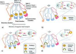
The developed automated LabDisk-based CRP MCIA is shown in Fig. 3. The procedure involves the loading of the following liquids in the inlets: inlet 1 is loaded with 40 μL of biotinylated anti-CRP detection Ab (0.17 μg mL−1) pre-conjugated to SA–HRP (diluted 1:3000), 4 μL of anti-CRP capture Ab-bound (diluted 1:10 in assay buffer), 8 μL blocked dummy-dynabeads® B and 30 μL of assay buffer. Subsequently, 40 μL of CRP (varying concentrations from 1–81 ng mL−1 in 1:100 diluted human serum or 0.1 M PBS buffer with pH 7.4 respectively). With the chosen dilution factor of 100, the CRP concentration in an undiluted sample corresponds to 0.1–8.1 μg mL−1, which lies well within the pathophysiological range for the high sensitivity CRP. Thereafter, inlets 2 and 3 are each loaded with 170 μL of wash buffer, whereas inlet 4 is loaded with 100 μL of luminol/H2O2 solution (1:1 v/v). The hands-on time for an operator for the entire process is about 5 min. Finally, the rotational protocol (ESI Table S1†) is started for the automated CRP MCIA.
|
| Fig. 3 Schematic of the automated LabDisk-based CRP MCIA. (a) Bead transport structure with inlets 1–4 for loading the IA reagents. The LabDisk is then accelerated to 20 Hz, which transports the reagents to the reaction chambers. (b) An interplay of magnetic and centrifugal forces, by constant acceleration/deceleration from 10 Hz to 20 Hz and back at 10 Hz s−1 for 15 min, mixes the reagents and anti-CRP Ab-bound dynabeads® resulting in the formation of sandwich immuno-complex. (c) The dynabeads® are then transported sequentially via gas-transfer-magnetophoresis (GTM)35 to the two consecutive chambers for the washing steps. (d) Finally, the washed dynabeads®-bound immune complexes are transported via GTM into the final detection chamber, where the chemiluminescent signal is generated by the enzyme–substrate reaction after mixing the reagents using centrifugal and magnetic forces. The chemiluminescent signal is measured by the integrated detector in the LabDisk player. | |
Data analysis
The data resulting from the readout of chemiluminescence signals were subjected to four-parameter logistic fit-based standard curve analysis using SigmaPlot software, version 11.2. The relevant analytical parameters (maximal half-effective concentration (EC50), R2 and Hillslope) were taken from the software analysis report.
The LOD and LOQ for the manual MCIA as well as the LabDisk-based MCIA were calculated by standard formula:
LOD = 3 × standard deviation (SD)Blank/Hillslope |
and
LOQ = 10 × standard deviation (SD)Blank/Hillslope.40 |
Results and discussion
Developed LabDisk-based CRP MCIA
The developed LabDisk-based MCIA provides a highly prospective format for the automated detection of CRP at PoC settings. It is ideal for our intended healthcare application aimed at the highly sensitive, rapid and automated diagnosis of neonatal sepsis, which has the relevant clinical cut-off value of CRP of 5 μg mL−1. In case of neonatal sepsis, two distinct CRP concentration ranges of 0.2–480 μg mL−1 (normal range) and 0.08–80 μg mL−1 (low range), have to be analyzed.9 Initially, a high sensitivity CRP assay is conduced and in the case of elevated CRP levels above 80 μg mL−1, CRP assays are applied for the normal range. The developed LabDisk MCIA can detect the entire pathophysiological range i.e. 3–80 μg mL−1 in human serum after appropriate sample dilution for high sensitivity assays (dilution of 1:100) or for the normal range (dilution above 1:100). The current state-of-the-art lacks the availability of a reliable, easy-to-use and automated CRP IAs that can be used in the clinical laboratories. The ready-to-use test strips, being used in emergencies, can only provide semi-quantitative results with limited analytical sensitivity.26 Therefore, there is an immense need for the development of automated IAs for CRP.41–43 The developed LabDisk-based MCIA transports the magnetic beads through adjacent chambers containing a defined liquid volume by the incremental rotation of the LabDisk with respect to a non-rotating permanent magnet. A LabDisk, comprising of the microfluidic structures, and a spinning device, equipped with a permanent magnet fixed above the disk, are the only components required for the developed MCIA, thereby making it a highly cost-effective IA format.
The incubation time and the number of washings required for the MCIA were optimized initially by manual MTP-based ELISA using 3,3′,5,5′-tetramethylbenzidine (TMB) as the enzyme substrate. It was observed that the developed MCIA shows optimum performance with a 15 min incubation time for the formation of the sandwich immune complex and by applying two successive washing steps with washing buffer (see ESI Fig. S2A and B†). The LabDisk-based MCIA detected CRP in the range of 1–81 ng mL−1 with linearity in the range of 3–27 ng mL−1 (Fig. 4a). The LOD and LOQ of the LabDisk-based MCIA in buffer (0.1 M PBS, pH 7.4) were 0.2 ng mL−1 and 0.3 ng mL−1, whereas in serum the LOD and LOQ were 1.5 ng mL−1 and 1.8 ng mL−1, respectively (ESI Table S2†). The EC50 of the developed LabDisk-based MCIA for CRP detection was 16.3 ng mL−1 in buffer (0.1 M PBS, pH 7.4) and 8.2 ng mL−1 in diluted serum, respectively. On the other hand, the LOD and LOQ of the manual MCIA (see ESI Fig. S2C†) were 0.1 ng mL−1 and 0.2 ng mL−1 in buffer (0.1 M PBS, pH 7.4) and 0.3 ng mL−1 and 0.7 ng mL−1 in diluted human serum, respectively. The EC50 of the manual MCIA was 8.3 ng mL−1 for CRP spiked in buffer (0.1 M PBS, pH 7.4) and 10.2 ng mL−1 in diluted human serum, respectively. The sensitivity of the automated LabDisk-based MCIA was observed to be slightly lower than that of the manual MCIA in diluted human serum, which might be due to the lower sensitivity of the non-integrating chemiluminescent detector being employed in the LabDisk player in comparison to the optical detector in commercial MTP reader.
|
| Fig. 4 Controls (a) automated LabDisk-based MCIA for CRP in buffer and diluted human serum. All experiments were done in triplicate and the error bars represent the standard deviation. (b) Experimental process controls employed to analyse the nonspecific interactions between IA components in the developed MCIA. Anti-CRP1 and anti-CRP2 refers to the capture and detection Ab, respectively. Controls 1–5 employ the developed assay procedure but leaving out various process steps as specified here: (1) no capture Ab & no CRP (2) no capture Ab & no detection Ab (3) no capture Ab (4) no BSA (5) No BSA & no CRP. | |
Experimental process controls were conducted to determine the blocking efficiency of the BSA and to rule out any non-specific interference due to the components of MCIA, i.e. BSA, CRP, capture Ab, biotinylated detection Ab and SA–HRP. Therefore the developed manual MCIA standard procedure was employed but leaving out various comprising process steps as specified in Fig. 4b. The results pertaining to the various experimental process controls for the developed MCIA demonstrate highly specific CRP detection without any interfering effects of the applied reagents (Fig. 4b).
As described previously in literature,44 the latex agglutination or nephelometry based IAs, being used in clinical labs for CRP analysis, can detect CRP in the range of μg mL−1. In contrast, other CRP IA formats, e.g. based on electrochemical detection,45 zinc sulfide nanoparticles,46 electro-generated chemiluminescence, determination,47 fluorescence microscopy48 or micromosaic IAs49 have higher sensitivities with detection ranges from ng mL−1 to μg mL−1. Furthermore, surface plasmon resonance-based assays and similar label-free technologies have detection ranges between ng mL−1 and g mL−1. However, most of these IA formats are non-automated, complex, labor-intensive, time-consuming and require multiple handling steps. Therefore, they require highly-skilled personnel and thus cannot be performed by untrained primary healthcare personnel. Being automated and having adequate analytical sensitivity as desired for the healthcare needs, the developed LabDisk-based MCIA is ideal for use at PoC settings. It has superior analytical performance in terms of desired simplicity, rapid analysis time and minimal process steps in comparison to conventional IA formats (ESI Table S3†). It only requires the loading of IA components at the inlets of the LabDisk followed by the starting of the centrifugation protocol, which enables the fully-automated determination of CRP concentrations in samples based on the developed MCIA procedure. The LabDisk-based MCIA completely obviates the need of bulky and expensive MTP reader, MTP washer and magnetic rack, which are essential in case of conventional ELISAs.
Determination of bio-analytical parameters
The analytical precision of the automated LabDisk-based MCIA was compared with that of the manual MCIA procedure for the detection of varying CRP concentrations within the linear range of the MCIA (3, 9 and 27 ng mL−1) in diluted human serum and buffer. The results obtained by the LabDisk-based MCIA were in agreement with those of manual MCIA performed on the MTP (Table 1), which shows high analytical precision.
Table 1 Correlation of LabDisk-based MCIA with manual MCIA
Sample |
CRP in ng mL−1 |
LabDisk-based MCIA |
Manual MCIA |
Determined concentration in ng mL−1 |
Recovery [%] |
Determined concentration in ng mL−1 |
Recovery [%] |
Buffer |
27.0 |
26.4 |
97.8 |
22.9 |
84.8 |
9.0 |
9.3 |
103.3 |
10.0 |
111.1 |
3.0 |
2.6 |
86.7 |
2.8 |
93.3 |
Serum |
27.0 |
21.3 |
78.9 |
29.4 |
108.9 |
9.0 |
9.3 |
103.3 |
8.7 |
96.7 |
3.0 |
2.5 |
83.3 |
3.3 |
110.0 |
For CRP spiked in buffer (0.1 M PBS, pH 7.4), the percentage recoveries were in the ranges of 86.7 to 103.3 and 84.8 to 111.1 for the LabDisk-based MCIA and the manual MCIA, respectively. Similarly, the percentage recoveries for the determination of CRP spiked in diluted human serum were in the range of 78.9 to 103.3 for the LabDisk-based MCIA and 96.7 to 110.0 for the manual MCIA.
Conclusion
The developed LabDisk-based MCIA enables the automated, rapid and highly-sensitive detection of CRP in the desired clinically relevant pathophysiological range. It detects CRP in 1:100 diluted human serum in just 30 min with the dynamic range of 3 to 81 ng mL−1, and LOD and LOQ of 1.5 ng mL−1 and 1.8 ng mL−1, respectively. Moreover, it has high analytical precision as demonstrated by the agreement of results with those obtained by manual MTP-based MCIA. Thus, it is a highly prospective IA format for the automated and rapid analyte detection at the point-of-care settings. The modular design of the microfluidic structures can be integrated at any radial location of a LabDisk, which provides increased flexibility for the integration of additional assay modules on the LabDisk. For instance, sample pre-treatment units (e.g. plasma separation unit50,51) or downstream applications (such as array technologies or immuno-PCR for the analysis of multiple targets in a sample) can also be readily integrated into the design scheme. We are further investigating the prestorage of immunoreagents and washing buffers into stick-packs,52 which will obviate the need for the dispensing of reagents, thereby making the developed IA highly-simplified for the end-users.
Acknowledgements
The research leading to these results has received funding from the European Union's Seventh Framework Programme (FP7/2007–2013) under grant agreement no. 258759.
Notes and references
- C. Gabay and I. Kushner, N. Engl. J. Med., 1999, 340, 448–454 CrossRef CAS PubMed.
- L. Marnell, C. Mold and T. W. Du Clos, Clin. Immunol., 2005, 117, 104–111 CrossRef CAS PubMed.
- S. Black, I. Kushner and D. Samols, J. Biol. Chem., 2004, 279, 48487–48490 CrossRef CAS PubMed.
- P. M. Ridker, Am. Heart J., 2004, 148, S19–S26 CrossRef CAS PubMed.
- M. B. Pepys and M. L. Baltz, Adv. Immunol., 1983, 34, 141–212 CAS.
- C. Chiesa, F. Natale, R. Pascone, J. F. Osborn, L. Pacifico, E. Bonci and M. de Curtis, Clin. Chim. Acta, 2011, 412, 1053–1059 CrossRef CAS PubMed.
- M. B. Pepys and G. M. Hirschfield, J. Clin. Invest., 2003, 111, 1805–1812 CrossRef CAS.
- G. L. Myers, N. Rifai, R. P. Tracy, W. L. Roberts, R. W. Alexander, L. M. Biasucci, J. D. Catravas, T. G. Cole, G. R. Cooper and B. V. Khan, Circulation, 2004, 110, e545–e549 CrossRef CAS PubMed.
- S. K. Vashist, J. Basic Appl. Sci., 2014, 28, 451–452 Search PubMed.
- S. S. Bassuk, N. Rifai and P. M. Ridker, Curr. Probl. Cardiol., 2004, 29, 439–493 Search PubMed.
- H.-K. Kuo, C.-J. Yen, J.-H. Chen, Y.-H. Yu and J. F. Bean, Int. J. Cardiol., 2007, 114, 28–33 CrossRef PubMed.
- R. Testa, A. R. Bonfigli, C. Sirolla, M. Marra, M. Boemi, D. Mari, E. Sacchi, A. Dolci, A. Catalano, A. Procopio and A. Ceriello, Nutr., Metab. Cardiovasc. Dis., 2008, 18, 220–226 CrossRef CAS PubMed.
- H. Morimoto, K. Sakata, M. Oishi, K. Tanaka, S. Nakada, K. Nogawa and Y. Suwazono, Diabetes Metab., 2013, 39, 27–33 CAS.
- M.-S. Lin, S.-R. Shih, H.-Y. Li, L.-M. Chuang, T.-C. Chang, J.-N. Wei, G.-J. Shieh, Y.-F. Chien, C.-H. Hua and J.-J. Hwang, Diabetes Res. Clin. Pract., 2007, 77, 286–292 CrossRef CAS PubMed.
- C. L. Raison, L. Capuron and A. H. Miller, Trends Immunol., 2006, 27, 24–31 CrossRef CAS PubMed.
- C. Spitzer, S. Barnow, H. Völzke, H. Wallaschofski, U. John, H. J. Freyberger, B. Löwe and H. J. Grabe, J. Psychiatr. Res., 2010, 44, 15–21 CrossRef PubMed.
- M. Ishibashi, Y. Takemura, H. Ishida, K. Watanabe and T. Kawai, Clin. Chem., 2002, 48, 1103–1106 CAS.
- Food and Drug Administration, U.S. Dep. Health Hum. Serv., 2001, 4–10 Search PubMed.
- W. Leung, C. P. Chan, T. H. Rainer, M. Ip, G. W. H. Cautherley and R. Renneberg, J. Immunol. Methods, 2008, 336, 30–36 CrossRef CAS PubMed.
- M. H. F. Meyer, M. Hartmann and M. Keusgen, Biosens. Bioelectron., 2006, 21, 1987–1990 CrossRef CAS PubMed.
- K. Kriz, F. Ibraimi, M. Lu, L. Hansson and D. Kriz, Anal. Chem., 2005, 77, 5920–5924 CrossRef CAS PubMed.
- L. Gervais and E. Delamarche, Lab Chip, 2009, 9, 3330–3337 RSC.
- W.-B. Lee, Y.-H. Chen, H.-I. Lin, S.-C. Shiesh and G.-B. Lee, Sens. Actuators, B, 2011, 157, 710–721 CrossRef CAS PubMed.
- N. Christodoulides, S. Mohanty, C. S. Miller, M. C. Langub, P. N. Floriano, P. Dharshan, M. F. Ali, B. Bernard, D. Romanovicz, E. Anslyn, P. C. Fox and J. T. McDevitt, Lab Chip, 2005, 5, 261–269 RSC.
- P. Ding, R. Liu, S. Liu, X. Mao, R. Hu and G. Li, Sens. Actuators, B, 2013, 188, 1277–1283 CrossRef CAS PubMed.
- G. A. Posthuma-Trumpie, J. Korf and A. van Amerongen, Anal. Bioanal. Chem., 2009, 393, 569–582 CrossRef CAS PubMed.
- J. Park, V. Sunkara, T.-H. Kim, H. Hwang and Y.-K. Cho, Anal. Chem., 2012, 84, 2133–2140 CrossRef CAS PubMed.
- R. Gorkin, J. Park, J. Siegrist, M. Amasia, B. S. Lee, J.-M. Park, J. Kim, H. Kim, M. Madou and Y.-K. Cho, Lab Chip, 2010, 10, 1758–1773 RSC.
- M. Madou, J. Zoval, G. Jia, H. Kido, J. Kim and N. Kim, Annu. Rev. Biomed. Eng., 2006, 8, 601–628 CrossRef CAS PubMed.
- S. Lai, S. Wang, J. Luo, L. J. Lee, S. T. Yang and M. J. Madou, Anal. Chem., 2004, 76, 1832–1837 CrossRef CAS PubMed.
- J. H. Jung, B. H. Park, Y. K. Choi and T. S. Seo, Lab Chip, 2013, 13, 3383–3388 RSC.
- J. H. Jung, S. J. Choi, B. H. Park, Y. K. Choi and T. S. Seo, Lab Chip, 2012, 12, 1598 RSC.
- M. Czugala, D. Maher, F. Collins, R. Burger, F. Hopfgartner, Y. Yang, J. Zhaou, J. Ducrée, A. Smeaton, K. J. Fraser, F. Benito-Lopez and D. Diamond, RSC Adv., 2013, 3, 15928 RSC.
- S. K. Vashist, G. Czilwik, T. van Oordt, F. von Stetten, R. Zengerle, E. Marion Schneider and J. H. T. Luong, Anal. Biochem., 2014, 456, 32–37 CrossRef CAS PubMed.
- O. Strohmeier, A. Emperle, G. Roth, D. Mark, R. Zengerle and F. von Stetten, Lab Chip, 2013, 13, 146–155 RSC.
- M. Focke, F. Stumpf, B. Faltin, P. Reith, D. Bamarni, S. Wadle, C. Müller, H. Reinecke, J. Schrenzel, P. Francois, D. Mark, G. Roth, R. Zengerle and F. von Stetten, Lab Chip, 2010, 10, 2519–2526 RSC.
- C. K. Dixit, S. K. Vashist, B. D. MacCraith and R. O'Kennedy, Analyst, 2011, 136, 1406–1411 RSC.
- N. Pamme, Lab Chip, 2006, 6, 24–38 RSC.
- M. Shikida, K. Takayanagi, H. Honda, H. Ito and K. Sato, J. Micromech. Microeng., 2006, 16, 1875–1883 CrossRef.
- S. K. Vashist, E. Marion Schneider, R. Zengerle, F. von Stetten and J. H. T. Luong, Biosens. Bioelectron., 2015, 66, 169–176 CrossRef CAS PubMed.
- J. S. Ahn, S. Choi, S. H. Jang, H. J. Chang, J. H. Kim, K. B. Nahm, S. W. Oh and E. Y. Choi, Clin. Chim. Acta, 2003, 332, 51–59 CrossRef CAS.
- C. Albrecht, N. Kaeppel and G. Gauglitz, Anal. Bioanal. Chem., 2008, 391, 1845–1852 CrossRef CAS PubMed.
- L. Borque, L. Bellod, A. Rus, M. L. Seco and F. Galisteo-González, Clin. Chem., 2000, 46, 1839–1842 CAS.
- M. Algarra, D. Gomes and J. C. G. Esteves da Silva, Clin. Chim. Acta, 2013, 415, 1–9 CrossRef CAS PubMed.
- J. H. Lee, K. H. Yoon, K. S. Hwang, J. Park, S. Ahn and T. S. Kim, Biosens. Bioelectron., 2004, 20, 269–275 CrossRef CAS PubMed.
- C. L. Cowles and X. Zhu, Biosens. Bioelectron., 2011, 30, 342–346 CrossRef CAS PubMed.
- S. Wang, E. Harris, J. Shi, A. Chen, S. Parajuli, X. Jing and W. Miao, Phys. Chem. Chem. Phys., 2010, 12, 10073–10080 RSC.
- M. S. Islam, H. G. Lee, J. Choo, J. M. Song and S. H. Kang, Talanta, 2010, 81, 1402–1408 CrossRef CAS PubMed.
- M. Wolf, D. Juncker, B. Michel, P. Hunziker and E. Delamarche, Biosens. Bioelectron., 2004, 19, 1193–1202 CrossRef CAS PubMed.
- R. Burger, N. Reis, J. G. da Fonseca and J. Ducrée, J. Micromech. Microeng., 2013, 23, 035035 CrossRef.
- S. Haeberle, T. Brenner, R. Zengerle and J. Ducrée, Lab Chip, 2006, 6, 776–781 RSC.
- T. van Oordt, Y. Barb, J. Smetana, R. Zengerle and F. von Stetten, Lab Chip, 2013, 13, 2888–2892 RSC.
Footnote |
† Electronic supplementary information (ESI) available. See DOI: 10.1039/c5ra12527h |
|
This journal is © The Royal Society of Chemistry 2015 |
Click here to see how this site uses Cookies. View our privacy policy here.