
A sandwich electrochemical immunoassay for Salmonella pullorum and Salmonella gallinarum based on a AuNPs/SiO2/Fe3O4 adsorbing antibody and 4 channel screen printed carbon electrode electrodeposited gold nanoparticles†
Jianfeng Fei,
Wenchao Dou* and
Guangying Zhao*
Food Safety Key Lab of Zhejiang Province, College of Food Science and Biotechnology Engineering, Zhejiang Gongshang University, Hangzhou 310018, PR China. E-mail: zhaogy-user@163.com; wdou@zjsu.edu.cn
First published on 24th August 2015
Abstract
A rapid and highly sensitive sandwich electrochemical immunoassay method was constructed for Salmonella pullorum and Salmonella gallinarum (S. pullorum and S. gallinarum) determination based on immune magnetic beads (MBs) and an enzyme labeled antibody. An abundance of gold nanoparticles (AuNPs) were attached to SiO2 coated Fe3O4 nanoparticles (Fe3O4/SiO2) via covalent binding between the –SH groups of Fe3O4/SiO2 and the AuNPs. Antibodies against S. pullorum and S. gallinarum were immobilized on Fe3O4/SiO2/AuNPs nanocomposites (AuMNPs) by automatic adsorption between thiol and the AuNPs. S. pullorum and S. gallinarum in the sample were captured by the AuMNPs and separated from the samples by applying an external magnetic field. The AuMNPs–Salmonella complexes (Ag/Ab1/AuMNPs) were re-dispersed in a buffer solution then exposed to horseradish peroxidase labeled anti-S. pullorum and S. gallinarum (HRP-Ab2) solution, forming a sandwich-type immune complex (HRP-Ab2/Ag/Ab1/AuMNPs). A 4 channel screen printed carbon electrode (4-SPCE) was modified by gold nanoparticles (AuNPs) through the electrodeposition method to prepare AuNPs/4-SPCE. After magnetically separating the sandwich immune complexes from solution, HRP-Ab2/Ag/Ab1/AuMNPs was anchored on AuNPs/4-SPCE by a magnet. A linear response to S. pullorum and S. gallinarum was obtained in the concentration range from 102 to 106 CFU mL−1, with a limit of detection of 3.2 × 101 CFU mL−1 (at an SNR of 3). This nanoparticle-based immunoassay method offers sensitive, highly specific, and reproducible detection of S. pullorum and S. gallinarum. Given its low detection limit, it represents promising potential in the detection of other food-borne pathogens by exchanging the antibody.
1. Introduction
Research on electrochemical immunoassays has attracted more attention from scientists in recent years. Such research can be easily conducted using simple electrochemical instruments that have the potential of miniaturization and automation.1,2 Electrochemical immunoassays that capitalize on the selectivity of antigen–antibody reactions have excellent detection limits and selectivity. Moreover, they are not affected by the sample components that might interfere with spectroscopic detection, such as particles, chromophores, and fluorophores.3 Thus, the electrochemical immunosensor is very suitable for detecting food-borne pathogens in complex samples without interference from the matrix with excellent selectivity, reproducibility and usability.4,5Antibody immobilization is vital in the successful development of an electrochemical immunosensor, and the present immobilization methods, such as chemical modification, self-assembly, or physical absorption, are usually quite complex and liable to make the antibody deactivate in real applications.6–8 So if the antibody modification procedure can be excluded, there will be a good prospect for the method. Horseradish peroxidase (HRP) can biocatalyze H2O2 in the presence of thionine, resulting in an obvious increase of the redox and reduction peak in Cyclic Voltammetry (CV).9 The above detection principle has been widely used in the development of novel electrochemical immunosensors. By enriching the amount of the above sandwich complexes accumulated on the working electrode, the sensitivity of the immunoassay would be greatly improved.10
More recently, magnetic nanoparticles (MNPs) have received increasing attention due to their high surface-to-volume ratios, allowing for the direct capture, easy separation and concentration of targets in complex samples in an external magnetic field. MNPs are superparamagnetic, which means that they are attracted to a magnetic field but retain no residual magnetism after the field is removed.11 Therefore, MNPs tagged to the biomaterial of interest can be removed from a matrix using a magnetic field, but they do not agglomerate after removal of the field. These advantages make MNPs desirable candidates for electrochemical immunoassay, as they can function as both an amplifier to increase the sensitivity of the electrochemical immunoassay and simultaneously as a concentration purification agent to reduce background interference.12,13 Although MNPs are excellent agents for a low-interference and sensitive electrochemical immunoassay, they suffer from several drawbacks such as a lack of surface tunability for biocompatible applications, which makes it difficult to couple them with biomolecules directly.14 However, if the proteins were immobilized directly on the surface of the MNPs, pure magnetic particles may undergo rapid biodegradation when they are directly exposed to complex environmental and biological systems.10 Therefore, a suitable coating is essential to prevent such limitations from occurring. Silica has been reported to be one of the best candidate shell materials for the fabrication of novel magnetic core–shell MNPs/SiO2, exhibiting the desirable intrinsic properties of the magnetic core and the silica shell.15
It is well known that gold nanoparticles (AuNPs) possess the properties of high stability and good biocompatibility.16 The surface of gold nanoparticles (AuNPs) can be coated with an antibody based on the automatic adsorption between the antibody and the AuNPs, and the AuNPs can retain the high bioactivity of the adsorbed biomolecules.17,18 Thus, combining MNPs with silicon dioxide and a AuNP shell (Fe3O4/SiO2/AuNPs) will have great potential application in biotechnology.
Fowl typhoid (FT) and pullorum disease (PD) are caused by Salmonella gallinarum (S. gallinarum) and Salmonella pullorum (S. pullorum) respectively. FT, as an acute or chronic septicemia infectious disease, primarily transmits by oral or respiratory routes, and affects adult poultries or grower groups. PD is an acute systemic disease and is usually found in young birds.19 The disease can be transmitted vertically and horizontally to others with contaminated poultries which usually results in a high mortality rate. A huge economic loss and serious threat for the development of the intensive poultry industry can be caused by S. gallinarum and S. pullorum. Therefore, establishing an effective and fast detection measure for these two pathogens is required. Multilocus enzyme electrophoresis and sequence analysis clearly state that S. pullorum and S. gallinarum both possess antigen O1, O9 and O12, and exhibit high cross-reactivity with each other, and they are generally regarded as biotypes of the same serovar, resulting in the fact that they can be simultaneously detected.20–23 Barrow et al. deemed that it was difficult and unnecessary to differentiate S. pullorum and S. gallinarum strictly.24 And Oliveira et al. used ELISA to assess the serological response of chickens to S. pullorum and S. gallinarum.25
The purpose of this study is to establish a sensitive and rapid amperometric immunoassay method for food-borne pathogen detection. S. gallinarum and S. pullorum were used as the analysis target models. MNPs were applied to increase the sensitivity of our developed method, which takes advantage of magnetic particles as pre-concentrators. In this study, the Fe3O4/SiO2/AuNPs core–shell magnetic nanoparticles were synthesized by anchoring AuNPs on Fe3O4 magnetic composite particles by strong bonding forces between –SH and AuNPs. The AuNPs acted as the intermediary materials to link Fe3O4/SiO2–SH and the antibody and get the immunomagnetic nanocomposites (Ab1/AuMNPs). The HRP labeled antibody against S. pullorum and S. gallinarum (HRP-Ab2) was used as the signal tag. S. pullorum and S. gallinarum bacteria in the sample were captured by Ab1/AuMNPs and separated from the analyte samples by applying an external magnetic field. The MNP–Salmonella complexes were re-dispersed in a buffer solution then exposed to HRP-anti-S. pullorum and S. gallinarum. The final sandwich immunocomplexes were then attached on the surface of the working electrodes of a 4 channel screen printed carbon electrode (4-SPCE) by an external magnetic field. Moreover, 4-SPCE was used to shorten the detection time and improve the reproducibility. In addition, AuNPs were electrodeposited on the working electrodes of 4-SPCE due to their signal amplification function. CV was employed to determine S. pullorum and S. gallinarum via changes of the reduction peak current in the substrate solution of H2O2 with the electron mediator of thionine included.
2. Experimental section
2.1. Reagents and solutions
S. pullorum and S. gallinarum (CMCC 50770) were employed as the target bacteria, and Escherichia coli (E. coli, ATCC 8739), Staphylococcus aureus (S. aureus, ATCC 27217), Enterobacter sakazakii (E. sakazakii, ATCC 29544), Bacillus subtilis (B. subtilis, ACCC 11060), Bacillus cereus (B. cereus, ATCC 10987) and Bacillus stearothermophilus (B. stearothermophilus, CICC 20137) were purchased from China Center of Industrial Culture Collection (Beijing, China) and conserved in the laboratory of the authors. Phosphate buffered saline (PBS, 0.01 M, pH 7.4) was used as the control group. Anti-S. pullorum and S. gallinarum were obtained from the China Institute of Veterinary Drug Control (Beijing, China). HRP labeled anti-S. pullorum and S. gallinarum (HRP-Ab2) were obtained from Shanghai Youke Biotechnology Co., Ltd. Chloroauric acid was obtained from Hangzhou Chemical Reagent Co., Ltd. Thionine (Thi) was obtained from Shanghai Zhongtai Chemical Reagent Co., Ltd. (Shanghai, China). Tetraethyl orthosilicate (TEOS) was obtained from Aladdin Industrial Inc. (Shanghai, China). 3-Mercaptopropyltriethoxysilane (MPTES) was obtained from Nanjing Chengong Organic Silicone material Co., Ltd. (Nanjing, China). And other reagents were all of analytical grade and the water used was doubly distilled.2.2. Apparatus
CHI 1030 and CHI 760C electrochemical workstations were provided by Shanghai ChenHua Instruments, Inc. (Shanghai, China). A 4 screen printed carbon electrode (4-SPCE) was developed by Rong Bin Biotechnology Co., Ltd. (Nanjing, China). The diameter of the disk-shaped working electrode was 0.25 cm, and the working electrode and counter electrode were made of a carbon ink whereas the reference electrode was made of silver/silver chloride (Ag/AgCl), and they were all printed on a plastic support. The nanostructures of the electrode were characterized by SU-8010 field emission scanning electron microscopy (Tokyo, Japan). All electrochemical experiments were performed at 25 ± 2 °C.2.3. Synthesis of Fe3O4 magnetic nanoparticles
The Fe3O4 nanoparticles were prepared according to the method of Ziyang Lu.26 1.35 mM FeSO4·7H2O was added to 70 mL double distilled water, which had been removed of oxygen by continuously blowing with nitrogen for 30 min. Under vigorous mechanical stirring and nitrogen protecting, 2.7 mM FeCl3·6H2O and 5 mL ammonia solution were added to the above double distilled water. After reacting for 80 min at 80 °C, Fe3O4 magnetic beads were isolated from the solution by a magnet and rinsed five times by double distilled water and diluted with water to a total volume of 60 mL.2.4. Synthesis of Fe3O4/SiO2–SH nanoparticles
The synthesis mechanism of the Fe3O4/SiO2–SH nanoparticles is displayed in Fig. 1A and the details are as follows: 60 mL ethanol solution and 9 mL ammonia solution were mixed with 30 mL Fe3O4 magnetic beads solution in a 100 mL flask. 1 mL TEOS was dropped to the mixture slowly. With the help of stirring, the reaction was carried out for 2 h at 18 °C, then 0.5 mL MPTES was added, and reacted for 12 h. Fe3O4/SiO2–SH nanoparticles were isolated from the solution by a magnet and rinsed five times by double distilled water and diluted with water to a total volume of 6 mL. | ||
| Fig. 1 Schematic diagram of the modification process of the electrochemical immunoassay method and the measure mechanism: (A) the synthesis process of the Fe3O4/SiO2–SH nanoparticles; (B) the synthesis process of Ab1/AuMNPs; (C) the process of S. pullorum and S. gallinarum being captured from the samples by Ab1/AuMNPs and the formation of HRP-Ab2/Ag/Ab1/AuMNPs; (D) HRP-Ab2/Ag/Ab1/AuMNPs dropped on AuNPs/4-SPCE, which is the principle of electrochemical detection. | ||
2.5. Synthesis of Fe3O4/SiO2/AuNPs/Ab1 nanocomposites
Gold nanoparticles (AuNPs) were obtained according to the Frens method.27 In brief, 1 mL of 1% HAuCl4 and 100 mL ultra-pure water were mixed in a 250 mL flask. 5 mL of 1% sodium citrate solution was added quickly to the mixture after boiling, and the boiling of the mixture was kept for another 15 min. As a result, the color of the solution turned to wine red, implying that the diameter of the gold nanoparticles was between 5 nm and 20 nm. And the colloidal gold solution was stored at 4 °C to prevent agglomeration.Fe3O4/SiO2/AuNPs (AuMNPs) nanocomposites were prepared by automatic adsorption between AuNPs and Fe3O4/SiO2–SH nanoparticles.28 Fig. 1B shows the procedure of preparation. 20 μL Fe3O4/SiO2–SH suspension (0.15 mg mL−1) was dropped into a 4 mL centrifuge tube with 1.5 mL colloidal gold solution, and incubated for 12–24 h at room temperature. In order to make each Fe3O4/SiO2–SH homogeneously combine with the AuNPs which can improve the stability of the experiment results, the centrifuge tube was shaken slowly every four hours. The AuMNPs were separated by a magnet, and rinsed three times with PBS, then re-suspended with 1 mL PBS.
Ab1/AuMNPs was obtained as follows: 40 μL anti-S. pullorum and S. gallinarum (100 μg mL−1) and 1 mL AuMNPs suspension was mixed and stirred at 4 °C for 12 h. Ab1/AuMNPs was blocked by 1 mL 0.2% BSA at 4 °C for 1 h, then rinsed three times with PBS, dispersed in 1 mL PBS, and stored at 4 °C for use.
2.6. Preparation of electrochemical immunoassay method and measurements
The AuNPs (25 nm) deposited 4-SPCE was prepared according to the previous report.29 The electrochemical reduction was performed with 4-SPCE by CV in a dispersion containing 25 mg L−1 HAuCl4 with magnetic stirring and N2 bubbling. The scan potential was performed between −1.5 and 0.5 V at a scan rate of 25 mV s−1. Then the electrode was rinsed with double distilled water and dried with blowing N2 at room temperature (25 ± 2 °C).The preparation of the immunoassay method and the mechanism of rapid detection of S. pullorum and S. gallinarum are displayed in Fig. 1C and D. S. pullorum and S. gallinarum were detected according to the following procedure: S. pullorum and S. gallinarum were captured by 20 μL Ab1/AuMNPs in 1 mL sample solution, then separated with a magnet and rinsed carefully three times. 20 μL HRP-Ab2 (7.8 μg mL−1) was dropped into the above isolates and incubated for 30 min, rinsed carefully three times and re-suspended with 20 μL PBS. Then 5 μL HRP-Ab2/Ag/Ab1/AuMNPs was dropped on AuNPs/4-SPCE and adsorbed by a magnet. 300 μL Hac–NaAc (pH = 6.5, 0.1 mol L−1) containing 1.0 mmol L−1 Thi and 0.7 mmol L−1 H2O2 was dropped on the above modified electrode. CV was conducted with a CHI 1030 at a scan rate of 25 mV s−1 between −0.6 V and −0.1 V. The detection of S. pullorum and S. gallinarum was performed by measuring the reduction peak current change (ΔIpc) of the CV before and after the immune reaction. Before the immunoreaction, the current response was recorded as I1. Due to the accelerated decomposition of hydrogen peroxide by HRP, the current response of the immunoassay method increased after the immunoreactions and was recorded as I2. Therefore, changes of the immunosensor current value (ΔIpc) were expressed as ΔIpc = I2 − I1. All experimental solutions were desecrated by nitrogen for at least 10 min before measurement, and a nitrogen atmosphere was kept during the whole electrochemical measurement. Three successive CV scans were performed for each measurement, and the last cycle was recorded.
3. Results and discussion
3.1. Comparison of 4-SPCE and SPCE
The reproducibility of 4-SPCE and SPCE were compared using CV. As shown in Fig. S1,† The RSD of 4-SPCE is 5.05% (n = 6) and the RSD of SPCE is 8.54%, indicating that 4-SPCE has a better reproducibility than SPCE. The reason may be that there are four working electrodes on one 4-SPCE, meanwhile the four working electrodes of 4-SPCE use the same auxiliary electrode and reference electrode, which avoids effects of external factors changing. And it can simultaneously examine four samples under the same test conditions. Conversely, different SPCEs can’t be operated at the exact same conditions and don’t have completely consistent auxiliary electrodes and reference electrodes, that is to say, external factors can’t be exactly the same. And for the high sensitivity of sensors a slight change will affect the reproducibility. Therefore, 4-SPCE is more stable and has a better reproducibility, so it was chosen to use in this work.3.2. Optimization of the dosage of Fe3O4/SiO2–SH
During the preparation of AuMNPs, two kinds of nanoparticles, Fe3O4/SiO2–SH and AuNPs, were linked by a coupling agent to form a strong chemical bond. The composites are stable by employing this method because the magnetic particles were coated with a large amount of the free thiol group (–SH) on the SiO2 shell with 3-mercaptopropyltriethoxysilane (MPTES) which has been found to exhibit a strong binding force to AuNPs. Each AuMNPs needs to combine with sufficient anti-S. pullorum and S. gallinarum, in order to get the best effect of enrichment for S. pullorum and S. gallinarum. An experiment with different dosages of Fe3O4/SiO2–SH from 0.075 to 0.375 mg mL−1 mixed with 1.5 mL AuNPs was carried out. Meanwhile Fe3O4/SiO2 and PBS were used as control groups (tube 1 and tube 2). As Fig. 2 shows, there is no obvious difference between tube 1 (Fe3O4/SiO2) and tube 2 (PBS), suggesting that AuNPs cannot react with Fe3O4/SiO2. The solutions from tube 6 to 7 become transparent, and the absorbance almost no longer changes, indicating that all the AuNPs have linked with Fe3O4/SiO2–SH. Therefore, 0.225 mg mL−1 Fe3O4/SiO2–SH (tube 5) was selected as the optimal condition. | ||
| Fig. 2 Optimization of the Fe3O4/SiO2–SH dosage: (1) 0.150 mg mL−1 Fe3O4/SiO2; (2) PBS; (3) 0.075 mg mL−1 Fe3O4/SiO2–SH; (4) 0.150 mg mL−1 Fe3O4/SiO2–SH; (5) 0.225 mg mL−1 Fe3O4/SiO2–SH; (6) 0.300 mg mL−1 Fe3O4/SiO2–SH; (7) 0.375 mg mL−1 Fe3O4/SiO2–SH, dropped into 1.5 mL colloidal gold solution. | ||
3.3. Characterization of Ab1/AuMNPs
An agglutination test was utilized to verify whether anti-S. pullorum and S. gallinarum had successfully linked with AuMNPs. 10 μL Ab1/AuMNPs and 10 μL S. pullorum and S. gallinarum (109 CFU mL−1) were dropped on glass slides, and the results were recorded after reacting for 1 min. Meanwhile E. coli and PBS were used as control groups. Ab1/AuMNPs uniformly disperse in the solution of E. coli and PBS as shown in Fig. 3B and C, but an agglomerate appears when S. pullorum and S. gallinarum are added (Fig. 3A), indicating that the antibody has successfully linked with AuMNPs and Ab1/AuMNPs has a good dispersibility. | ||
| Fig. 3 The agglutination test results of (A) 10 μL S. pullorum and S. gallinarum, (B) 10 μL E. coli, and (C) 10 μL PBS, respectively mixed with 10 μL IMB. | ||
3.4. Characterization of the AuMNPs nanocomposite and the AuNPs layer
The morphology of bare 4-SPCE, AuNPs/4-SPCE and AuMNPs was characterized using SEM. As shown in Fig. 4A, bare 4-SPCE is covered by smooth and uniform nanoparticles with a diameter of about 50 nm. Fig. 4B shows that AuNPs with a diameter of about 25 nm are successfully electrodeposited on the working electrode. AuNPs were introduced into the fabrication of the immunoassay method to enhance the electrochemical signals and ensure the sensitivity of the test results. Fig. 4C shows AuNPs successfully loaded on the surface of Fe3O4/SiO2–SH, the size of which are about 250 nm. Fig. 4D displays the UV-vis spectra of Fe3O4/SiO2–SH, AuNPs and AuMNPs. AuNPs show an absorption peak at about 520 nm (red curve). And there is no obvious absorption peak from 400 to 700 nm (black curve). But an absorption peak appears at about 560 nm (blue curve) after AuNPs immobilize with Fe3O4/SiO2–SH, suggesting that the AuNPs are successfully loaded on Fe3O4/SiO2–SH. | ||
| Fig. 4 FE-SEM images of (A) bare SPCE and (B) AuNPs/SPCE, (C) TEM image of AuMNPs, and (D) UV-vis absorption spectra of Fe3O4/SiO2 (black), AuNPs (red) and AuMNPs (blue). | ||
3.5. Electrochemical characteristics of the stepwise modified electrodes
To investigate the effect of each component on the electrode, the redox behavior of a reversible redox couple was recorded by CV after each modified step. Curves were recorded in 1.0 mM Thi. Fig. 5 displays a pair of reversible redox peaks of Thi at the bare 4-SPCE (curve A). After electrodepositing in HAuCl4, the peak currents of the redox peaks of 4-SPCE (curve B) significantly increase. But the redox current (curve C) gradually decreases when Ab1/AuMNPs is dropped on AuNPs/4-SPCE. Compared with curve C, the redox current of Ag/Ab1/AuMNPs (curve D) significantly decreases. This result indicates that S. pullorum and S. gallinarum are firmly captured to Ab1/AuMNPs through the specific binding affinity between the antigen and antibody. And the formed electronic barriers hindered electron transfer toward the electrode surface, resulting in the decrease of the peak current. After the addition of HRP-anti-S. pullorum and S. gallinarum, the reduction peak current value (curve E) greatly increases, implying the enzyme labeled antibody is bound onto Ag/Ab1/AuMNPs through the immune interaction, and HRP catalyzes the reduction of H2O2 with the assistance of an electron mediator, which promotes electron transfer between the enzyme and the electrode. The immunoassay method response is based on the following redox process:| HRP (red) + H2O2 → HRP (ox) + H2O | (1) |
| Thionine (red) + HRP (ox) → HRP (red) + thionine (ox) | (2) |
| Thionine (ox) + 2e− → thionine (red) | (3) |
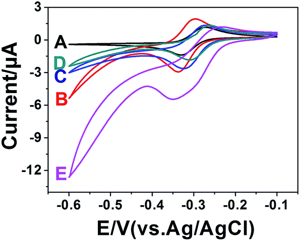 | ||
| Fig. 5 Current curves of different modified electrodes: (A) bare 4-SPCE, (B) AuNPs/4-SPCE, (C) anti-S. pullorum and S. gallinarum/AuMNPs/4-SPCE, (D) S. pullorum and S. gallinarum/anti-S. pullorum and S. gallinarum/AuMNPs/4-SPCE, (E) HRP-anti-S. pullorum and S. gallinarum/S. pullorum and S. gallinarum/anti-S. pullorum and S. gallinarum/AuMNPs/4-SPCE. | ||
3.6. EIS characterization
Electrochemical impedance spectroscopy (EIS) was employed to monitor the interface properties of the carbon electrode surface during stepwise modifications.30,31 Different stages of the modified electrode were characterized in the test base solution containing 0.1 mM KCl and 5.0 mM [Fe(CN6)]3−/4−. As seen from Fig. S2,† the Ret of AuNPs/4-SPCE (curve B) significantly decreases compared with the bare electrode (curve A), due to the gold nanoparticles not only having a large specific surface area, but also having a highly efficient electron transport property and electrocatalytic activity. The gold nanoparticles greatly reduced the resistance and accelerated the rate of electron transfer. When Ab1/AuMNPs was dropped onto AuNPs/4-SPCE, a larger semicircle (curve C) was observed, indicating that the Ret greatly increased. After S. pullorum and S. gallinarum were incubated with Ab1/AuMNPs and dropped onto AuNPs/4-SPCE, the semicircle (curve D) became larger, and the antigen and Ab1/AuMNPs formed a barrier which impeded electron transfer. Similar situations occurred when HRP-Ab2/Ag/Ab1/AuMNPs was dropped onto the AuNPs/4-SPCE (curve E). This result suggested that every step of the modification was successful.3.7. Optimization of the experimental conditions
To achieve the best performance, the experimental conditions were optimized. The results are given in Fig. S3.† the following experimental conditions were found to give the best results: (A) a concentration of H2O2 of 0.7 mmol L−1; (B) a sample pH value of 6.5; (C) an incubation time between anti-S. pullorum and S. gallinarum and S. pullorum and S. gallinarum of 30 min; (D) an incubation temperature between anti-S. pullorum and S. gallinarum and S. pullorum and S. gallinarum of 32 °C; (E) an incubation time between S. pullorum and S. gallinarum and HRP-anti-S. pullorum and S. gallinarum of 30 min; and (F) an incubation temperature between S. pullorum and S. gallinarum and HRP-anti-S. pullorum and S. gallinarum of 30 °C.3.8. Calibration curve of the immunoassay method
Under these optimal conditions different concentrations of S. pullorum and S. gallinarum (from 100 to 108 CFU mL−1) were detected. As Fig. 6A shows, with an increasing concentration of S. pullorum and S. gallinarum, the amount of HRP labeled antibody that reacted with the immobilized S. pullorum and S. gallinarum increased, therefore, ΔIpc increased. The plot of ΔIpc versus the logarithm of the S. pullorum and S. gallinarum concentration shows a linear relationship in the concentration range from 102 to 106 CFU mL−1, and the linear regression equation is ΔIpc (μA) = 0.3418x + 0.4698, and R2 = 0.9953. The limit of detection (LOD), which was defined as three times the standard deviation of the blank sample measurements, was estimated to be 3.2 × 101 CFU mL−1 (Fig. 6A inset). As Table 1 shows, this sensor performance shows potential in reducing the detection limit and is more convenient as compared to other systems for bacteria detection. In many of the past reports, a sample solution was dropped on the surface of SPCE to detect pathogenic bacteria, and the volume of the sample solution was always less than 30 μL,32 resulting in the fact that the detection limit can never be lower than 102 CFU mL−1, because 30 μL 102 CFU mL−1 pathogen suspension only contains three bacteria in theory. And this problem is well solved in this developed method. | ||
| Fig. 6 The performances of the immunoassay method: (A) ΔIpc of different logarithmic concentrations of S. pullorum and S. gallinarum (inset: linear relation between the reduction peak current change (ΔIpc) and the S. pullorum and S. gallinarum concentration); the specificity of the immunoassay method for S. pullorum and S. gallinarum; (B) the modified electrodes incubated with S. pullorum and S. gallinarum, E. sakazakii, B. cereus, B. subtilis, E. coli, and B. stearothermophilus, PBS (0.01 M, pH 7.4) under the best reaction conditions, and mixed bacteria liquid A and B (S. pullorum and S. gallinarum mixed with E. sakazakii and B. subtilis), respectively. | ||
| Material/method used | Analytical range (CFU mL−1) | LOD (CFU mL−1) | Reproducibility | Reference |
|---|---|---|---|---|
| a AuNPs (gold nanoparticles), PAMAM (poly(amidoamine)), MWCNTs (multi wall carbon nanotubes), Chi (chitosan), GCE (glassy carbon electrode), OCMCS (O-carboxymethylchitosan surface), MBs-pSAb (magnetic beads modified with primary antibodies), S (Salmonella), sSAb-AuNPs (AuNPs modified with secondary antibodies), MSNTs (magnetic silica nanotubes), IDAM (interdigitated array microelectrodes), MNPs (magnetic nanoparticles), QDs (quantum dots). | ||||
| OCMCS/Fe3O4/GCE (EIS) | 103 to 107 | 1.0 × 10³ | 6.3% | 33 |
| MBs-pSAb/S/sSAb-AuNPs/SPCE (DPV) | 103 to 106 | 1.43 × 10² | 2.4% | 1 |
| MSNTs/IDAM (EIS) | 103 to 107 | 103 | — | 34 |
| MNPs/QDs (—) | 2.5 × 10³ to 1.95 × 10⁸ | 5.0 × 10² | — | 35 |
| AuNPs/PAMAM/MWCNT/Chi/GCE (EIS) | 103 to 106 | 5.0 × 10² | 3.8% | 36 |
| Fe3O4/SiO2/AuNPs/AuNPs/4-SPCE (CV) | 102 to 106 | 3.2 × 101 | 5.3% | This work |
3.9. Specificity, reproducibility and stability of the immunoassay method
The specificity and interference are very important for the immunoassay method to distinguish the target bacteria from other food-borne pathogens in samples. To prove the specificity of the constructed immunoassay method, experiments were conducted using E. sakazakii, E. coli, B. subtilis, B. cereus, B. stearothermophilus, S. pullorum and S. gallinarum, all of the bacteria solution concentrations were 106 CFU mL−1, and PBS was used as a blank control. And E. sakazakii and B. subtilis were mixed with S. pullorum and S. gallinarum, respectively. The results are displayed in Fig. 6B, and the current increase induced by S. pullorum and S. gallinarum (ΔIpc = 2.3273 ± 0.1393 μA) is significantly larger than the current increase induced by other bacteria and PBS. The largest mean value and standard deviation was 0.7823 ± 0.0241 μA, suggesting that the immunoassay method had a high specificity for S. pullorum and S. gallinarum. And the ΔIpc value caused by the mixed bacteria solution containing E. sakazakii and B. subtilis just had an inconspicuous change, indicating that the immunoassay method had a high anti-interference ability. The specificity of the immunoassay method was attributed to the highly specific antigen–antibody immunoreactions.The long-term storage stability of the prepared immunoassay method was also measured. Ab1/AuMNPs were stored at 4 °C when they were not in use, and intermittently measured every five days with three modified electrodes; they retained 93.95% of their initial signal after a storage period of 30 days. Therefore, the modified sensors towards S. pullorum and S. gallinarum had good stability.
The reproducibility of the immunoassay method was investigated by independently monitoring the reduction peak current values of five modified electrodes under the same experimental conditions. And the relative standard deviation (RSD) obtained at a concentration of 106 CFU mL−1 was 5.33%. Therefore, the modified sensors towards S. pullorum and S. gallinarum had satisfying reproducibility. Different modified electrodes for the determination of Salmonella were compared, and the data are displayed in Table 1. The performance of this sensor shows potential in reducing the detection limit and being more stable as compared to others for bacteria detection.
3.10. Detection of S. pullorum and S. gallinarum in real samples
In order to better verify the application of the newly developed immunoassay method in practical sample detection, a series of food samples (chickens) were bought from a market, and the real samples were tested for S. pullorum and S. gallinarum by the China national food safety standard (GB/T 17999.8-2008) for the detection of S. pullorum and S. gallinarum. And we found that all of them were not affected by S. pullorum and S. gallinarum. A blind method was used and performed by two teams. The detailed steps were as follows: one team randomly added a proper dose of S. pullorum and S. gallinarum into the negative samples and mixed them with the other samples. Another team used the new sensors and the China national food safety standard (GB/T 17999.8-2008) in the assays. The two teams were not allowed to interact during the whole process. The results are shown in Table 2, and the numbers in Table 2 are the numbers of true positive or negative results detected by the corresponding methods. Accuracy is defined as the compliance between the results got by the developed method and the reference standard method for identical samples. By comparing the results of the electrochemical immunoassay method with the standard culture method, accuracy was 93.3%, the true positive rate was 94.2% and the true negative rate was 87.5%. We find that this sensor reveals good agreement with the standard method, indicating that there was acceptable accuracy and reliability of the immunoassay method. This result revealed that the immunoassay method held great promise as a reliable tool for the detection of S. pullorum and S. gallinarum in real samples.| Immunoassay method | ||||
|---|---|---|---|---|
| Positive | Negative | Total | ||
| GB | Positive | 49 | 3 | 52 |
| Negative | 1 | 7 | 8 | |
| Total | 50 | 10 | 60 | |
4. Conclusions
A rapid and highly sensitive electrochemical immunoassay method based on Fe3O4/SiO2/AuNPs and 4-SPCE has been successfully constructed for S. pullorum and S. gallinarum detection in this work. AuNPs were used as bridging materials between biomolecules and Fe3O4/SiO2–SH; the AuNPs can easily immobilize the antibody onto Fe3O4/SiO2/AuNPs and retain a high bioactivity of the adsorbed biomolecules. Electrodeposited AuNPs on the working electrodes increased the current signal of this method. This biosensor showed a wide linear range, a low detection limit and high specificity. It can also be used for the detection of S. pullorum and S. gallinarum in real samples. Importantly, this assay strategy remarkably improved the detection limit of the immunoassay method, provided a sensing platform for the detection of S. pullorum and S. gallinarum, and shortened and simplified the whole analytical process using AuMNPs and 4-SPCE. This immunoassay method can be used to develop other biosensors for pathogenic bacteria and would become a useful tool for pathogenic microorganism screening in clinical diagnostics, food safety and environmental monitoring.Acknowledgements
This project was supported by the Food Science and Engineering most important discipline of Zhejiang province (JYTSP20141062), Zhejiang Public Innovation Platform Analysis and testing projects (2015C37023), the Talent training provincial superior paper funded project (1110JY1412001P), Postgraduate Scientific and Technological Innovation Project of Zhejiang Gongshang University (3100XJ1514146) and plans for college students in Zhejiang Province science and technology innovation activities (acrobatic tender grass talent programme) project (1110JQ4212048G). Project supported by the fund of the National Natural Science Fund (30571623).Notes and references
- A. S. Afonso, B. Pérez-López, R. C. Faria, L. H. C. Mattoso, M. Hernández-Herrero, A. X. Roig-Sagués, M. Maltez-da Costa and A. Merkoçi, Electrochemical detection of Salmonella using gold nanoparticles, Biosens. Bioelectron., 2013, 40(1), 121–126 CrossRef CAS PubMed.
- F. Salam and I. E. Tothill, Detection of Salmonella typhimurium using an electrochemical immunosensor, Biosens. Bioelectron., 2009, 24(8), 2630–2636 CrossRef CAS PubMed.
- N. Gan, P. Xiong, J. Wang, T. Li, F. Hu, Y. Cao and L. Zheng, A Novel Signal-Amplified Immunoassay for the Detection of C-Reactive Protein Using HRP-Doped Magnetic Nanoparticles as Labels with the Electrochemical Quartz Crystal Microbalance as a Detector, J. Anal. Methods Chem., 2013, 2013, 482316 Search PubMed.
- H. Wu, Y. Zuo, C. Cui, W. Yang, H. Ma and X. Wang, Rapid Quantitative Detection of Brucella melitensis by a Label-Free Impedance Immunosensor Based on a Gold Nanoparticle-Modified Screen-Printed Carbon Electrode, Sensors, 2013, 13(7), 8551–8563 CrossRef CAS PubMed.
- J. Huang, G. Yang, W. Meng, L. Wu, A. Zhu and J. Xa, An electrochemical impedimetric immunosensor for label-free detection of Campylobacter jejuni in diarrhea patients’ stool based on O-carboxymethylchitosan surface modified Fe3O4 nanoparticles, Biosens. Bioelectron., 2010, 25(5), 1204–1211 CrossRef CAS PubMed.
- H. Qi, M. Li, R. Zhang, M. Dong and C. Ling, Double electrochemical covalent coupling method based on click chemistry and diazonium chemistry for the fabrication of sensitive amperometric immunosensor, Anal. Chim. Acta, 2013, 792, 28–34 CrossRef CAS PubMed.
- Y. Wang, X. Li, W. Cao, Y. Li, H. Li, B. Du and Q. Wei, Facile fabrication of an ultrasensitive sandwich-type electrochemical immunosensor for the quantitative detection of alpha fetoprotein using multifunctional mesoporous silica as platform and label for signal amplification, Talanta, 2014, 129, 411–416 CrossRef CAS PubMed.
- R. Luo, W. Zhang, W. Cheng, D. Zhao, Y. Li, X. Lin, F. Dong and S. Ding, A Novel Electrochemical Immunosensor for Detection of AngiotensinII at a Glass Carbon Electrode Modified by Carbon Nanotubes/Chitosan Film, Int. J. Electrochem. Sci., 2013, 8(3), 3186–3196 CAS.
- D. Tang, R. Yuan and Y. Chal, Ultrasensitive electrochemical immunosensor for clinical immunoassay using thionine-doped magnetic gold nanospheres as labels and horseradish peroxidase as enhancer, Anal. Chem., 2008, 80(5), 1582–1588 CrossRef CAS PubMed.
- X. Zhao, Y. Cai, T. Wang, Y. Shi and G. Jiang, Preparation of Alkanethiolate-Functionalized Core/Shell Fe3O4@Au Nanoparticles and its Interaction with Several Typical Target Molecules, Anal. Chem., 2008, 80(23), 9091–9096 CrossRef CAS PubMed.
- H.-H. Yang, S.-Q. Zhang, X.-L. Chen, Z.-X. Zhuang, J.-G. Xu and X.-R. Wang, Magnetite-containing spherical silica nanoparticles for biocatalysis and bioseparations, Anal. Chem., 2004, 76(5), 1316–1321 CrossRef CAS PubMed.
- S. Guo and S. Dong, Biomolecule-nanoparticle hybrids for electrochemical biosensors, TrAC, Trends Anal. Chem., 2009, 28(1), 96–109 CrossRef CAS PubMed.
- D. Tang, B. Su, J. Tang, J. Ren and G. Chen, Nanoparticle-Based Sandwich Electrochemical Immunoassay for Carbohydrate Antigen 125 with Signal Enhancement Using Enzyme-Coated Nanometer-Sized Enzyme-Doped Silica Beads, Anal. Chem., 2010, 82(4), 1527–1534 CrossRef CAS PubMed.
- R.-P. Liang, G.-H. Yao, L.-X. Fan and J.-D. Qiu, Magnetic Fe3O4@Au composite-enhanced surface plasmon resonance for ultrasensitive detection of magnetic nanoparticle-enriched α-fetoprotein, Anal. Chim. Acta, 2012, 737, 22–28 CrossRef CAS PubMed.
- Y. Deng, D. Qi, C. Deng, X. Zhang and D. Zhao, Superparamagnetic High-Magnetization Microspheres with an Fe3O4@SiO2 Core and Perpendicularly Aligned Mesoporous SiO2 Shell for Removal of Microcystins, J. Am. Chem. Soc., 2008, 130(1), 28–29 CrossRef CAS PubMed.
- X. Huang, H. Tu, D. Zhu, D. Du and A. Zhang, A gold nanoparticle labeling strategy for the sensitive kinetic assay of the carbamate–acetylcholinesterase interaction by surface plasmon resonance, Talanta, 2009, 78(3), 1036–1042 CrossRef CAS PubMed.
- S. Lou, J.-Y. Ye, K.-Q. Li and A. Wu, A gold nanoparticle-based immunochromatographic assay: the influence of nanoparticulate size, Analyst, 2012, 137(5), 1174–1181 RSC.
- M. J. Pollitt, G. Buckton, R. Piper and S. Brocchini, Measuring antibody coatings on gold nanoparticles by optical spectroscopy, RSC Adv., 2015, 5(31), 24521–24527 RSC.
- P. A. Barrow and O. C. F. Neto, Pullorum disease and fowl typhoid—new thoughts on old diseases: a review, Avian Pathol., 2011, 40(1), 1–13 CrossRef CAS PubMed.
- A. J. Baumler, R. M. Tsolis, T. A. Ficht and L. G. Adams, Evolution of host adaptation in Salmonella enterica, Infect. Immun., 1998, 66, 4579–4587 CAS.
- C. Hu, W. Dou and G. Zhao, Enzyme immunosensor based on gold nanoparticles electroposition and streptavidin–biotin system for detection of S. pullorum & S. gallinarum, Electrochim. Acta, 2014, 117, 239–245 CrossRef CAS PubMed.
- A. J. Baumler, B. M. Hargis and R. M. Tsolis, Tracing the origins of Salmonella outbreaks, Science, 2000, 287, 50–52 CrossRef CAS.
- J. P. Christensen, J. E. Olsen and M. Bisgaard, Ribotypes of Salmonella enterica serovar Gallinarum biovars gallinarum and pullorum, Avian Pathol., 1993, 22(4), 725–738 CrossRef CAS PubMed.
- P. A. Barrow, A. Jr. Berchieri and O. Al-Haddad, Serological Response of Chickens to Infection with Salmonella gallinarum–S. pullorum Detected by Enzyme-Linked Immunosorbent Assay, Avian Dis., 1992, 36(2), 227–236 CrossRef CAS.
- G. Oliveira, J. Â. Berchieri, H. J. Montassier and A. C. Fernandes, Assessment of serological response of chickens to Salmonella Gallinarum and Salmonella Pullorum by Elisa, Rev. Bras. Ciênc. Avíc., 2004, 6(2), 111–115 Search PubMed.
- Z. Lu, G. Wang, J. Zhuang and W. Yang, Effects of the concentration of tetramethylammonium hydroxide peptizer on the synthesis of Fe3O4/SiO2 core/shell nanoparticles, Colloids Surf., A, 2006, 278(1–3), 140–143 CrossRef CAS PubMed.
- J. Kimling, M. Maier, B. Okenve, V. Kotaidis, H. Ballot and A. Plech, Turkevich method for gold nanoparticle synthesis revisited, J. Phys. Chem. B, 2006, 110(32), 15700–15707 CrossRef CAS PubMed.
- N. Gan, H. Jin, T. Li and L. Zheng, Fe(3)O(4)/Au magnetic nanoparticle amplification strategies for ultrasensitive electrochemical immunoassay of alfa-fetoprotein, Int. J. Nanomed., 2011, 6, 3259–3269 CrossRef CAS PubMed.
- D. Wang, W. Dou, G. Zhao and Y. Chen, Immunosensor based on electrodeposition of gold-nanoparticles and ionic liquid composite for detection of Salmonella pullorum, J. Microbiol. Methods, 2014, 106, 110–118 CrossRef CAS PubMed.
- R. Feng, Y. Zhang, H. Yu, D. Wu, H. Ma, B. Zhu, C. Xu, H. Li, B. Du and Q. Wei, Nanoporous PtCo-based ultrasensitive enzyme-free immunosensor for zeranol detection, Biosens. Bioelectron., 2013, 42, 367–372 CrossRef CAS PubMed.
- X. Li, Q. Guo, W. Cao, Y. Li, B. Du and Q. Wei, Enhanced electrochemiluminescence from luminol at carboxyl graphene for detection of α-fetoprotein, Anal. Biochem., 2014, 457, 59–64 CrossRef CAS PubMed.
- M. Labib, A. S. Zamay, O. S. Kolovskaya, I. T. Reshetneva, G. S. Zamay, R. J. Kibbee, S. A. Sattar and T. N. Zamay, Aptamer-based viability impedimetric sensor for bacteria, Anal. Chem., 2012, 84(21), 8966–8969 CAS.
- J. Huang, G. Yang, W. Meng, L. Wu, A. Zhu and J. Xa, An electrochemical impedimetric immunosensor for label-free detection of Campylobacter jejuni in diarrhea patients’ stool based on O-carboxymethylchitosan surface modified Fe3O4 nanoparticles, Biosens. Bioelectron., 2010, 25(5), 1204–1211 CrossRef CAS PubMed.
- P.-D. Nguyen, T. B. Tran, D. T. X. Nguyen and J. Min, Magnetic silica nanotube-assisted impedimetric immunosensor for the separation and label-free detection of Salmonella typhimurium, Sens. Actuators, B, 2014, 197, 314–320 CrossRef CAS PubMed.
- H. Kuang, G. Cui, X. J. Chen, H. H. Yin, Q. Q. Yong, L. G. Xu, C. F. Peng, L. B. Wang and C. L. Xu, A One-Step Homogeneous Sandwich Immunosensor for Salmonella Detection Based on Magnetic Nanoparticles (MNPs) and Quantum Dots (QDs), Int. J. Mol. Sci., 2013, 14(4), 8603–8610 CrossRef PubMed.
- J. Dong, H. Zhao, M. Xu, Q. Ma and S. Ai, A label-free electrochemical impedance immunosensor based on AuNPs/PAMAM–MWCNT–Chi nanocomposite modified glassy carbon electrode for detection of Salmonella typhimurium in milk, Food Chem., 2013, 141(3), 1980–1986 CrossRef CAS PubMed.
Footnote |
| † Electronic supplementary information (ESI) available. See DOI: 10.1039/c5ra12491c |
| This journal is © The Royal Society of Chemistry 2015 |