Xiaofan Du,
Daming Dong*,
Xiande Zhao,
Leizi Jiao,
Pengcheng Han and
Yun Lang
Beijing Research Center of Intelligent Equipment for Agriculture, Beijing Academy of Agriculture and Forestry Sciences, China. E-mail: damingdong@hotmail.com; duxiaofan444@163.com; Fax: +86-010-51503626; Tel: +86-010-51503654
First published on 3rd September 2015
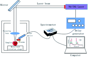
The detection of pesticide residues on fruit surfaces is highly relevant to people’s lives. Based on our previous research, we further explored the detection of chlorpyrifos residues on apple surfaces by laser induced breakdown spectroscopy (LIBS) in this paper. We observed the characteristic peaks of P at 213.62 nm, 214.91 nm, 253.56 nm, and 255.33 nm and the characteristic peak of Cl at 837.59 nm. We studied the influence of pesticide concentration and argon on the intensity of the LIBS signals. The intensity of the LIBS signal showed a linear relationship with the pesticide concentration, and argon could enhance the intensity of the LIBS signal. In the case of purging with argon, the characteristic peak of P was observed in the LIBS spectrum of a chlorpyrifos solution of 1:
1000 dilution. Then we studied the differences in the LIBS signals of pesticide residues among different matrixes and pesticides. Finally, we performed the quantitative detection of the pesticide residues with LIBS, which will provide a reference for quantitative detection. Our work gives a new method for the fast detection of pesticide residues on fruit.
Conventional detection methods for pesticide residues on the surfaces of agricultural products including gas chromatography, high-performance liquid chromatography, gas chromatography coupled with mass spectrometry, and high-performance liquid chromatography coupled with mass spectrometry1–3 are time-consuming and laborious and can hardly achieve real-time detection.2,3 Moreover, some of the organic solvents used in these methods are toxic and unfriendly to the environment.1 Considering the above shortcomings of the traditional methods, some scholars have studied optical methods to analyze agricultural products, such as surface-enhanced Raman spectroscopy.4–6 Shende et al. studied the detection of a chlorpyrifos-methyl solution in orange juice using surface-enhanced Raman spectroscopy coupled with solid-phase extraction and realized the detection of chlorpyrifos-methyl at 50 ppb.4 Shende et al. studied the detection of organophosphorus pesticides on fruit surfaces by surface-enhanced Raman spectroscopy and successfully detected 0.1% fonofos on apple surfaces within approximately 5 min, with a sensitivity of 10 ppm.5 Liu et al. studied the detection of pesticide residues on fruit surfaces by surface-enhanced Raman spectroscopy coupled with gold nanostructures and achieved limits of detection (LODs) for carbaryl, phosmet, and azinphos-methyl of 4.51 ppm, 6.51 ppm, and 6.66 ppm on apple surfaces and 5.35 ppm, 2.91 ppm, and 2.94 ppm on tomato surfaces, respectively.6 Although the limit of detection (LOD) of surface-enhanced Raman spectroscopy is low, it cannot realize quick online detection.
Since the method using ruby maser-induced plasma was proposed by Brech in 1962,7,8 LIBS has been applied in solid, liquid, and gas fields.9,10 Moreover, LIBS can realize the fast real-time detection of multiple components without a sample preparation procedure and LIBS is developing rapidly.11–13 Kim et al. studied the detection method for nutrient elements and agricultural products contaminated by pesticides with LIBS and quantitatively analyzed the nutrient elements in rice and spinach.12 The LODs of Mg, Ca, Na, and K were 29.63 mg kg−1, 102.65 mg kg−1, 36.36 mg kg−1, and 44.46 mg kg−1 in spinach, respectively. The LODs of Mg, Ca, Na, and K were 7.54 mg kg−1, 1.76 mg kg−1, 4.19 mg kg−1, and 6.70 mg kg−1 in unpolished rice, respectively. They also distinguished the samples contaminated by pesticides from uncontaminated samples and the mistaken determination percentage was less than 2%. Multari et al. studied the method of LIBS to distinguish samples contaminated by different pesticides in complex matrixes (tissue fats and fatty oils).14 The detection concentration of pesticides ranged from 0.005 to 0.100 μg g−1. The detection method distinguished not only the samples of different pesticides, but also the samples of different concentrations. Our previous study described the detection of chlorpyrifos residues on apple surfaces by LIBS.15 We distinguished the high concentration samples (1:
1 and 1
:
20) accurately. However, it was difficult to distinguish the samples with lower concentrations (1
:
100 and 1
:
1000).
In this paper, based on the previous study, we further studied the precise detection method of pesticide residues on fruit surfaces with LIBS in three aspects. Firstly, we explored the influence of the concentration of pesticides and purging with argon on the intensity of the LIBS signal. Secondly, we studied the differences of the LIBS signal among different matrixes and pesticides. Thirdly, we quantitatively analyzed the pesticide residues on apple surfaces. This paper provides the technical support for the application and development of LIBS in the detection of pesticide residues on agricultural products.
The chemical name of omethoate is O,O-dimethyl-S-methylcarbamoylmethyl phosphorothioate (C5H12NO4PS). Omethoate was produced by Zhengzhou Labor Agrochemicals Co., Ltd and the content of the active ingredients was 40%. We dissolved omethoate in deionized water according to a dilution ratio of 1:
100.
A delay generator (Fig. 1) is integrated into the equipment to produce the delay time from laser emission to spectrometer collection. A motorized rotation stage is used to adjust the position of the samples. Through the adjustment, the focus of the lens just falls on the sample surfaces. The laser generator produced by Beamtech Optronics Co., Ltd can deliver monochromatic light at 1064 nm. The maximum energy of the laser beam is 200 mJ and the frequency is 20 Hz. The laser energy was set as 160 mJ in the experiment. The spectrometer (HR2000+) was produced by Ocean Optics Company. The spectral range of the spectrometer is from 200 nm to 1000 nm. The resolution is 0.2 nm and the signal-to-noise ratio is 250:
1.
The delay time was set as 2 μs, which was the optimal value obtained in the experiment. Compared with our previous studies, this study optimized the focusing lens group used to collect the plasma signal. We also optimized the focusing system of the laser and enhanced the laser energy. All these optimizations have improved the detection performance of the system that will be presented in the Results and analysis section.
Then, we analyzed the pesticide LIBS signal from four aspects. Table 1 shows the experimental design.
Experiments | Matrix | Pesticide | Concentration | |
---|---|---|---|---|
Characteristics of the pesticide LIBS signal | Apple | Chlorpyrifos | 1![]() ![]() |
|
Influencing factors of the pesticide LIBS signal | Pesticide concentration | Apple | Chlorpyrifos | 1![]() ![]() ![]() ![]() ![]() ![]() ![]() ![]() |
Purging with argon | Apple | Chlorpyrifos | 1![]() ![]() |
|
Different matrixes | Apple | Chlorpyrifos | 1![]() ![]() |
|
Pear | Chlorpyrifos | 1![]() ![]() |
||
Different pesticides | Apple | Chlorpyrifos | 1![]() ![]() |
|
Apple | Omethoate | 1![]() ![]() |
||
Quantitative analysis of the pesticide LIBS signal | Apple | Chlorpyrifos | 1![]() ![]() ![]() ![]() ![]() ![]() ![]() ![]() ![]() ![]() ![]() ![]() |
Fig. 2 shows the LIBS spectra of P and Cl. The red lines represents the spectra of the apple sample contaminated with chlorpyrifos solution (1:
100). The black lines represents the spectra of the clean apple. We observed the characteristic peaks of P at 213.62 nm and 214.91 nm (Fig. 2a), which were the same as our previous results. In addition, we found the other characteristic peaks of P at 253.56 nm and 255.33 nm (Fig. 2b), which were not found in our previous study. As Fig. 2c shows, our previous study showed that peak A was located at 393.33 nm and peak B was located at 396.89 nm, which were regarded as the characteristic peaks of S. After careful analysis, we found that peaks A and B were not the characteristic peaks of S. We also found the characteristic peak of the Cl element at 837.59 nm (Fig. 2d), which was also observed in our previous study.
In addition, we found the LIBS characteristic peaks of P in the apple sample which was contaminated with the chlorpyrifos solution of 1:
500 (Fig. 3a and b). However, in the previous study, the LIBS characteristic peaks of P were found in the apple samples which were contaminated with chlorpyrifos solution which had a concentration higher than 1
:
100. We also found the LIBS characteristic peak of Cl in the apple sample which was contaminated with the chlorpyrifos solution of 1
:
100 (Fig. 3c), while the characteristic peak of Cl was only found in the apple sample which was contaminated with the chlorpyrifos solution of 1
:
1 in the previous study. We attributed this change to the optimization of the laser energy and the focusing lens group.
The characteristic peaks of P in the apple matrix contaminated with chlorpyrifos solution are stronger than those in the pear matrix contaminated with chlorpyrifos solution. We attributed this difference to the difference in physical properties between an apple and a pear. The peel of the Red Fuji apple was very smooth and the flesh was firm. Therefore, the chlorpyrifos solution smeared on the surface of the apple was not easily absorbed. On the contrary, the peel of the Hosui pear was coarse and the flesh was sparse. Therefore, the chlorpyrifos solution smeared on the surface of the pear was easily absorbed. The difference in physical properties led to the difference in intensity of the LIBS signal of the pesticide residues on the surfaces of different fruits. From the analysis, we concluded that the intensity of the pesticide LIBS signal varied with the matrix.
According to the above analysis, we can conclude that the LIBS signal is closely related to the matrixes and pesticides used.
All the correlation coefficients (R2) are larger than 0.88, which indicates that the fitted curves are reliable. From the curves we know that the intensity of the LIBS spectra has a linear relationship with the solution concentration. Therefore, we can use the calibration curves to detect chlorpyrifos quantitatively. It should be noted that the slopes of the calibration curves at different wavelengths are different. We think this may be due to the following reasons: (1) in terms of the spectral characteristics, at each wavelength there is a different detection limit and signal-to-noise ratio, and if the concentrations in the calibration curve are near the detection limit, this may lead to different slopes in the calibration curves. (2) The pre-processing method (e.g. background subtraction etc.) may have caused the slopes of the calibration curves at different wavelengths to vary.
In order to explore the detection ability of LIBS for pesticide residues, we calculated the LODs of the characteristic peaks. First, we subtracted the intensity of the background from the original LIBS signal to acquire the actual intensity of the characteristic peak. Then we calculated the LODs by the equation which is defined as 2σ/k, where σ is the standard deviation of the background and k is the slope of the calibration curve.12,20–22 Table 2 shows the LODs of the characteristic peaks. We can see that the LODs of P at 213.62 nm, 214.91 nm, 253.56 nm and 255.33 nm are 4.3 mg kg−1, 2.1 mg kg−1, 1.5 mg kg−1, and 6.9 mg kg−1, respectively, and the LOD of Cl at 837.59 nm is 3.0 mg kg−1. The Chinese standard for chlorpyrifos residue on apple is 1 mg kg−1.23 Although the LODs we calculated are a little bigger than the Chinese standard, we can observe the characteristics of P in the apple sample contaminated with chlorpyrifos of 1:
1000 purged with argon, which is equivalent to 1 mg kg−1. So the method of LIBS is reliable for the detection of pesticide residues.
Elements | Wavelength (nm) | LOD |
---|---|---|
P I | 213.62 nm | 4.3 mg kg−1 |
P I | 214.91 nm | 2.1 mg kg−1 |
P I | 253.56 nm | 1.5 mg kg−1 |
P I | 255.33 nm | 6.9 mg kg−1 |
Cl I | 837.59 nm | 3.0 mg kg−1 |
If a wide range of concentrations of pesticide solution is used, the calibration curve and the LOD will be more accurate. This section is only an exploration of quantitative detection using LIBS.
The quantitative analysis of pesticide residues and the calculation of the LOD provide a reference for the quantitative LIBS detection of pesticide residues on the surfaces of agricultural products.
Overall, this paper provides a reference for the detection of pesticide residues on the surfaces of agricultural products as well as a reference for the application of LIBS.
This journal is © The Royal Society of Chemistry 2015 |