Evaluation of toxicity and biodegradability for cholinium-based deep eutectic solvents

Ibrahim Juneidiab, Maan Hayyan*bc and Mohd Ali Hashimab
aDepartment of Chemical Engineering, University of Malaya, Kuala Lumpur 50603, Malaysia
bUniversity of Malaya Centre for Ionic Liquids (UMCiL), University of Malaya, Kuala Lumpur 50603, Malaysia. E-mail: maan_hayyan@yahoo.com; maan@um.edu.my
cDepartment of Civil Engineering, University of Malaya, Kuala Lumpur 50603, Malaysia

Received 26th June 2015 , Accepted 25th September 2015

First published on 25th September 2015


Abstract

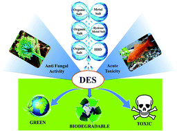
This study presented the toxicological and biodegradable assessment of different cholinium-based deep eutectic solvents (DESs). They were formed from choline chloride (ChCl) and N,N-diethyl ethanol ammonium chloride (EAC) as salts and four hydrogen bond donors, namely ethylene glycol (EG), glycerol (Gly), urea (U), malonic acid (MA), in addition to a metal salt, i.e. zinc chloride (ZnCl2), and a hydrated metal salt, i.e. zinc nitrate hexahydrate (ZnN). The toxicity towards Aspergillus niger of pure and aqueous DESs was evaluated by observing the inhibition zone using an agar well diffusion assay, and the minimum inhibition concentration (MIC) using a broth dilution assay, respectively. The MIC values of the DESs varied from 1 to 650 mg mL−1, whereas the inhibition zones changed according to the DES dose amount. Another test for acute toxicity was performed by evaluating the lethal concentration at 50% (LC50) of the same DESs on Cyprinus carpio fish. The LC50 of DESs ranged from practically harmless (e.g. ChCl[thin space (1/6-em)]:[thin space (1/6-em)]EG-DESaq) to highly toxic (e.g. EAC[thin space (1/6-em)]:[thin space (1/6-em)]ZnCl2-DESaq). The toxicity profile of the DESs depended on their concentration, type of individual components, and interaction with living organisms. Moreover, the DESs recorded higher toxicity compared to their individual components on fungi. However, lower toxicity was found for the DESs tested on Cyprinus carpio. Types I (organic salts and metal salt) and II (organic salt and hydrate metal salt) eutectics exhibited significantly higher toxicity than type III (organic salts and HBD). This was due to the presence of the innate toxicity of the metal salts. The biodegradability was appraised by a closed bottle test in which all the DESs were found to be readily biodegradable. To the best of our knowledge, there are no previous studies reported regarding the toxicity of cholinium-based DESs on freshwater fish or fungi and the biodegradability of EAC-based DESs. Therefore, this investigation can be used as a benchmark for future development of DESs.
Introduction

Developing low-cost, environment-friendly, sustainable and compatible solvents for chemical reactions has been one of the great interests in green technology. Deep eutectic solvents (DESs) as equivalents of ionic liquids (ILs) have attracted significant attention in recent years as compatible solvents for chemical reactions.1 DESs are usually obtained by the complexation of a quaternary ammonium salt with a metal salt or hydrogen bond donor (HBD) to form a eutectic solvent with a much lower melting point than any of the individual components.2,3

DESs and ILs share a few interesting characteristics (low melting point, low volatility, high thermal stability, and high solubility).4 This has led to some authors to consider them as a new generation of ILs.5,6 However, ILs carry some limitations which are higher cost (around 10 times higher than organic solvents), possess similar or higher toxicity than organic solvents, and has generally low biodegradability.7 On the contrary, due to its low-cost, biodegradable, and environmental-friendly starting material, DESs are believed to be more effective and environmental-friendly.8,9 Cholinium based DESs (which is also known as choline) are receiving considerable attention as a solvent for many processes and applications such as catalysis, electrochemistry,10 material chemistry, nanomaterials,11 biomass treatment and enzyme-catalysed reactions.1,4,12–14 N,N-Diethyl ethanol ammonium chloride (EAC) and 2-hydroxy-N,N,N-trimethylethanaminium chloride known as (choline chloride) (ChCl)-based DESs were previously reported in different studies along with their physical properties.15–18 Most of the earlier studies focused on ChCl-based DESs. However, EAC as salt has superior advantages compared to other salts. It was proven that maximum solubility of NaCl was achieved in some of EAC-based DESs.19 The densities, refractive indices, viscosities, electrolyte applicability, electrical conductivity and other physical properties for different cholinium-based DESs (e.g. glycerol and ethylene glycol) were well studied and explained.20–22

Several researchers reported the green characteristics of DESs. Their assumption was based on the pharmaceutically acceptable profile that was reported in the material safety data sheets individual components.23,24 However, the first studies on the toxicity of DESs conducted by our group have proved DESs exhibit some toxic profile.25,26 It was found that phosphonium-based DESs possessed some antibacterial activity and cytotoxicity towards Brine shrimp and different strains of bacteria. In addition, it was found that the toxicity of the DESs tested was much higher than their individual components on Brine shrimp.25 However, no inhibition was observed for ChCl-based DESs on all of the bacteria strains tested. Later, Wen et al.27 studied the toxicity of ChCl and choline acetate (ChAc) as the salt, and urea (U), acetamide (A), glycerol (Gly), ethylene glycol (EG) as the HBDs on E. coli, garlic, and Hydra. The tested DESs showed inhibition of the bacteria due to their interactions with bacterial cellular membranes. Moreover, the effect of DESs on fish and human cell line were evaluated using three types of ChCl-based DESs.28 The cytotoxicity of the DESs tested ranged from low to moderate, while the results on wheat phytotoxicity implied that the DESs tested are non-toxic with seed germination. Recently, another study of our group29 on the toxicity and cytotoxicity in vitro cell lines and in vivo animal model of ChCl-based DESs with glycerine (Gl), EG, TEG, and U as the HBDs was conducted. It was found that the cytotoxicity effect of DESs was varied depending on the types of cell lines. However, both cytotoxicity and selectivity can be influenced by different combinations of salt/HBD and their molar ratio. Cvjetko Bubalo et al.30 reported that the percentage of yeast cells that survived for 24 h within aqueous ChCl[thin space (1/6-em)]:[thin space (1/6-em)]EG-DES are almost identical to those within a conventional buffer. So far, only two studies have tested the biodegradability of DESs where both focused on ChCl-based DESs. However, different results were obtained. Radošević et al.28 reported that all DESs tested (ChC[thin space (1/6-em)]:[thin space (1/6-em)]Gly, ChCl[thin space (1/6-em)]:[thin space (1/6-em)]Glc, ChCl[thin space (1/6-em)]:[thin space (1/6-em)]OA) were classified as (68–96%) readily biodegradable. Meanwhile, Wen et al.27 found that only two DESs (ChCl[thin space (1/6-em)]:[thin space (1/6-em)]U and ChCl[thin space (1/6-em)]:[thin space (1/6-em)]A) were biodegradable among the group tested.

The common freshwater fish, carp (Cyprinus carpio) is a typical cyprinid which can tolerate low oxygen levels for extended periods of time. It was used before in numerous ecotoxicological studies on different materials and was suggested by OECD 203 fish: acute toxicity test.31,32 Cyprinus carpio fish are important consumers in the aquatic food chain and play a vital role in keeping the balance of the aquatic ecosystem.32 The toxicity assessment DESs on Cyprinus carpio in particular is very useful for future involvement of DESs in the industry. Cyprinus carpio is the largest freshwater teleost family33 and most extensively cultured fish species in Asia with 70% total freshwater aquaculture production.34 The fungi strain Aspergillus niger (A. niger) was selected based on their common usage in fermentation processes, enzyme production, and biochemical applications.35,36 Lately, choline based DESs were considered to be enzyme activators and stabilizers. Cvjetko Bubalo et al.30 found that cholinium-based DESs are promising candidates for green whole-cell biocatalysis. Therefore, providing the toxicity assessments of DESs on these particular fungi strain may open the door for future applications using DESs as solvents in fungi biofermentation processes and enzyme production.

Based on the aforementioned, the toxicity and biodegradability of DESs should be studied in order for DESs to be safely implemented in industrial applications.26,37 This study was conducted to evaluate the toxicity and biodegradability of cholinium-based DESs. The tested DESs are presented in Tables 1 and 2 with their individual names, structures, and abbreviations. The DESs ratios selected are the most common ratios reported in literatures and used in diverse applications.11,15–19,38,39

Table 1 Structures, names, and abbreviations of the individual components of the tested DESs
Ammonium salts
image file: c5ra12425e-u1.tif

Hydrogen bond donors/metal salt/hydrate metal salt
image file: c5ra12425e-u2.tif


Table 2 The names of the tested DESs and their molar ratio
DES name Molar ratio
ChCl[thin space (1/6-em)]:[thin space (1/6-em)]EG 1[thin space (1/6-em)]:[thin space (1/6-em)]2
ChCl[thin space (1/6-em)]:[thin space (1/6-em)]Gly 1[thin space (1/6-em)]:[thin space (1/6-em)]2
ChCl[thin space (1/6-em)]:[thin space (1/6-em)]U 1[thin space (1/6-em)]:[thin space (1/6-em)]2
EAC[thin space (1/6-em)]:[thin space (1/6-em)]EG 1[thin space (1/6-em)]:[thin space (1/6-em)]2
EAC[thin space (1/6-em)]:[thin space (1/6-em)]Gly 1[thin space (1/6-em)]:[thin space (1/6-em)]2
EAC[thin space (1/6-em)]:[thin space (1/6-em)]MA 1[thin space (1/6-em)]:[thin space (1/6-em)]1
EAC[thin space (1/6-em)]:[thin space (1/6-em)]ZnN 1[thin space (1/6-em)]:[thin space (1/6-em)]1
EAC[thin space (1/6-em)]:[thin space (1/6-em)]ZnCl2 1[thin space (1/6-em)]:[thin space (1/6-em)]2


Smith et al.37 presented a new classification for all existed DESs and divided them into four main categories which are:

• Organic salts and metal (type I).

• Organic salt and hydrate metal salt (type II).

• Organic salt and HBD (type III).

• Metal salt with HBD (type VI).

According to this classification, the tested DESs represent three out of four types. EAC[thin space (1/6-em)]:[thin space (1/6-em)]ZnCl2 is a combination of metal and organic salt, EAC[thin space (1/6-em)]:[thin space (1/6-em)]ZnN is a combination of hydrate metal salt and organic salt, while the rest are a combination of organic salts and HBDs. It is important to clarify that the toxicity of DESs were assessed by aqueous and non-aqueous (pure DES) systems. Considering water dilution may lead to a partial destruction of the DESs structure, we differentiated the designations given for each system. Pure DES, denoted as e.g. (EAC[thin space (1/6-em)]:[thin space (1/6-em)]Gly-DES) and aqueous DES denoted as e.g. (EAC[thin space (1/6-em)]:[thin space (1/6-em)]Gly-DES)aq. The antifungal activity for pure DESs was determined by measuring the inhibition zone diameters using agar well diffusion method. Meanwhile, aqueous DESs toxicity was determined by evaluating the minimum inhibitory concentration (MIC) using broth dilution method. The toxicity on freshwater organisms was assessed by calculating the 50% of the lethal concentration (LC50) on Cyprinus carpio fish and the biodegradability using wastewater microorganisms in a closed bottle test. The data obtained would expand knowledge about the environmental fate of DESs and could be beneficial to design a more environmentally friendly and biodegradable solvent.

Material and method

Biological and chemical material

EAC, ChCl, sodium acetate, glycerol (Gly), urea (U), were purchased from Sigma-Aldrich. Ethylene glycol (EG), 1-butyl-1-methylpyrrolidinium bis(trifluoromethylsulfonyl)imide [BMPyr][NTf2] and all other reagents used were of analytical grade and purchased from Merck. Aspergillus niger (A. niger) strain was purchased from German Collection of Microorganisms and Cell Cultures, Mascheroder Weg 1 B, Braunschweig, Germany. DESs were prepared by the procedures described earlier in our previous studies.40,41 The DESs were prepared according to the molar ratios explained in the Table 2.

Toxicity towards fungi

Broth dilution method. The toxicity of IL towards fungi strain was determined by measuring the MIC which is the lowest drug concentration that showed complete growth inhibition (100%). The MIC was measured for each DESs using the broth dilution method as described in previous studies.42 Aspergillus niger was cultured into potato dextrose broth (PDB) for 3 days at 28 °C. The fungal spores were collected and suspended in sterile water with the concentration adjusted to 1.45 × 10⁴ cfu mL−1 (colony forming unit), and were kept for future use. A series of tubes (25 × 200 mm) were prepared and supplemented with 5 mL of PDB together with different concentrations of sterilized DESs or its components (0–4500 mM) which correspond to (0–650 mg DES mL−1 broth) depending on the molecular weight of the sterilized DESs. Free DES medium was prepared as a positive control where the negative controls were without microorganisms. Each tube was then inoculated with 1 mL of fungal inoculum and incubated in the dark for 3 days at a temperature of 28 °C. Each tube was sub-cultured at least thrice to ensure viability. The MIC was determined by visual examination at the lowest concentration where there was no visible growth obtained.43 The test was conducted for the DESs and the individual materials, and the results were referred to the aqueous DES denoted as e.g. (EAC[thin space (1/6-em)]:[thin space (1/6-em)]Gly-DES)aq. Values are presented as the mean of MIC values ± standard deviation (SD) of three replicates in mg mL−1.
Agar well diffusion method. The experiment was conducted according to the agar well diffusion method.44,45 The fungal inoculum was taken from the 10 day-old culture grown on potato dextrose agar (PDA) medium. The fungal strain spores were adjusted with a spectrophotometer (A595 nm) to a final concentration of 1.45 × 10⁴ cfu mL−1. A. niger was then cultured in Petri dishes consisting of PDA, then sterilized pipette tips were used to cut a well with 8 mm diameter in the agar. This is followed by pouring different doses (10–500 mg) of the tested pure DESs. The plates were kept in 4 °C for 3–4 h to a better penetration of DESs in agar. The plates were then allowed to incubate at 28 °C for 48 h. The inhibition zone diameters caused by DESs were measured by digital caliper. Finally, the results were referred to the pure DES denoted as e.g. (EAC[thin space (1/6-em)]:[thin space (1/6-em)]Gly-DES). Values are presented as the mean of the inhibition zone ± standard deviation (SD) of three replicates in millimeters.
Acute toxicity on Cyprinus carpio and the determination of LC50. Reconstituted water was prepared according to ISO 1982 whereby the total water hardness was 180–190 mg CaCO3 per litre for each container with pH of 7–7.5 and conductivity of 7–7.5 μS cm−1. The acute toxicity tests were conducted according to the Environmental Protection Agency (EPA).46 The test was conducted according to OECD Guideline 203 (ref. 47) with small modifications. The fish (Cyprinus carpio) were chosen based on their important role in ecotoxicology as a renowned model vertebrate for testing the acute toxicity. It is widespread and presently cultured in various countries.48,49 They weighed between 1 and 2 g, measured between 3 and 4 cm, and were exposed to the test substance for approximately 96 h. Mortalities were recorded at 1 h, 24 h, 48 h, 72 h and 96 h, and then removed from the container. Different concentration were added gradually starting from the lowest concentration (0% mortality) to the highest concentration (100% mortality). The tests were conducted to determine the concentration when 50% of the fish were killed (LC50).

Reconstituted water was distributed equally (10 L) to each container (static system). The testing was conducted in three phases.

Phase 1: single addition of each individual component to the water aquarium, denoted by their abbreviations, Table 1.

Phase 2: simultaneous addition of the two components in the same DESs ratio, denoted as e.g. (EAC[thin space (1/6-em)]:[thin space (1/6-em)]EG-Mix)aq.

Phase 3: direct addition of the pure formed DESs, denoted as e.g. (EAC[thin space (1/6-em)]:[thin space (1/6-em)]EG-DES)aq.

Different range of doses was added for each substance in order to cover the maximum and minimum lethal doses. The tested DESs and their mixture components were equally measured according to the DES ratio. The solution was then mixed properly to ensure the solubility of the DESs. Saturated oxygen level was provided through a central air pump, which distributed oxygen equally through pipes to each container. The pH of the solution was measured accordingly at 1 h, 24 h, 48 h, 72 h and 96 h, and before the fish is added to each container (ten fishes). In addition, a temperature control system was provided to keep the temperature within the preferable range (20 °C–24 °C). Oxygen concentration was maintained above 90% of the saturation. The containers were kept under daylight for 16 h and were not fed during the study. Finally, controlled blank containers were added to the test. Mortalities were recorded at 1 h, 24 h, 48 h, 72 h and 96 h and subsequently removed from the container. The total number of mortalities after 96 h at different DESs concentrations was used to calculate the LC50 using US-EPS's Probit analysis program. The toxicity of the DESs toward Cyprinus carpio was represented by LC50 values which indicate the concentrations that kill 50% of the population. The results were referred to the aqueous solutions of the DESs.

Biodegradability in water

The biodegradability test was conducted according to the closed bottle test described in OECD Guideline 301 D and used in several studies.27,50 The pass level for ready biodegradability is 60% removal of theoretical oxygen demand (ThOD) in a 10 day window within 28 days period. The mineral medium was prepared accordingly by mixing 1 mL of each of the following stock solutions in 1 L of water. The first stock solution was composed of 8.5 mg L−1 KH2PO4, 21.75 g L−1 K2HPO4, 33.4 g L−1 Na2HPO4·2H2O and 0.5 g L−1 NH4Cl. The second solution composed of 27.5 g L−1 of CaCl2, while the third solution contained 22.5 g L−1 of MgSO4·7H2O and the last solution had 0.25 g L−1 FeCl3·6H2O. The mineral medium was then aerated and allowed to stand for future use. The inoculum (5 mL inoculum for each one litre solution) was collected from secondary effluent treatment plant (Indah Water Sewage Treatment Plant, Malaysia). Then, it was added into the prepared mineral medium (3 L) together with different test substances (5 mg L−1). The mixture (3 L) for each DES was then divided into ten different Scott bottles (250 mL each). The bottles were tightly closed and kept in the dark at 24 ± 1 °C. Another 10 bottles were added for both control (activated sludge only, 5.0 mg L−1) and reference (sodium acetate) and tested in parallel as suggested by the OECD Guideline 301. For comparison purposes, the IL [BMPyr][NTf2] was tested. Two bottles were taken every seven days (up to 28 days) to determine the dissolved oxygen using EXTECH dissolve oxygen (DO) meter Model SDL150. The biodegradability was then calculated by dividing the biochemical oxygen demand (BOD) by the ThOD, (BOD; expressed as mg of O2 per mg of the tested material).50

Statistical analysis

LC50 (96 h) for the fish with reference tests and their 95% confidence limits were determined by probit analysis (OECD 1984a) using a computer software (USEPA Probit analysis program, version 1.5) with small modification in order to produce the exact LC50 using the same software. Mean values were calculated using at least three replicates for each tested concentration and their 95% confidence intervals (CI) (lower limit; upper limit). The tested DESs were categorized from practically harmless to highly toxic using different LC50 values according to the acute toxicity rating developed by Passino and Smith (1987).51

Results and discussion

Antimicrobial activities

Broth dilution method. The toxicity of different aqueous DESs and their components at different concentrations (i.e. 10–6000 mM) equivalent to (i.e. 1–650 mg mL−1) were tested with a concentration gap 100 mM except for (EAC[thin space (1/6-em)]:[thin space (1/6-em)]ZnN-DES)aq, and (EAC[thin space (1/6-em)]:[thin space (1/6-em)]ZnCl2-DES)aq which was 10 mM due to their high toxicity. A. niger was used as the test model. In general, ChCl-based DESs show lower toxicity compared to EAC-based DESs (see Table 3) with various ranges of toxicity. In all of the aqueous DESs tested (except ChCl[thin space (1/6-em)]:[thin space (1/6-em)]U-DESaq and ChCl[thin space (1/6-em)]:[thin space (1/6-em)]Gly-DESaq), a linear relation between the toxicity and the growth inhibition was observed. The highest toxicity on A. niger strain was shown by (EAC[thin space (1/6-em)]:[thin space (1/6-em)]ZnCl2-DES)aq and (EAC[thin space (1/6-em)]:[thin space (1/6-em)]ZnN-DES)aq (MIC, <2.2 mg mL−1) and followed by EAC[thin space (1/6-em)]:[thin space (1/6-em)]MA-DESaq (MIC, 64.42 mg mL−1). A moderate toxicity was observed for ChCl[thin space (1/6-em)]:[thin space (1/6-em)]U-DESaq (MIC, 138.5 mg mL−1). However, the impact of ChCl[thin space (1/6-em)]:[thin space (1/6-em)]EG-DESaq, ChCl[thin space (1/6-em)]:[thin space (1/6-em)]Gly-DESaq, EAC[thin space (1/6-em)]:[thin space (1/6-em)]EG-DESaq and EAC[thin space (1/6-em)]:[thin space (1/6-em)]Gly-DESaq were negligible at the first series tested, which necessitated to increase the range of the concentration tested up to 6000 mM to achieve the MIC values.
Table 3 MIC of aqueous DESs and their individual components towards A. nigera
Solvent MIC mg mL−1
a The minimum inhibitory concentration (MIC) expressed as the mean value of three replicates ± SD (mg DES mL−1 broth).
ChCl[thin space (1/6-em)]:[thin space (1/6-em)]EG-DESaq 325.3 ± 34
ChCl[thin space (1/6-em)]:[thin space (1/6-em)]Gly-DESaq 550.4 ± 51
ChCl[thin space (1/6-em)]:[thin space (1/6-em)]U-DESaq 138.5 ± 23
EAC[thin space (1/6-em)]:[thin space (1/6-em)]EG-DESaq 314.8 ± 44
EAC[thin space (1/6-em)]:[thin space (1/6-em)]Gly-DESaq 495.4 ± 63
EAC[thin space (1/6-em)]:[thin space (1/6-em)]MA-DESaq 64.4 ± 14
EAC[thin space (1/6-em)]:[thin space (1/6-em)]ZnN-DESaq <2.2
EAC[thin space (1/6-em)]:[thin space (1/6-em)]ZnCl2-DESaq <1.3
ChCl 628.2 ± 54
EAC 614.6 ± 41
EG 322.7 ± 32
Gly 534.1 ± 71
U 126.1 ± 24
MA 31.2 ± 8
ZnN <2.9
ZnCl2 <1.36


It is worth to mention that at low concentrations (8–45 mg mL−1) of ChC[thin space (1/6-em)]:[thin space (1/6-em)]U-DESaq and ChCl[thin space (1/6-em)]:[thin space (1/6-em)]Gly-DESaq, higher A. niger spore growth compared to the blank was observed virtually. This indicates that these DESs might be used as a supplementary material for fungal growth. Studies52,53 reported that U can be used as a nitrogen source and fertilizer for fungi, incorporating it into the cell through specific transporters. In addition, the same studies mentioned that some mould fungi such as Chrysosporium keratinophilum favour growth in the presence of U. Nevertheless, an inhibition was observed as the concentration increased beyond 400 mM which was also detected in this study for all of the DESs tested and its components.

In order to have a better analysis for this study, the MIC values were investigated for some of the individual components of DESs, namely ChCl, EAC, EG, G, MA and U, and compared to their formed DESs. ChCl and EAC possess very high MIC values, which means a very low toxicity. These results corresponds to a previous study for ammonium salt on A. niger fungi which reported that the MIC value was above the range tested (800 μmol L−1).54 DESs possess anti-fungal property higher than their individual components. For instance, the MIC was 322.7 mg mL−1 for EG and 628.2 mg mL−1 for ChCl, whereas for the formed DES ChCl[thin space (1/6-em)]:[thin space (1/6-em)]EG-DESaq was 325.3 mg L−1. This is in line with DESs toxicity on E. coli reported earlier.27 It was found that DESs exhibited higher inhibition index at different concentrations compared to their individual components. This phenomenon could be due to the charge delocalization through the hydrogen bonding between the DESs components, and it was found that chemicals with delocalized charges are usually more toxic than those with localized charges.25,27,55 Moreover, the HBD might denature the proteins in a living cell.56 The relatively high toxicity of EAC[thin space (1/6-em)]:[thin space (1/6-em)]MA-DESaq is due to their acidity (pH 4.14), as the concentration of DESs increase the solution becomes more acidic which is an unfavourable environment for A. niger growth. It was observed that EAC[thin space (1/6-em)]:[thin space (1/6-em)]ZnCl2 and EAC[thin space (1/6-em)]:[thin space (1/6-em)]ZnN possess very high toxic effect by presenting the lowest MIC (i.e. <2.0 mg mL−1). This result could be ascribed to the innate toxicity of the metal salt which make types I, II, and IV DESs more toxic than type III which contains amides and polyols such as U, Gly, EG.57 EAC[thin space (1/6-em)]:[thin space (1/6-em)]ZnCl2 and EAC[thin space (1/6-em)]:[thin space (1/6-em)]ZnN made up of organic salt (EAC) and metal (ZnCl2)/hydrate metal (ZnN). Their combination resulted in ionic interaction, which exhibits a high toxic effect on the cell surface, inhibition of respiration growth and germination of spores.58 The same study has reported that the MIC for zinc on A. niger and E. coli less than to 0.5 mM.59

Considering partial destruction of the DESs structure, agar well diffusion test for pure DESs was also conducted. The results presented in Table 4. Agar well diffusion assays also confirmed the results obtained in broth dilution method. The activity of DESs was found to follow the same toxicity pattern. EAC[thin space (1/6-em)]:[thin space (1/6-em)]ZnCl2-DES exhibited the highest toxicity with strong 18 ± 4 mm inhibition zone diameter at the lowest DES dose tested (10 mg). This is followed by EAC[thin space (1/6-em)]:[thin space (1/6-em)]ZnN-DES and EAC[thin space (1/6-em)]:[thin space (1/6-em)]MA-DES with 11 ± 2 and no inhibition at the same concentration, respectively. The ChCl[thin space (1/6-em)]:[thin space (1/6-em)]U-DES showed no inhibition for the doses between 10 to 150 mg. However, inhibition of 14 ± 2 mm was observed at 200 mg. On the other hand, the least antifungal activity was exhibited by ChCl[thin space (1/6-em)]:[thin space (1/6-em)]Gly-DES and EAC. The inhibition zone was only observed after addition of 300 mg dose of their formed DESs.

Table 4 Antifungal activity of different doses of pure DESs against A. niger growth in vitro (agar well diffusion method)a
Amount (mg) Zone of inhibition in millimeter (mm)/agar disc
10 50 100 150 200 300 400
a Antifungal activity detected as the mean zones of inhibition ± SD of three replicates in millimeter (mm), No IN = no inhibition, Full IN = full inhibition, N.D = not determined.
ChCl[thin space (1/6-em)]:[thin space (1/6-em)]EG-DES No IN No IN No IN No IN No IN 10 ± 3 28 ± 4
ChCl[thin space (1/6-em)]:[thin space (1/6-em)]Gly-DES No IN No IN No IN No IN No IN 4 ± 1 20 ± 4
ChCl[thin space (1/6-em)]:[thin space (1/6-em)]U-DES No IN No IN No IN No IN 14 ± 2 25 ± 3 N.D
EAC[thin space (1/6-em)]:[thin space (1/6-em)]EG-DES No IN No IN No IN No IN No IN 14 ± 4 33 ± 5
EAC[thin space (1/6-em)]:[thin space (1/6-em)]Gly-DES No IN No IN No IN No IN No IN 7 ± 3 24 ± 2
EAC[thin space (1/6-em)]:[thin space (1/6-em)]MA-DES No IN 8 ± 1 14 ± 3 22 ± 3 29 ± 4 37 ± 4 N.D
EAC[thin space (1/6-em)]:[thin space (1/6-em)]ZnN-DES 11 ± 2 25 ± 4 36 ± 5 Full IN N.D N.D N.D
EAC[thin space (1/6-em)]:[thin space (1/6-em)]ZnCl2-DES 18 ± 4 31 ± 5 Full IN Full IN N.D N.D N.D


In our previous studies E. coli as bacterium was used together with some other bacteria strains for testing the toxicity of some DESs, ChCl-based DESs showed no inhibition to all bacterial strains tested (E. coli, S. aureus, P. aeuriginosa, and B. subtilis).26 However, some inhibition was observed for phosphonium-based DESs.25 Wen et al.27 reported the toxicity of cholonium-based DESs on E. coli. similar to the finding of this study on both fungi strains. They observed that DESs toxicity increased on E. coli as the concentration increased which indicated that the toxicity of DESs towards fungi was due to higher concentrations.60 There were two common DESs used in the study of Wen et al.,27 i.e. ChCh[thin space (1/6-em)]:[thin space (1/6-em)]U and ChCl[thin space (1/6-em)]:[thin space (1/6-em)]EG, with determined LC50 on E. coli 295.9 mM and 532 mM, respectively. However, these results were not comparable with our findings due to the different organisms used. Nevertheless, both studies shared a similarity in which the ChCl[thin space (1/6-em)]:[thin space (1/6-em)]EG was reported as one of the least toxic DESs toward E. coli and A. niger.

It can be observed from the obtained results that most DESs showed no inhibition at low concentrations. As a result, it was necessary to add relatively large doses of some DESs to expose their antifungal activity. In consistence with this finding, Cardellini et al.61,62 reported the first toxicity studies of pure DESs toward yeast fungi strain. They evaluated the toxicity of some novel DESs on Saccharomyces cerevisiae cells via an FTIR-bioassay, the DESs studied composed of zwitter ionic trimethylglycine and carboxylic acids as well as mixtures of (1S)-(+)-10-camphorsulfonic acid (CSA) and differently structured sulfobetaines (SB). The results indicated the green profile of the tested DESs. They proved that the toxicity resulted was due to the high concentration of these compounds which led to dehydrating effect on the cells. The normalized spectra from cells treated with DESs and CaCl2 (dehydrating agent known not to be toxic) were found to be almost identical.

The results obtained in this study are comparable with other studies on IL toxicity using different strains of fungi as a model test. Petkovic et al.43 used filamentous fungi to evaluate the toxicity of different ILs. Similar to the MIC values for most of the DESs tested, ILs showed active growth in media containing relatively high IL concentrations, up to 2 M for cholinium chloride alkanoates.63 The antifungal activities of eight yeasts and filamentous fungi were evaluated for imidazolium-based IL. Some of them showed no inhibition at the highest concentration screened (i.e. 2.0 mM).64 IL proved to have toxic effects on the fungi and different types of bacteria. The antimicrobial activity is governed by the molecular structure of each IL.42 In general, the antimicrobial activity of IL increases with the extension of substituent alkyl chain.63 Consequently, lipophilicity was increased. In general, primarily, imidazolium, pyridinium, and quaternary ammonium-based head groups are the most antimicrobial IL reported.65

Aspergillus nidulans canidia was used as a model test for the toxicity of alkyl tributyl phosphonium chloride.43 It was found that the toxicity occurred as a result of the interaction with the plasma membrane and the cell wall. In addition, the availability for a chemical uptake by the tested species depends on both the species tested and the chemical form and mobility.59 It might be difficult to explain the toxicity mechanisms of DESs on fungi. However, in general, the major toxicity mechanism often results in strong coordinating abilities, which will cause several effects, such as blocking biologically functional groups, interaction with the membrane structure, disruption of cell wall, and denaturation of enzymes. Consequently, every aspect of fungi growth and metabolism will be affected.66 More specifically, the mechanism of DESs toxicity towards yeast fungi was studied by Cardellini et al.61,62 They found that DESs challenged the cells by inducing dehydrating effects similar to that produced by CaCl2. This indicated that biological action for their tested DESs is rather independent of the compound structure. On the other hand, consistent correlation between the toxicity of the pure and aqueous DESs and their individual components were observed in our study, indicating that different types of DESs can affect the cell growth in different ways.

Fig. 1 shows microscopic investigation for MIC sample (3700 mM, 325.3 mg L−1) and the lower concentration sample (3600 mM) was conducted using the light microscope (Leica DM1000 LED) to ensure the inhibition effect. Fig. 1B represents the microscopic image of the sample taken from the (3600 mM) tube where the fungi show growth and colony formation. Fig. 1C represents the (3600 mM, 316.5 mg L−1) tube where no growth or colonies can be observed.


image file: c5ra12425e-f1.tif
Fig. 1 Optical images illustrating the antifungal activates of ChCl[thin space (1/6-em)]:[thin space (1/6-em)]EG-DESaq. (A) Test tube series for minimum inhibitory concentration of ChCl[thin space (1/6-em)]:[thin space (1/6-em)]EGDES for Aspergillus niger. Light microscopic picture for sample taken from (B) 3600 mM (316.5 mg L−1) concentration tube showing good fungal growth and (C) 3700 mM (325.3 mg L−1) tube showing 100% growth inhibition (MIC).

Acute toxicity on fish

The LC50 values of the ammonium salts tested, ChCl and EAC, and HBDs, U, Gly, EG, ZnCl2, ZnN and MA, are evaluated separately in addition to the aqueous solution of the DESs individual components and their formed DESs. The results represented in Fig. 2 and the exact values in Table A1 (ESI). The fish in the control (Blank) aquarium showed no behavioral changes. Conversely, fish that were exposed to DESs showed different behaviour based on the types of DESs and concentrations. The difference between phase 2 and 3 is the order of mixing. In phase 2 the salt and HBD were added simultaneously to the test solutions (fish aquarium) considering the same molar ratio for the formed DESs. Whereas, in phase 3, the salt and HBD were added at specific molar ratio to form DESs, followed by addition of the formed DESs to fish aquarium. This massive study was conducted to observe the synergistic effect between DESs components. However, bearing in mind that water may lead to dissociation of the DESs mixture, the study was extended to include phase 2 in order to determine whether the hydrogen bonding or the complexation created after DESs formation have any effect on the toxicity after dissolving in water. Therefore, it was necessary to include p-phase 2 followed by comparing the result with phase 3. This provided a more reliable analysis and broader evaluation to the fate of DESs toxicity in aqueous solutions. However, it was not possible to test the toxicity of the pure DESs on fish due to the acute toxicity guidelines constrain.
image file: c5ra12425e-f2.tif
Fig. 2 LC50 of aqueous DESs, their mixtures and individual components determined on Cyprinus carpio fish in water with 95% CI error bar.

The investigated aqueous DESs ranged from slightly toxic LC50, 10–100 mg L−1 to relatively harmless LC50, >1000 mg L−1 according to the acute toxicity rating scale by Fish and Wildlife service (FSW) (see Table 5). EAC[thin space (1/6-em)]:[thin space (1/6-em)]ZnCl2-DESaq was the most toxic (LC50, 33.9 mg L−1) followed by EAC[thin space (1/6-em)]:[thin space (1/6-em)]ZnN-DESaq (111 mg L−1), while EAC[thin space (1/6-em)]:[thin space (1/6-em)]MA-DESaq showed slightly lower toxicity (LC50, 190 mg L−1). However, DESs formed by EG and Gly as HBDs (ChCl[thin space (1/6-em)]:[thin space (1/6-em)]EG-DESaq, ChCl[thin space (1/6-em)]:[thin space (1/6-em)]Gly-DESaq, EAC[thin space (1/6-em)]:[thin space (1/6-em)]EG-DESaq and EAC[thin space (1/6-em)]:[thin space (1/6-em)]Gly-DESaq) were relatively harmless, obtaining an LC50 range from 5400 to 12[thin space (1/6-em)]500 mg L−1. A moderate toxicity was exhibited by ChCl[thin space (1/6-em)]:[thin space (1/6-em)]U-DESaq (LC50, 5310.47 mg L−1). The effective concentration was higher (less toxic) for ChCl-based DESs than EAC-based DESs.

Table 5 Acute toxicity rating scale by Fish and Wildlife service (FSW)
Relative toxicity Aquatic LC50 (mg L−1)
Super toxic 0.01–0.1
Highly toxic 0.1–1
Moderately toxic 1–10
Slightly toxic 10–100
Practically nontoxic 100–1000
Relatively harmless >1000


Interestingly, all of the aqueous DESs (phase 3) were clearly less toxic on Cyprinus carpio than their individual components (see Fig. 2). The individual components mixtures (phase 2) of the DES solutions were prepared by adding them simultaneously to the distilled water. They showed higher toxicity in all of the DESs tested, for example, the LC50 for ChCl[thin space (1/6-em)]:[thin space (1/6-em)]EG-DESaq is 7342.4 mg L−1 whereas ChCl[thin space (1/6-em)]:[thin space (1/6-em)]EG-Mixaq is 4974.1 mg L−1. Other DESs followed the same pattern as well. The difference in toxicity between DESs and their individual components could be attributed to the hydrogen bonding or the complexation created after the formation of DESs. This is in agreement with previous studies for ChCl[thin space (1/6-em)]:[thin space (1/6-em)]Uaq toxicity on the growth of Hydra and garlic roots.27,67 It was found that Hydra lived longer inside the DES solutions compared to its mixture solution or individual components. This is similarly observed for the growth of garlic roots. The higher toxicity of the components mixture solution was ascribed to the salt but this effect was weakened by the extensive hydrogen bonding network after DESs formation, in which the hydrogen bonding network throughout the DES was sufficiently strong to prevent the DESs from dissociation in aqueous solution.27,68 It was also reported that the hydrogen-bond network in DESs lowers the chemical potential of the components of DESs and thereby affected the toxicological profile.25

In this study, the salts and HBD or metal salts were tested separately. Both ammonium salts showed very benign behaviour whereas metal salts or HBD showed higher toxicity in all cases. This indicated that the toxicity shown by the tested DESs was ascribed to the metal salts or HBDs. However, the strong association during the formation of DESs decreased their reactivity, making them inert in most cases, and thus, decreased their toxicity.69 The LC50 for ZnCl2 and ZnN as individual materials, their DESs and aqueous solutions were very low, ranging from 27–111 mg L−1. Zinc is known as a heavy metal and most of its derivatives are very toxic. Many studies reported that the toxicity of zinc compounds is harmful to fish such as the rainbow fish and other organisms.70–72 However, the pH of the solvent plays a vital role as well. It was observed that EAC[thin space (1/6-em)]:[thin space (1/6-em)]MA-DESaq possesses a high acidity value (pH, 4.14). Consequently, it possesses a relatively low LC50. According to literature, a pH of 6–8 is the preferable range for C. carpio growth.

Up to date, there is no specific investigation on the toxicity of cholinium-based DESs on fish. Therefore, the comparison of our results with the literature data is limited. However, some investigations were carried out for DESs using different marine organisms such as Vibrio fischeri,73 Brine shrimp25 and Hydra.27 Other toxicity studies were carried on E. coli bacteria, garlic, and cell line toxicity.29,73 However, several attempts to justify the ecotoxicity were provided on conventional IL and cholinium salts by several research groups using Daphnia magna as the model test.74,75 A similar conclusion was reported. Firstly, the ILs ecotoxicity varied widely with different organisms across the trophic levels in which the fish are less sensitive to ILs toxicity.76,77 Secondly, the chemical structures of ILs anion and cation core may cause completely different effects. For example, the cholinium cation has higher toxicity compared to other quaternary ammonium,76,78 and a clear contribution from anions was noticed on algae toxicity (Scenedesmus vacuolatus).77 Thirdly, many researchers reported that the extension of the cation alkyl side chain is responsible for the increment on the ecotoxicity until a certain number of carbons.76,79,80 However, it is noteworthy that the functionalization of cation side chain decreases the toxicity of ILs. Another group reported that the longer alkyl chain of the imidazolium ring of a more electronegative atom drastically reduces the acute toxicity of Daphnia magna.73 As a result, it can be concluded that, similar to ILs, the toxicity of DESs highly depended on the salt and HBD in which it will most likely result in a toxic DES. However, the strong hydrogen bonding in DESs has a positive effect of lowering their toxicity by incorporating the components into eutectic mixtures and making them inert rather than by physical mixing (aqueous) which showed almost no change in their toxicity. This is in agreement with a study conducted on the Hydra where it lived longer inside DESs compared to their individual components.27,68,81

Similar conclusion was reported by Gork et al.14 and Lindberg et al.82 to explain the positive enzyme activity of ChCl-based DESs despite the presence of chloride and hydrogen bonding. They hypothesized that strong hydrogen bonds between DES components lowered their reactivity which helped in dampening their toxic effects. The DESs were found to possess higher toxicity than their individual components toward fungi. Similar results were observed earlier by Wen et al.27 for E. coli bacteria. In contrast, our results showed that DESs possess lower toxicity than their individual components on the Cyprinus carpio fish, which was the same behaviour observed earlier on Hydra.68

The higher toxicity of the aqueous solution can be ascribed to the existence of cholinium salts, but incorporating it into DESs can weaken its toxicity by the establishment of an extensive hydrogen bonding network throughout the DES. This network is sufficiently strong to prevent the DESs from a serious dissociation in aqueous solution. On the other hand, the cytotoxicity for ChCl and phosphonium-based DESs were found to be higher than their individual components on Brine shrimp and bacteria.25,26 Later, Radošević et al. confirmed this hypothesis by comparing the EC50 values determined for ChCl-based DESs with their individual compounds (e.g. ChCl and oxalic acid) on the CCO fish cell line and MCF-7 human tumour cell line. Both authors attributed this behaviour to the charge delocalization occurring through hydrogen bonding which makes the DESs more toxic compared to their components. Therefore, DESs and their individual components exhibit different toxicological profile based on the method followed and tested organisms, in which each system exhibits different response, and the results, cannot be generalized over other systems or organisms.

The use of DESs in the biological or biochemical applications is common with aqueous solutions. Therefore, information on lethal concentration and minimum inhibitory concentration of DESs in aqueous medium is essential. For instance, in enzyme catalysed reactions, DESs can be applied as additive and co-solvent for lipase hydrolysis in aqueous medium. This can potentially be a stepping stone for further application of DESs as solvent for enzyme productions, in which non-toxic DESs can be used as solvents for fungi biotransformation process in the future.

Biodegradability in water

It is very important to evaluate the biodegradability of DESs since it plays an important role in securing the future of the environment. Therefore, the aerobic biodegradability of different ammonium chloride DESs was evaluated using the closed bottle test. The test is considered successful if the reference compound achieved 60% of the ThOD in 14 days, and the chemicals are considered to be readily biodegradable if they reach 60% of biodegradability level or higher within 28 days. For this study, sodium acetate as a reference compound achieved 71.2% within the 14 day-window which verifies the validity of this test.50 In addition, [BMPyr][NTf2] was tested as a representative of ionic liquids, which is considered among the most important and commonly used ILs.83 Its physical properties are well characterized including viscosity, thermal stability, pH and density. However, it is less soluble in water than other ILs or tested DESs but considering the small amount used in this procedure (5 mg L−1), the IL was soluble.

As shown in Fig. 3, the biodegradability was more than 60% for all of the tested DESs in water, therefore all of them can be referred to as ‘readily biodegradable’. The highest biodegradability is shown by ChCl[thin space (1/6-em)]:[thin space (1/6-em)]Gly-DESaq (91%) followed by ChCl[thin space (1/6-em)]:[thin space (1/6-em)]U 85%. The lowest result was observed with EAC[thin space (1/6-em)]:[thin space (1/6-em)]MA-DESaq (61%). ChCl-based aqueous DESs were more biodegradable (77–91%) in comparison to EAC-based except for EAC[thin space (1/6-em)]:[thin space (1/6-em)]ZnN-DESaq (80%). Comparing among the HBDs, Gly, U, and EG were more liable for biodegradation than ZnCl2 and MA. The biodegradability of the conventional IL [BMPyr][NTf2] was tested. Most of the DESs showed either similar or higher biodegradability except for EAC[thin space (1/6-em)]:[thin space (1/6-em)]ZnCl2-DESaq and EAC[thin space (1/6-em)]:[thin space (1/6-em)]M-DESaq.


image file: c5ra12425e-f3.tif
Fig. 3 Biodegradability of aqueous DESs determined by closed bottle test.

The high biodegradation level for the DESs tested can be credited to the ammonium salts and HBDs that form these DESs. ChCl is readily biodegradable. According to OECD-criteria (MITI-I test; BOD measurements), it reached 93% within 14 days.28 Similar results were shown for different HBDs where 92% biodegradation was reported after 30 days for glycerol.84 In addition, the BOD5/COD ratio was 0.86 and the fact that it was greater than 0.5 further supports the ready biodegradability of glycerol. EG is readily biodegradable under both aerobic and anaerobic conditions in standard tests using sewage sludge.85 Urea is biodegradable on an average of 93–98% according to EPA.46 Moreover, MA is 86–87% biodegradable according to the Material Safety Data Sheet. The potential of cholinium-based solvents to be biodegradable was recently confirmed by Hou et al.78 They evaluated the biodegradability of 18 cholinium amino acid ILs via closed bottle tests and CO2 headspace tests using wastewater microorganisms.

Differences in aerobic biodegradability between ChCl and EAC-based DESs can be noticed. This could be accounted on the structures of ChCl and EAC (see Table 1). The presence of less carbon atoms of methyl groups (CH3) in ChCl might be more favourable for biodegradation than ethyl groups in EAC (C2H5).86,87 The structures of HBDs play an important role in the biodegradation level. A slight improvement in biodegradation can be noticed with the existence of the hydroxyl group, which was also reported previously in different ILs.87 For instance, due to the presence of three hydroxyl groups in glycerol, the level of biodegradation was marginally higher (ChCl[thin space (1/6-em)]:[thin space (1/6-em)]Gly-DESaq 91% and EAC[thin space (1/6-em)]:[thin space (1/6-em)]Gly-DESaq 74%) than that in EG with two hydroxyl groups (ChCl[thin space (1/6-em)]:[thin space (1/6-em)]EG-DESaq 77% EAC[thin space (1/6-em)]:[thin space (1/6-em)]EG-DESaq 70% respectively).28

A relatively high biodegradability was showed by ChCl[thin space (1/6-em)]:[thin space (1/6-em)]U-DESaq (85%) and IL, this is due to the carboxylic acid and amide groups which showed excellent degradability. These results are in agreement with Boethling's rules of thumb where the presence of carboxylate groups or hydrolysable bonds such as ester and amide generally increase the aerobic biodegradability.78 ChCl[thin space (1/6-em)]:[thin space (1/6-em)]MA-DESaq was just about the passing level for ready biodegradability (61%). This relatively poor biodegradation stemmed from its acidity after its addition to the mineral solution with pH of 4.14, which is not a favoured growth environment for all types of microorganisms.

Recently, only two studies have covered the biodegradability of DESs. However, they obtained contradicting results. According to Radošević et al.,28 all the tested aqueous DESs (ChCl[thin space (1/6-em)]:[thin space (1/6-em)]Gly, ChCl[thin space (1/6-em)]:[thin space (1/6-em)]glucose, and ChCl[thin space (1/6-em)]:[thin space (1/6-em)]oxalic acid) were readily biodegradable (68–96%). Moreover, ChCl[thin space (1/6-em)]:[thin space (1/6-em)]Gly-DESaq already reached 84% biodegradation level after 7 days. The author attributed the high level of DESs biodegradation to the components that form them which has been reported in many papers as readily biodegradable. In contrast, Wen et al.27 tested the biodegradability of 8 DESs combined from ChCl and choline acetate (ChAc) as the salt and U, Gly and EG as HBD with 1[thin space (1/6-em)]:[thin space (1/6-em)]1 molar ratio. The result showed that all the tested DESs were not readily biodegradable (less than 60%) except for ChCl[thin space (1/6-em)]:[thin space (1/6-em)]U and ChCl[thin space (1/6-em)]:[thin space (1/6-em)]A. This difference might be caused due to the different reaction of conditions used, different sources of the wastewater microorganisms obtained and different molar ratio of the DESs used.

Several studies reported on the biodegradability of ILs. Recently, Hou et al.74 studied the potential of 18 cholinium-based ILs as biodegradable solvents using the closed bottle test and CO2 headspace tests. It was found that all ILs tested were readily biodegradable (62–87%). In addition, choline as a cation was completely decomposed within 5 days. These results are in agreement with our DESs tested as ChCl and EAC are cholinium-based. Another study reviewed by Coleman and Gathergood showed that different ranges of biodegradation were based on the anion and cation structure and the existing functional groups.50

It is worth mentioning that the DESs tested are considered ‘readily biodegradable’. Previously, in classical ILs, a conflict between the toxicity and biodegradability always occurs; toxic ILs were found to be the most biodegradable whereas greener ILs pose lack of biodegradability.78 This conflict between the toxicity and biodegradability did not occur in this study. All the tested DESs were biodegradable regardless of their toxicological profile. The results of this study strongly indicate that DESs are sufficiently compatible as solvents to be implemented in industrial applications.

Conclusion

The present work assessed the toxicity profile of the cholinium-based DESs on different living organisms, fungi (A. niger) and fish (C. carpio). Moreover, it was evaluated the biodegradability of these DESs in water through the use of closed bottle tests. The toxicity and the biodegradability of DESs depended on their individual component types and structures. Some of the DESs tested in this study possessed a significant anti-fungal activity and acute toxicity towards fish. Comparatively, ChCl-based DESs were more deleterious than the EAC-based. Type III (organic salt and HBD) DESs were the least toxic whereas type I (organic salts and metal salt) was the most toxic DESs. In addition, the toxicity of DESs and their individual components varied based on the organism tested. The hydrogen bonding or complexation after DESs formation proved to affect their toxicity level toward organisms even after dissolving in aqueous solution. However, all the aqueous DESs tested were readily biodegradable. This particular type of solvents will provide guidance to design and develop truly green solvents with both low-toxicity and high biodegradability.

Acknowledgements

The authors would like to express their thanks to University of Malaya HIR-MOHE (D000003-16001), Centre for Ionic Liquids (UMCiL).

References

  1. B. Tang and K. H. Row, Monatsh. Chem., 2013, 144, 1427–1454 CrossRef CAS.
  2. A. P. Abbott, D. Boothby, G. Capper, D. L. Davies and R. K. Rasheed, J. Am. Chem. Soc., 2004, 126, 9142–9147 CrossRef CAS PubMed.
  3. A. P. Abbott, G. Capper, D. L. Davies, R. K. Rasheed and V. Tambyrajah, Chem. Commun., 2003, 70–71,  10.1039/b210714g.
  4. H. Zhao and G. A. Baker, J. Chem. Technol. Biotechnol., 2013, 88, 3–12 CrossRef CAS PubMed.
  5. P. Dominguez de Maria and Z. Maugeri, Curr. Opin. Chem. Biol., 2011, 15, 220–225 CrossRef CAS PubMed.
  6. W. L. Hough, M. Smiglak, H. Rodríguez, R. P. Swatloski, S. K. Spear, D. T. Daly, J. Pernak, J. E. Grisel, R. D. Carliss and M. D. Soutullo, New J. Chem., 2007, 31, 1429–1436 RSC.
  7. M. Cvjetko Bubalo, K. Radošević, I. Radojčić Redovniković, J. Halambek and V. Gaurina Srček, Ecotoxicol. Environ. Saf., 2014, 99, 1–12 CrossRef CAS PubMed.
  8. F. Del Monte, D. Carriazo, M. C. Serrano, M. C. Gutierrez and M. L. Ferrer, ChemSusChem, 2014, 7, 999–1009 CrossRef CAS PubMed.
  9. J. Garcia-Alvarez and C. Vidal, J. Am. Chem. Soc., 2013, 245 Search PubMed.
  10. L. Wei, Z. Y. Zhou, S. P. Chen, C. D. Xu, D. Su, M. E. Schuster and S. G. Sun, Chem. Commun., 2013, 49, 11152–11154 RSC.
  11. A. Abo-Hamad, M. Hayyan, M. A. AlSaadi and M. A. Hashim, Chem. Eng. J., 2015, 273, 551–567 CrossRef CAS PubMed.
  12. H. Zhao, C. Zhang and T. D. Crittle, J. Mol. Catal. B: Enzym., 2013, 85–86, 243–247 CrossRef CAS PubMed.
  13. E. Durand, J. Lecomte, B. Baréa, G. Piombo, E. Dubreucq and P. Villeneuve, Process Biochem., 2012, 47, 2081–2089 CrossRef CAS PubMed.
  14. J. T. Gorke, F. Srienc and R. J. Kazlauskas, Chem. Commun., 2008, 1235–1237,  10.1039/b716317g.
  15. L. Bahadori, M. H. Chakrabarti, F. S. Mjalli, I. M. AlNashef, N. S. A. Manan and M. A. Hashim, Electrochim. Acta, 2013, 113, 205–211 CrossRef CAS PubMed.
  16. U. B. Patil, A. S. Singh and J. M. Nagarkar, RSC Adv., 2014, 4, 1102–1106 RSC.
  17. A. Hayyan, F. S. Mjalli, I. M. AlNashef, Y. M. Al-Wahaibi, T. Al-Wahaibi and M. A. Hashim, J. Mol. Liq., 2013, 178, 137–141 CrossRef CAS PubMed.
  18. M. Hayyan, T. Aissaoui, M. A. Hashim, M. A. AlSaadi and A. Hayyan, J. Taiwan Inst. Chem. Eng., 2015, 50, 24–30 CrossRef CAS PubMed.
  19. F. S. Ghareh Bagh, F. S. Mjalli, M. A. Hashim, M. K. O. Hadj-Kali and I. M. AlNashef, J. Chem. Eng. Data, 2013, 58, 2154–2162 CrossRef CAS.
  20. R. B. Leron, A. N. Soriano and M.-H. Li, J. Taiwan Inst. Chem. Eng., 2012, 43, 551–557 CrossRef CAS PubMed.
  21. M. Chakrabarti, N. Brandon, F. Mjalli, L. Bahadori, I. Al Nashef, M. A. Hashim, M. A. Hussain, C. Low and V. Yufit, J. Solution Chem., 2013, 42, 2329–2341 CrossRef CAS.
  22. K. R. Siongco, R. B. Leron, A. R. Caparanga and M.-H. Li, Thermochim. Acta, 2013, 566, 50–56 CrossRef CAS PubMed.
  23. A. P. Abbott, D. Boothby, G. Capper, D. L. Davies and R. K. Rasheed, J. Am. Chem. Soc., 2004, 126, 9142–9147 CrossRef CAS PubMed.
  24. B. S. Singh, H. R. Lobo and G. S. Shankarling, Catal. Commun., 2012, 24, 70–74 CrossRef CAS PubMed.
  25. M. Hayyan, M. A. Hashim, M. A. Al-Saadi, A. Hayyan, I. M. AlNashef and M. E. S. Mirghani, Chemosphere, 2013, 93, 455–459 CrossRef CAS PubMed.
  26. M. Hayyan, M. A. Hashim, A. Hayyan, M. A. Al-Saadi, I. M. AlNashef, M. E. Mirghani and O. K. Saheed, Chemosphere, 2013, 90, 2193–2195 CrossRef CAS PubMed.
  27. Q. Wen, J.-X. Chen, Y.-L. Tang, J. Wang and Z. Yang, Chemosphere, 2015, 132, 63–69 CrossRef CAS PubMed.
  28. K. Radošević, M. Cvjetko Bubalo, V. Gaurina Srček, D. Grgas, T. Landeka Dragičević and I. Radojčić Redovniković, Ecotoxicol. Environ. Saf., 2015, 112, 46–53 CrossRef PubMed.
  29. M. Hayyan, C. Y. Looi, A. Hayyan, W. F. Wong and M. A. Hashim, PLoS One, 2015, 10, e0117934 Search PubMed.
  30. M. Cvjetko Bubalo, M. Mazur, K. Radošević and I. Radojčić Redovniković, Process Biochem., 2015 DOI:10.1016/j.procbio.2015.07.015.
  31. S.-H. Wu, A. R. Caparanga, R. B. Leron and M.-H. Li, Thermochim. Acta, 2012, 544, 1–5 CrossRef CAS PubMed.
  32. X. Li, L. Liu, Y. Zhang, Q. Fang, Y. Li and Y. Li, Chemosphere, 2013, 93, 428–433 CrossRef CAS PubMed.
  33. J. S. Nelson, Fishes of the World, John Wiley & Sons, 2006 Search PubMed.
  34. A. Hedayati, A. M. Y. Mohammad Forouhar Vajargah, S. Abarghoei and M. Hajiahmadyan, J. Coastal Life Med., 2014, 2, 841–844 CAS.
  35. J. C. Malaquias, D. Regesch, P. J. Dale and M. Steichen, Phys. Chem. Chem. Phys., 2014, 16, 2561–2567 RSC.
  36. R. Sharma, R. Rawat, R. S. Bhogal and H. S. Oberoi, Process Biochem., 2015, 50, 696–704 CrossRef CAS PubMed.
  37. E. L. Smith, A. P. Abbott and K. S. Ryder, Chem. Rev., 2014, 114, 11060–11082 CrossRef CAS PubMed.
  38. L. Bahadori, N. S. Abdul Manan, M. H. Chakrabarti, M. A. Hashim, F. S. Mjalli, I. M. AlNashef, M. A. Hussain and C. T. J. Low, Phys. Chem. Chem. Phys., 2013, 15, 1707–1714 RSC.
  39. J. T. Gorke, F. Srienc and R. J. Kazlauskas, Chem. Commun., 2008, 1235–1237,  10.1039/b716317g.
  40. A. Hayyan, F. S. Mjalli, I. M. AlNashef, Y. M. Al-Wahaibi, T. Al-Wahaibi and M. A. Hashim, J. Mol. Liq., 2013, 178, 137–141 CrossRef CAS PubMed.
  41. A. Hayyan, M. A. Hashim, M. Hayyan, F. S. Mjalli and I. M. AlNashef, J. Cleaner Prod., 2014, 65, 246–251 Search PubMed.
  42. J. Pernak, I. Goc and I. Mirska, Green Chem., 2004, 6, 323–329 RSC.
  43. M. Petkovic, J. Ferguson, A. Bohn, J. Trindade, I. Martins, M. B. Carvalho, M. C. Leitao, C. Rodrigues, H. Garcia, R. Ferreira, K. R. Seddon, L. P. N. Rebelo and C. Silva Pereira, Green Chem., 2009, 11, 889–894 RSC.
  44. H. Hajfarajollah, B. Mokhtarani, K. A. Noghabi, A. Sharifi and M. Mirzaei, RSC Adv., 2014, 4, 42751–42757 RSC.
  45. S. Magaldi, S. Mata-Essayag, C. Hartung de Capriles, C. Perez, M. T. Colella, C. Olaizola and Y. Ontiveros, Int. J. Infect. Dis., 2004, 8, 39–45 CrossRef CAS PubMed.
  46. U. S. E. P. Agency, 2011.
  47. J. H. Oh and J. S. Lee, J. Nanosci. Nanotechnol., 2014, 14, 3753–3757 CrossRef CAS PubMed.
  48. R. Aydın and K. Köprücü, Pestic. Biochem. Physiol., 2005, 82, 220–225 CrossRef PubMed.
  49. J. Schwaiger, O. Spieser, C. Bauer, H. Ferling, U. Mallow, W. Kalbfus and R. Negele, Aquat. Toxicol., 2000, 51, 69–78 CrossRef CAS.
  50. D. Coleman and N. Gathergood, Chem. Soc. Rev., 2010, 39, 600–637 RSC.
  51. D. R. M. Passino and S. B. Smith, Environ. Toxicol. Chem., 1987, 6, 901–907 CrossRef CAS PubMed.
  52. C. Abreu, M. Sanguinetti, S. Amillis and A. Ramon, Fungal Genet. Biol., 2010, 47, 1023–1033 CrossRef CAS PubMed.
  53. A. Azizullah, A. Nasir, P. Richter, M. Lebert and D.-P. Häder, Environ. Exp. Bot., 2011, 74, 140–150 CrossRef CAS PubMed.
  54. L. Massi, F. Guittard, S. Géribaldi, R. Levy and Y. Duccini, Int. J. Antimicrob. Agents, 2003, 21, 20–26 CrossRef CAS.
  55. J. S. Modica-Napolitano and J. R. Aprille, Adv. Drug Delivery Rev., 2001, 49, 63–70 CrossRef CAS.
  56. J. Gorke, F. Srienc and R. Kazlauskas, Biotechnol. Bioprocess Eng., 2010, 15, 40–53 CrossRef CAS.
  57. E. L. Smith, A. P. Abbott and K. S. Ryder, Chem. Rev., 2014, 114, 11060–11082 CrossRef CAS PubMed.
  58. C. Torri, C. Samorì, A. Adamiano, D. Fabbri, C. Faraloni and G. Torzillo, Bioresour. Technol., 2011, 102, 8707–8713 CrossRef CAS PubMed.
  59. H. Babich and G. Stotzky, Appl. Environ. Microbiol., 1978, 36, 906–914 CAS.
  60. U. De Corato, E. Viola, G. Arcieri, V. Valerio, F. A. Cancellara and F. Zimbardi, Crop Prot., 2014, 55, 109–118 CrossRef CAS PubMed.
  61. F. Cardellini, R. Germani, G. Cardinali, L. Corte, L. Roscini, N. Spreti and M. Tiecco, RSC Adv., 2015, 5, 31772–31786 RSC.
  62. F. Cardellini, M. Tiecco, R. Germani, G. Cardinali, L. Corte, L. Roscini and N. Spreti, RSC Adv., 2014, 4, 55990–56002 RSC.
  63. M. Petkovic, J. L. Ferguson, H. Q. N. Gunaratne, R. Ferreira, M. C. Leitao, K. R. Seddon, L. P. N. Rebelo and C. S. Pereira, Green Chem., 2010, 12, 643–649 RSC.
  64. L. Myles, R. Gore, M. Spulak, N. Gathergood and S. J. Connon, Green Chem., 2010, 12, 1157–1162 RSC.
  65. J. N. Pendleton and B. F. Gilmore, Int. J. Antimicrob. Agents, 2015, 46, 131–139 Search PubMed.
  66. G. M. Gadd, New Phytol., 1993, 124, 25–60 CrossRef CAS PubMed.
  67. Z.-L. Huang, B.-P. Wu, Q. Wen, T.-X. Yang and Z. Yang, J. Chem. Technol. Biotechnol., 2014, 89, 1975–1981 CrossRef CAS PubMed.
  68. B.-P. Wu, Q. Wen, H. Xu and Z. Yang, J. Mol. Catal. B: Enzym., 2014, 101, 101–107 CrossRef CAS PubMed.
  69. E. Durand, J. Lecomte, B. Barea, E. Dubreucq, R. Lortie and P. Villeneuve, Green Chem., 2013, 15, 2275–2282 RSC.
  70. R. Lloyd, Ann. Appl. Biol., 1960, 48, 84–94 CrossRef CAS PubMed.
  71. J.-S. Park, J. Kuang, H.-J. Gwon, Y.-M. Lim, S.-I. Jeong, Y.-M. Shin, M. Seob Khil and Y.-C. Nho, Radiat. Phys. Chem., 2013, 88, 60–64 CrossRef CAS PubMed.
  72. J. Skidmore, Q. Rev. Biol., 1964, 227–248 CAS.
  73. S. P. Ventura, C. S. Marques, A. A. Rosatella, C. A. Afonso, F. Goncalves and J. A. Coutinho, Ecotoxicol. Environ. Saf., 2012, 76, 162–168 CrossRef CAS PubMed.
  74. S. Stolte, S. Steudte, O. Areitioaurtena, F. Pagano, J. Thöming, P. Stepnowski and A. Igartua, Chemosphere, 2012, 89, 1135–1141 CrossRef CAS PubMed.
  75. P. Nockemann, B. Thijs, K. Driesen, C. R. Janssen, K. Van Hecke, L. Van Meervelt, S. Kossmann, B. Kirchner and K. Binnemans, J. Phys. Chem. B, 2007, 111, 5254–5263 CrossRef CAS PubMed.
  76. M. Matzke, J. Arning, J. Ranke, B. Jastorff and S. Stolte, in Handbook Green Chem., Wiley-VCH Verlag GmbH & Co. KGaA, 2010,  DOI:10.1002/9783527628698.hgc069.
  77. S. Stolte, M. Matzke, J. Arning, A. Boschen, W.-R. Pitner, U. Welz-Biermann, B. Jastorff and J. Ranke, Green Chem., 2007, 9, 1170–1179 RSC.
  78. X.-D. Hou, Q.-P. Liu, T. J. Smith, N. Li and M.-H. Zong, PLoS One, 2013, 8, e59145 CAS.
  79. J. M. Ma, L. L. Cai, B. J. Zhang, L. W. Hu, X. Y. Li and J. J. Wang, Ecotoxicol. Environ. Saf., 2010, 73, 1465–1469 CrossRef CAS PubMed.
  80. T. P. Thuy Pham, C.-W. Cho and Y.-S. Yun, Water Res., 2010, 44, 352–372 CrossRef PubMed.
  81. E. Durand, J. Lecomte, B. Baréa, G. Piombo, E. Dubreucq and P. Villeneuve, Process Biochem., 2012, 47, 2081–2089 CrossRef CAS PubMed.
  82. D. Lindberg, M. de la Fuente Revenga and M. Widersten, J. Biotechnol., 2010, 147, 169–171 CrossRef CAS PubMed.
  83. M. Shamsipur, A. A. M. Beigi, M. Teymouri, S. M. Pourmortazavi and M. Irandoust, J. Mol. Liq., 2010, 157, 43–50 CrossRef CAS PubMed.
  84. Henkel, Ultimate biodegradability in the Closed Bottle Test, 2001 Search PubMed.
  85. W. H. Evans and E. J. David, Water Res., 1974, 8, 97–100 CrossRef CAS.
  86. R. S. Boethling, E. Sommer and D. DiFiore, Chem. Rev., 2007, 107, 2207–2227 CrossRef CAS PubMed.
  87. L. Ford, J. R. Harjani, F. Atefi, M. T. Garcia, R. D. Singer and P. J. Scammells, Green Chem., 2010, 12, 1783–1789 RSC.

Footnote

Electronic supplementary information (ESI) available. See DOI: 10.1039/c5ra12425e

This journal is © The Royal Society of Chemistry 2015