Enhanced actuated strain of titanium dioxide/nitrile-butadiene rubber composite by the biomimetic method

Dan Yangab, Shuo Huangbc, Yibo Wuab, Mengnan Ruanbc, Shuxin Liab, Yuwei Shangab, Xiuguo Cuia, Yang Wanga and Wenli Guo*ab
aDepartment of Material Science and Engineering, Beijing Institute of Petrochemical Technology, Beijing, 102617, China. E-mail: gwenli@bipt.edu.cn
bBeijing Key Lab of Special Elastomeric Composite Materials, Beijing, 102617, China
cMaterials Science and Engineering, Beijing University of Chemical Technology, Beijing 100029, China

Received 25th June 2015 , Accepted 24th July 2015

First published on 27th July 2015


Abstract

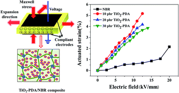
In order to improve compatibility between the dielectric filler and polymeric matrix, we used bio-inspired polydopamine (PDA) to modify titanium dioxide (TiO2) nano-particles. The PDA coated TiO2 (TiO2-PDA) nano-particles were incorporated into nitrile-butadiene rubber (NBR) which contains a large amount polar groups to obtain a dielectric elastomer composite with a large actuated strain under a low electric field. The relatively soft insulating PDA layer on the TiO2 nano-particles led to the composites filled with TiO2-PDA nano-particles displaying better filler dispersion, much lower elastic modulus, lower dielectric loss, and higher electric breakdown field compared with the composites filled with pristine TiO2 nano-particles, resulting in a high electromechanical sensitivity (β). At last, an actuated strain of 5.2% at a relatively safe electric field of 12.5 kV mm−1 without any pre-strains was obtained by the 10 phr TiO2-PDA/NBR composite, a 140% increase in actuated strain compared with the actuated strain (0.69%) of pure NBR at 20 kV mm−1 without any pre-strains. This biomimetic method is simple, efficient, nontoxic, and easy to control, which can be used in other dielectric fillers to improve electromechanical properties of dielectric elastomers.


1 Introduction

Dielectric elastomers (DEs) represent a new and promising electromechanical transducer technology because of their unparalleled combination of high energy density, high efficiency, large deformation, quick response, compactness, silent operation, and ability to hold an induced displacement at a constant voltage without consuming electrical energy.1,2 Over the past few decades, a wide range of applications have been proposed based on DEs by virtue of their unique properties, for example, biomimetic soft robots, responsive prosthetics, tactile displays, bio-inspired electrochromism sensor, positioners, and flat-panel speakers.3–8 However, unlike other electroactive materials, such as carbon nanotubes,9 ionic polymer–metal composites,10 and conductive polymers,11 DEs need a high operating voltage (several kilovolts) to induce a large deformation,2 which could be harmful to humans and damage equipment, limiting the commercial viability of DE technology.12,13 Therefore, getting a large actuated strain at a low driving voltage is a big challenge for dielectric elastomer actuators (DEAs).14

A dielectric elastomer actuator is composed by a dielectric elastomer film and two compliant electrodes coated on both surfaces of the film. When a high voltage is applied to the compliant electrodes, the DEA can quickly change its shape by Maxwell pressure P:15–17

 
P = −εrε0E2 (1)
where ε0 and εr are the permittivity of free space and the relative permittivity (dielectric constant) of the polymer, respectively, and E is the applied electric field. The thickness strain Sz is given by the ratio of electrostatic pressure to the elastic modulus Y of the elastomer. Based on the assumption of constant modulus and free boundary conditions for a dielectric elastomer film, the thickness strain Sz can be approximated by18
 
Sz = P/Y = −εrε0E2/Y (2)

According to eqn (2), the operating voltage can be lowered by decreasing the elastic modulus, increasing the dielectric constant of elastomer materials, or reducing the thickness of dielectric film. In current, the thickness of dielectric elastomer film can be reduced to 10–20 μm in large-scale processing and reduction thickness of dielectric film can also uphold the dielectric loss and breakdown strength, however, the driving voltage is still hundred volts and further reductions could increase importance of inhomogeneities, produce localized areas of high electrical field, and premature electrical breakdown.2 In previous studies, some researchers introduced plasticizer into the elastomer matrix to decrease elastic modulus of DE. However, the plasticizer will be evaporated, migrated, or extracted from elastomer matrix, because of instability of host–guest structure, resulting in poor mechanical strength of DE.19,20 Increasing dielectric constant is an easy method to improve actuated strain of dielectric elastomer, which can not only lead high electrical energy density, but also reduce the driving voltage.21

A commonly used method for increasing dielectric constant of the polymer by introducing organic dipole groups to the polymer chain,21,22 or adding conductive particles23,24 and high-dielectric-constant ceramic powder.25,26 Grafting of organic dipoles groups can improve dielectric constant of polymer matrix on a molecular level avoid problems of agglomeration and yield elastomer films that are homogeneous down to the molecular level. However, the polar group significantly reduces the dielectric strength of polymer and increases sensitivity to moisture.27 Adding a little conductive fillers can dramatically increase the dielectric constant of the polymeric matrix if the concentration of conductive fillers approaches the percolation threshold.23,24 However, the composite should suffer from low dielectric strength and high dielectric loss, resulting in limited application.28 The dielectric constant of a polymer can also be effectively improved by adding high-dielectric-constant ceramic powders when the amount of filler up to 40–50 vol%. However, large amount inorganic filler will lead the elastic modulus increase significantly and severely destroy the flexibility of composite, resulting in a decreased electromechanical sensitivity (β = εr/Y). In addition, as the surface characteristics between the inorganic ceramic fillers and the organic polymeric matrix is much different, it is difficult to naturally disperse the inorganic fillers uniformly in the polymeric matrix, which is not beneficial for obtaining a desirable DE with high actuated strain.29,30 Hence, in order to obtain a high performance dielectric elastomer actuator, the interface between the filler and polymeric matrix cannot be negligible. S. M. Khaled et al.31 used methacrylic acid as a functionalization agent to modify TiO2 to get a good distribution polymethyl methacrylate (PMMA)-based composites. Zhimin Dang et al.32 modified TiO2 nanoparticles by silane couple agent and further incorporated them into silicone rubber matrix to improve the dielectric and mechanical properties of silicone rubber. Although above surface modification of the particles can improve the dispersion of particles, they are complicated and time consuming. After modification, a process to remove residual chemicals is necessary.

In 2007, Lee et al.33 introduced a facile surface modification approach inspired by the adhesion mechanism of mussels. 3,4-Dihydroxy-L-phenylalanine (DOPA), as the main ingredient of these adhesive proteins, contains many catechol groups and ethylamino groups can form a hydrophilic poly(dopamine) (PDA) layer on the surface of various substrates, including organic and inorganic materials.34,35 Recently, PDA was used as a general building block for surface treatments of BaTiO3 to improve the compatibility between the BaTiO3 filler and polymeric matrix.29,36,37 This method is simple, efficient, nontoxic, and easy to control.

Nitrile-butadiene rubber (NBR), which contains a large amount of strong polar acrylonitrile (ACN) groups with a high dielectric constant (>10), can be used as a good candidate material for dielectric elastomer. In our work, high-dielectric-constant titanium dioxide (TiO2) particles modified by PDA (denoted as TiO2-PDA) are incorporated into the NBR to improve its dielectric properties and actuated strain for the good interaction between polar groups of PDA and ACN groups of NBR. The purpose of surface modification of TiO2 particles is to enhance the matrix–filler (host–guest) compatibility, improve the particle distribution, and avoid the introduction of dielectric loss by Maxwell–Wagner polarization. The effects of the poly(dopamine) layer on the particle dispersion, elastic modulus, dielectric properties, and actuated strain of NBR composites were investigated.

2 Experimental

2.1 Materials

A commercial grade of NBR (NBR3305, acrylonitrile content 32.5–34.5 wt%, produced by PetroChina Lanzhou Petrochemical Company, China) was used as raw material. Rutile TiO2 particles with an average diameter of 30 nm were purchased from Wan Jing New Material Co., Ltd (China). Dopamine and tris(hydroxymethyl)-amino-methane (Tris) were purchased from Alfa Aesar Company, USA. The crosslinking agent dicumyl peroxide (DCP) was purchased from Beijing Chemical Reagents Co., Ltd (China).

2.2 Surface modification of TiO2 particles by polydopamine

The TiO2 particles were surface modified with aqueous solutions of dopamine concentrations of 2.0 g L−1. Tris was added into the dopamine solution to adjust pH to a predetermined value of 8.5. The solution was stirred for 24 hours at room temperature and the color of the solution changed from light pink to dark brown with the spontaneous deposition of an adherent PDA film. Then the TiO2 nano-particles coated with PDA were taken out, washed with deionized water, and dried at 60 °C in vacuum. The obtained samples were denoted as TiO2-PDA. The process of dopamine self-polymerization on the surface of TiO2 particles and the possible mechanism for the in situ spontaneous oxidative polymerization of dopamine is illustrated in Fig. 1.35,38
image file: c5ra12311a-f1.tif
Fig. 1 (a) Illustration of procedure for preparing TiO2-PDA particle and (b) possible mechanism of dopamine oxidative self-polymerization.

2.3 Preparation of dielectric composites

The dielectric composites consisting of 0, 10, 20, and 30 phr (parts per hundred parts of rubber) of TiO2 or TiO2-PDA particles, 2 phr of DCP, and 100 phr of NBR were prepared through mechanical mixing on a 6-inch two-roll mill. The cured elastomers were obtained at the pressure of 25 MPa for their optimum cure time as determined by a rotorless curemeter (GT-M2000-FA, Goteah Testing Machines Inc., Taiwan) at 160 °C.

2.4 Characterization methods

The chemical compositions of the surface of the TiO2 and TiO2-PDA nano-particles were determined by X-ray photoelectron spectroscopy (XPS). XPS measurements were carried out on an ESCALAB 250 XPS system (Thermo Electron Corporation, USA) with an Al Kα X-ray source (1486.6 eV photons). The core-level signals were obtained at a photoelectron take-off angle of 45° with respect to the sample surface.

The morphologies of the TiO2 and TiO2-PDA nano-particles were studied by using a high resolution transmission electron microscope (HR-TEM) (Hitachi H9000, Japan) operating at a voltage of 300 kV.

The morphologies of the NBR composites filled with TiO2 and TiO2-PDA nano-particles were investigated by scanning electron microscopy (SEM) using the FEI NanoSEM 430 scanning electron microscope.

The crystalline structure of the TiO2 and TiO2-PDA nano-particles were studied by X-ray diffraction (XRD) (D8 Focus, Bruker, Germany) using Cu Kα radiation with a wavelength of 1.54056 Å, and the diffraction patterns were recorded in the 2θ range of 10–75°.

Thermogravimetric analysis (TGA) was performed in the temperature range of 30–590 °C with heating rate of 20 °C min−1 by using a TA Q500 thermogravimetric analyzer. The TGA experiments were carried out under a nitrogen atmosphere.

The elastic modulus of the samples was determined by the slope of the stress–strain curve at 5% strain, which were obtained by using a tensile apparatus (RG2000-100, Shenzhen Reger. Instrument Co., Ltd, China) at 25 °C according to Chinese Standards GB/T528-1998. The crosshead speed was 50 mm min−1.

The dielectric properties of the samples were measured by an impedance analyzer (E4980A, Agilent, U.S.A.) over the frequency range of 100 to 106 Hz at room temperature.

The samples for actuated strain tests had a thickness of 0.15 mm and a diameter of 5 cm. Prior to use, compliant graphite electrodes composed of graphite, silicone oil, and curing agent with a diameter of 11 mm were applied to each side of the dielectric elastomer film by an airbrush. As it is difficult to accurately measure the change in thickness, we instead measured the change in planar area SP to evaluate the thickness actuated strain Sz. Based on the law of volume constancy,

 
(1 + Sz)(1 + SP) = 1 (3)

Rearrangement of eqn (3) gives an expression for the planar strain:

 
image file: c5ra12311a-t1.tif(4)

Actuated strain tests were performed by using circular membrane actuators at the air condition for considering practical application of dielectric elastomer actuators, in which the dielectric elastomer films were laid flat between two circular frames without any pre-strains. Voltages from 0 kV mm−1 were loaded on the electrode area to obtain the planar strain until electric breakdown occurred. During actuation, the video images of the biaxial extension of the electrode area were captured by a camera (Canon Ixus 210, Japan) fitted with a wide-angle lens, and the relative change in planar area was determined by analyzing the captured video images with Adobe Photoshop graphics editing software. The planar strain can be calculated according to

 
SP = (AA0)/A0 × 100% (5)
where A is the actuated planar area and A0 is the original area.

Every experimental data point of elastic modulus, dielectric properties, and electromechanical strain in this work is the average of the results obtained from at least five samples under the same condition.

3 Results and discussion

3.1 Surface modification of TiO2 particles by polydopamine

The chemical composition of the surface of presence of TiO2 and TiO2-PDA nano-particles was determined by XPS. The XPS wide-scan spectrum of the TiO2 and TiO2-PDA nano-particles is showed in Fig. 2. Both the wide-scan spectra of the TiO2 and TiO2-PDA surface contain C 1s, Ti 2p3, and O 1s peaks. Nevertheless, there is a new peak component at binding energy (BE) of about 400 eV in the wide-scan spectra of the TiO2-PDA, which is attributed to the nitrogen-containing species in PDA. The N 1s core-level spectra of the TiO2-PDA particles can be curve-fitted with two peak components: one peak at 399.5 eV attributed to the amine (–N–H) groups and the other at 398.5 eV attributed to the imine ([double bond, length as m-dash]N–) groups. The (–N–H) groups are formed through the amine group of dopamine, while the imine ([double bond, length as m-dash]N–) groups are formed by the indole groups through structure evolution during the dopamine self-polymerization. The XPS results suggest that PDA has been successfully adhered to the TiO2 particles through oxidative self-polymerization of dopamine.
image file: c5ra12311a-f2.tif
Fig. 2 X-ray photoelectron spectroscopy wide-scan spectra and N 1s core-level spectra of TiO2 and TiO2-PDA nano-particles.

HR-TEM was used to observe the morphologies of TiO2 and TiO2-PDA nano-particles. The HR-TEM micrographs of TiO2 and TiO2-PDA nano-particles are showed in Fig. 3. Comparing with Fig. 3(a), we can easily find a distinct layer of PDA with thickness of 3–5 nm coated on the TiO2 particles in Fig. 3(b), confirming the successfully deposition of PDA on TiO2 particles again. In addition, the TGA curves of TiO2, TiO2-PDA nano-particles, and pure PDA are showed in Fig. 4. As the samples are heated to 590 °C, the TiO2, TiO2-PDA, and pure PDA show a weight loss of 0.78%, 1.78%, and 42.91%, respectively. The weight loss of TiO2-PDA is between the weight of pure PDA and pristine TiO2, demonstrating the PDA successfully deposition on surface of TiO2 particles again.


image file: c5ra12311a-f3.tif
Fig. 3 HR-TEM images of (a) TiO2 particles and (b) TiO2-PDA particles.

image file: c5ra12311a-f4.tif
Fig. 4 TG curves of TiO2, TiO2-PDA nano-particles, and pure PDA.

The crystalline structures of the TiO2 and TiO2-PDA nano-particles were detected by XRD, and the XRD patterns are shown in Fig. 5. From Fig. 5, we can obviously find that there is no difference between the TiO2 and TiO2-PDA nano-particles. The PDA layer on the TiO2 nano-particles surface has no effect on the XRD pattern, indicating the PDA layer is amorphous.


image file: c5ra12311a-f5.tif
Fig. 5 XRD patterns of TiO2 and TiO2-PDA nano-particles.

3.2 Microstructure of dielectric composites

The typical SEM micrographs of the fractured surfaces of the composites of NBR filled with different amounts of TiO2 and TiO2-PDA nano-particles are shown in Fig. 6. From Fig. 6(a) and (c), we can find many particles are exposed on the fractured surfaces and there are some serious agglomerations of TiO2 particles in the composites filled with pristine TiO2, indicating poor compatibility between the TiO2 particles and NBR matrix. This can be explained by the large difference of surface energy between the TiO2 particles and NBR matrix. However, the composites filled with TiO2-PDA nano-particles exhibit better dispersion comparing with the composites filled with pristine TiO2 particles. Besides, the fractured surfaces of the composites filled with TiO2-PDA nano-particles are smoother than those of the composites filled with TiO2 nano-particles, an indication of compatibility between filler and matrix becomes well by dopamine treating. This result further confirms that PDA exhibits super strong adhesion, not only to the surface of inorganic fillers but also to NBR macromolecules.
image file: c5ra12311a-f6.tif
Fig. 6 SEM micrographs of NBR composites filled with (a) 10 phr TiO2, (b) 10 phr TiO2-PDA, (c) 30 phr TiO2, and (d) 30 phr TiO2-PDA nano-particles.

3.3 Mechanical properties and elastic modulus of dielectric composites

Fig. 7 and Table 1 show the stress–strain curves and the stresses at 100%, 200%, and 300% strain and tensile strength of the NBR composites filled with different amounts of TiO2 and TiO2-PDA nano-particles, respectively. Usually, dispersion of inorganic filler into a polymer matrix leads to the elongation at break of composites decrease with increasing content of filler. From Fig. 7, we can find the elongation at break of 10 phr TiO2/NBR composite and 10 phr TiO2-PDA/NBR composite are lower than that of pure NBR because the filler particles act as physical links in the NBR matrix, thus hindering the mobility of the elastomer chains. However, the elongation at break of 30 phr TiO2/NBR composite and 30 phr TiO2-PDA/NBR composite are larger than that of pure NBR. This could be the crosslink density of composites decreased ascribed to the interference of TiO2 nano-particles on the crosslinking process, as reported in several studies.4,6 Generally, a large elongation at break will bring a large tensile strength. Hence, the tensile strength of 10 phr TiO2/NBR composite and 10 phr TiO2-PDA/NBR composite are lower than that of the pure NBR, while the tensile strength of 30 phr TiO2/NBR composite and 30 phr TiO2-PDA/NBR composite are higher than that of the pure NBR. In addition, the elongation at break of TiO2-PDA/NBR composites is higher than that of TiO2/NBR composites filled at the same content of filler, mainly because of strong interfacial interaction between TiO2-PDA particles and rubber matrix, and uniform dispersion of particles with fewer defects. As the elongation at break of TiO2-PDA/NBR composites is higher, which allow the composites develop a larger tensile strength.
image file: c5ra12311a-f7.tif
Fig. 7 Stress–strain curves of NBR composites filled with different contents of TiO2 particles and TiO2-PDA particles.
Table 1 Stresses at 100%, 200%, and 300% strain and tensile strength of NBR with different contents of TiO2 and TiO2-PDA nano-particles
Property Stress at 100% strain (MPa) Stress at 200% strain (MPa) Stress at 300% strain (MPa) Tensile strength (MPa)
NBR 0.847 1.258 1.591 1.757
10 phr TiO2/NBR composite 0.894 1.349 1.552
10 phr TiO2-PDA/NBR composite 0.866 1.343 1.678
30 phr TiO2/NBR composite 0.904 1.429 1.937 2.213
30 phr TiO2-PDA/NBR composite 0.88 1.374 1.884 2.287


The elastic modulus of the composites filled with different amounts of TiO2 and TiO2-PDA nano-particles are shown in Fig. 8. From Fig. 8, we can find the elastic modulus of composites increase with increasing content of filler whatever the TiO2 or TiO2-PDA nano-particles. The increased elastic modulus is ascribed to the inorganic particles, which usually cause a gradual increase in the stiffness of polymer matrix with increasing amount of filler.25,39 However, we can find the elastic modulus of composites filled with TiO2-PDA nano-particles is lower than that of composites filled with TiO2 nano-particles at the same content of filler. This might be due to the elastic modulus of TiO2-PDA nano-particles is lower than that of TiO2 nano-particles, because the elastic modulus of organic polymeric PDA is much lower than that of rigid TiO2 nano-particles. The relatively soft PDA layer coating on the TiO2 nano-particles will increase the volume fraction of whole polymeric phase in the composites. Assuming that the TiO2 nano-particles were spherical and 30 g TiO2 particles were incorporated into 100 g NBR matrix, we can calculate the volume of relatively soft PDA layer (VPDA) coated on TiO2 nano-particles roughly according to the following equations:40

 
image file: c5ra12311a-t2.tif(6)
where r and VsTiO2 are the radius and volume of signal TiO2 nano-particles, respectively, nTiO2 is the number of TiO2 nano-particles in the matrix, mTiO2 is weight of all the TiO2 nano-particles in the matrix (i.e. 30 g), h is the thickness of PDA (i.e. 4 nm), and ρTiO2 is the density of TiO2 nano-particles (i.e. 4.25 g cm−3). It was found in the ca.100 cm3 NBR, the polymeric volume introduced by PDA coating is about 6 cm3 while the volume of TiO2 nano-particles (VTiO2) is about 7.5 cm3.


image file: c5ra12311a-f8.tif
Fig. 8 Elastic modulus of NBR composites filled with different amounts of TiO2 and TiO2-PDA nano-particles.

3.4 Dielectric properties of dielectric composites

The dielectric constant and dielectric loss tangent of NBR composites filled with different contents of TiO2 and TiO2-PDA nano-particles at frequency of 1 kHz are shown in Fig. 9. As expected, the semiconductor nature of TiO2 nano-particles is beneficial to movement of electrical charge, leading to the dielectric constant of all composites increase with increasing content of TiO2 particles whether they are coated with PDA or not. The increased dielectric constant can be ascribed to the increase in electron polarization and interface polarization at the interface between the NBR matrix and TiO2 particles.26,32 The electron polarization and interface polarization will increase not only the dielectric constant, but also the dielectric loss of the composites. We can find the dielectric loss tangent of all composites are higher than that of pure NBR in Fig. 9. However, the dielectric loss tangent of all composites are lower than 0.05 at all filler contents, indicating that most of electric energy can be converted to mechanical energy with just a little energy dissipation through dielectric loss during actuation. In addition, the dielectric constant and dielectric loss tangent of the composites filled with TiO2-PDA are lower than those of the composite filled with TiO2 at the same contents of filler, ascribe to the insulating dopamine coating on TiO2 nano-particles prevents the conductive particles interacting with each other, thus acting as an electrical potential barrier. A portion of the electrons cannot jump from one side of the potential gap to the other, thus minimizing the leakage of current and decreasing of dielectric constant and dielectric loss.32,41 This coating method was also used to achieve excellent dielectric composites with low dielectric loss and high electric breakdown strength in previous studies.42
image file: c5ra12311a-f9.tif
Fig. 9 Dielectric constant and dielectric loss tangent of NBR composites filled with different contents of TiO2 and TiO2-PDA nano-particles at frequency of 1 kHz.

Fig. 10 shows dependence of dielectric constant and dielectric loss tangent of NBR composites on frequency at different contents of TiO2-PDA particles. From Fig. 10(a), we can see that the dielectric constant decrease with increasing frequency. The dielectric constant of the composites at low frequency (1 kHz) is higher than that at high frequency (10 MHz). However, the dielectric constant decreases very slowly when the frequency is less than 105 Hz. The reason could be the polarization of the composites has not enough relaxation time to catch up with the frequency change of the loaded electric field.26,43 However, the dielectric loss tangent displays an obvious change from 102 to 106 Hz, as shown in Fig. 10(b).


image file: c5ra12311a-f10.tif
Fig. 10 Frequency dependence of (a) dielectric constant and (b) dielectric loss tangent of NBR composites at different contents of TiO2-PDA nano-particles.

3.5 Actuated strain of dielectric composites

The actuated strains of NBR composites with different contents of TiO2 and TiO2-PDA nano-particles are plotted against the applied electric field are shown in Fig. 11. From Fig. 11, we can find the NBR composites filled with 10 phr TiO2 and TiO2-PDA nano-particles exhibit the highest actuated strain at the same electric field in TiO2/NBR and TiO2-PDA/NBR composites respectively, ascribe to the highest electromechanical sensitivity (εr/Y) (see Fig. 12). This can be explained by that the dielectric constant of NBR composite increased whereas the elastic modulus changed a little with the addition of 10 phr dielectric filler because of no obvious filler network exists in system. When content of dielectric filler increases to 30 phr, the filler network formed in the composite lead to the elastic modulus increased sharply, leading electromechanical sensitivity (εr/Y) decreased. In addition, we can find the NBR composites filled with TiO2-PDA nano-particles exhibit higher actuated strain than those of NBR composites filled with pristine TiO2 nano-particles at the same electric field. This can be ascribed to much decreased elastic modulus and little lower dielectric constant of TiO2-PDA/NBR composites compared with TiO2/NBR composites, leading to a larger electromechanical sensitivity β = εr/Y. At last, the NBR composites filled with 10 phr TiO2-PDA displays the largest actuated strain of 5.2% at 12.5 kV mm−1 without any pre-strains, a 140% increase in actuated strain comparing with the actuated strain (0.69%) of pure NBR at 20 kV mm−1 without any pre-strains. In addition, we can find from Fig. 12 that the electromechanical sensitivity (εr/Y) of 10 phr TiO2-PDA/NBR composite (11.48) is just higher a little than that of 20 phr TiO2-PDA/NBR composite (11.45), however, the actuated strain of 10 phr TiO2-PDA/NBR composite is almost 20% larger than that of 20 phr TiO2-PDA/NBR composite. The reason could be the dielectric loss tangent of 20 phr TiO2-PDA/NBR composite (0.0336 at 1 kHz) is higher than that of 10 phr TiO2-PDA/NBR composite (0.0315 at 1 kHz) (see Fig. 9), leading to more electric energy convert into heat energy during the actuation process.44 In addition, more inorganic particles in the NBR matrix leading a poor dispersion, resulting in a part of electric energy dissipated through mechanical loss.44
image file: c5ra12311a-f11.tif
Fig. 11 Dependence of actuated strain of NBR composites filled with different contents of (a) TiO2 and (b) TiO2-PDA nano-particles on electric field.

image file: c5ra12311a-f12.tif
Fig. 12 Electromechanical sensitivity of NBR composites filled with different contents of TiO2 and TiO2-PDA nano-particles.

Usually, the electrical breakdown field of composites decreases after filled with inorganic filler. In our work, the electrical breakdown field of NBR composites filled with TiO2 decreased with increasing content of TiO2, in agreement with previous studies. Two reasons could be responsible for this phenomenon. First, with increasing filler in the system, more and more charge species are generated in the interface between the polymer matrix and filler, leading to the generation of interfacial charge to break down the system.30,45 Second, agglomerations of filler particles in the system will introduce defect centers that distort and enhance the local electric field.30,46 However, the electrical breakdown field of NBR composites filled with 30 phr TiO2-PDA is higher than that of NBR composites filled with 10 phr TiO2-PDA and 20 phr TiO2-PDA particles. It is reasonable to think that the mobility of polymer chains that are tightly bonded to the TiO2-PDA filler decreases with increasing content of PDA. The transfer of charge carriers through loose polymer chains not bonded to the filler is suppressed, thus increasing the electrical breakdown field.36 In addition, insulating dopamine coating on TiO2 nano-particles prevents the conductive particles from interacting with each other, leading to the electrical breakdown field increased.

The history dependence, as an important property of DEAs, was also studied. Fig. 13 shows the time dependence of actuated strain of 10 phr TiO2-PDA/NBR composite actuator under an electric field of 5 kV mm−1 for 10 cycles without any pre-strains. Each application period of 10 seconds was followed by an off-interval of 3 seconds. We can find there is a small fluctuation in strain, maybe arising from viscoelastic loss of composite and poor compatibility between the matrix and particles.


image file: c5ra12311a-f13.tif
Fig. 13 Cyclic electric field loadings on 10 phr TiO2-PDA/NBR composite actuator without any pre-strains.

Table 2 compares various advanced DEA materials reported in the literature with the 10 phr TiO2-PDA/NBR composite in terms of the maximum actuated strain and actuated strain at 12.5 kV mm−1. We can find the actuated strain of TiO2-PDA/NBR composites at low electric fields is relatively high comparing with pure NBR and most of DEs reported in previous studies under the conditions of no pre-strain and low electric field (see Table S1 containing almost all DEA materials), demonstrating the advantage of TiO2-PDA/NBR composites.

Table 2 Comparison of actuated performance of 10 phr TiO2-PDA/NBR composite with that of various advanced DEA materials reported in the literature without any pre-strains
Material Maximal actuated straina (%) Breakdown strengtha (kV mm−1) Actuated straina at 12.5 kV mm−1 Ref.
a Estimated from graphical data in cited reference, when no tabulated value was provided.
TiO2/PDMS/DMSO 13 30 <4% 19
PANI@PDVB/PDMS 12 54 <1% 39
PEG/PDMS 11.5 40 <2% 46
14PANI/15PolyCuPc/85PU 9.3 20 <3% 47
SEBS-MA grafted PANI 1.4 27 <0.4% 48
NBR/TiO2/DOP 3.04 20 <0.25% 49
DAN/TPU 2.6 20 <1.5% 20
PDBII 25.5 42 <8% 14
m-BT/SR4 26 12 13
SeRM/BT 10 23 <2% 50
Azo-g-PDMS 17 67.5 <3% 51
NBR 2.15 20 0.69  
TiO2-PDA/NBR 5.2 12.5 5.2%  


4 Conclusion

We successfully coated a PDA layer on TiO2 particles through a biomimetic method. The organic insulating dopamine layer not only improved dispersion of TiO2 particles in the NBR matrix, but also decreased dielectric loss and enhanced electric breakdown strength. In addition, the relatively soft PDA increased the volume fraction of whole polymeric phase of composites, resulted in a low elastic modulus and high electromechanical sensitivity. At last, an actuated strain of 5.2% at a relatively safe electric field of 12.5 kV mm−1 without any pre-strains was obtained by 10 phr TiO2/NBR composite, a 140% increase in actuated strain comparing with the actuated strain (0.69%) of pure NBR at 20 kV mm−1 without any pre-strains. Our biomimetic method is simple, efficient, nontoxic, and easy to control, which can be used in other dielectric filler to improve actuated strain of dielectric elastomers.

Acknowledgements

This work was supported by the Youth Backbone Personal Project of Beijing (No. 2014000020124G085), National Natural Science Foundation of China (No. 51373026), Innovation Promotion Project of Beijing Municipal Commission of Education, China (TJSHG201310017034), Beijing Higher Education Young Elite Teacher Project, and Project of Petrochina (No. PRIKY14041).

References

  1. S. Hunt, T. G. Mckay and I. A. Anderson, Appl. Phys. Lett., 2014, 104, 113701 Search PubMed .
  2. F. B. Madsen, L. Yu, A. E. Daugaard, S. Hvilsted and A. L. Skov, RSC Adv., 2015, 5, 10254–10259 RSC .
  3. W. Hu, S. N. Zhang, X. Niu, C. Liu and Q. Pei, J. Mater. Chem. C, 2014, 2, 1658–1666 RSC .
  4. F. Carpi and D. de Rossi, IEEE Trans. Dielectr. Electr. Insul., 2005, 12, 835–843 CrossRef CAS .
  5. F. Carpi, G. Gallone, F. Galantini and D. de Rossi, Adv. Funct. Mater., 2008, 18, 235–241 CrossRef CAS PubMed .
  6. J. K. Nelson and Y. Hu, J. Phys. D: Appl. Phys., 2005, 38, 213–222 CrossRef CAS .
  7. Z. Li, L. A. Fredin, P. Tewari, S. A. DiBenedetto, M. T. Lanagan, M. A. Ratner and T. J. Marks, Chem. Mater., 2010, 22, 5154–5164 CrossRef CAS .
  8. Y. Jang and T. Hirai, Soft Matter, 2011, 7, 10818–10823 RSC .
  9. R. H. Baughman, C. Cui, A. A. Zakhidov, Z. Iqbal, J. N. Barisci, G. M. Spinks, G. G. Wallace, A. Mazzoldi, D. de Rossi and A. G. Rinzler, Science, 1999, 284, 1340–1344 CrossRef CAS .
  10. S. Nemat-Nasser and Y. Wu, J. Appl. Phys., 2003, 93, 5255–5267 CrossRef CAS PubMed .
  11. L. Bay, K. West, P. Sommer-Larsen, S. Skaarup and M. Benslimane, Adv. Mater., 2003, 15, 310–313 CrossRef CAS PubMed .
  12. M. Molberg, Y. Leterrier, C. J. G. Plummer, C. Walder, C. Lowe, D. M. Opris, F. A. Nuesch, S. Bauer and J. A. E. Manson, J. Appl. Phys., 2009, 106, 054112 CrossRef PubMed .
  13. D. Yang, F. Ge, M. Tian, N. Ning, L. Zhang, C. Zhao, K. Ito, T. Nishi, H. Wang and Y. Luan, J. Mater. Chem. A, 2015, 3, 9468–9479 CAS .
  14. W. Lei, R. Wang, D. Yang, G. Hou, X. Zhou, H. Qiao, W. Wang, M. Tian and L. Zhang, RSC Adv., 2015, 5, 47429–47438 RSC .
  15. D. M. Opris, M. Molberg, C. Walder, Y. S. Ko, B. Fischer and F. A. Nüesch, Adv. Funct. Mater., 2011, 21, 3531–3539 CrossRef CAS PubMed .
  16. P. Brochu and Q. Pei, Macromol. Rapid Commun., 2010, 31, 10–36 CrossRef CAS PubMed .
  17. J. Liang, A. Betts, D. Kennedy and S. Jerrams, Mater. Sci. Eng., C, 2015, 49, 754–760 CrossRef PubMed .
  18. R. Pelrine, R. Kornbluh, Q. Pei and J. Joseph, Science, 2000, 287, 836–839 CrossRef CAS .
  19. H. Zhao, D. R. Wang, J. W. Zha, J. Zhao and Z. M. Dang, J. Mater. Chem. A, 2013, 1, 3140–3145 CAS .
  20. N. Ning, B. Yan, S. Liu, Y. Yao, L. Zhang, T. W. Chan, T. Nishi and M. Tian, Smart Mater. Struct., 2015, 24, 032002 CrossRef .
  21. B. Kussmaul, S. Risse, G. Kofod, R. Waché, M. Wegener, D. N. McCarthy, H. Krüger and R. Gerhard, Adv. Funct. Mater., 2011, 21, 4589–4594 CrossRef CAS PubMed .
  22. S. Risse, B. Kussmaul, H. Krüger and G. Kofod, Adv. Funct. Mater., 2012, 22, 3958–3962 CrossRef CAS PubMed .
  23. Z. M. Dang, L. Wang, Y. Yin and Q. Zhang, Adv. Mater., 2007, 19, 852–857 CrossRef CAS PubMed .
  24. C. C. Wang, J. F. Song, H. M. Bao, Q. D. Shen and C. Z. Yang, Adv. Funct. Mater., 2008, 18, 1299–1306 CrossRef CAS PubMed .
  25. G. Gallone, F. Carpi, D. de Rossi, G. Levita and A. Marchetti, Mater. Sci. Eng., C, 2007, 27, 110–116 CrossRef CAS PubMed .
  26. Z. M. Dang, H. P. Xu and H. Y. Wang, Appl. Phys. Lett., 2007, 90, 012901 CrossRef PubMed .
  27. T. Chen, J. Qiu, K. Zhu, J. Li, J. W. Wang, S. Q. Li and X. Wang, J. Phys. Chem. B, 2015, 119, 4521–4530 CrossRef CAS PubMed .
  28. J. K. Yuan, Z. M. Dang, S. H. Yao, J. W. Zha, T. Zhou, S. T. Li and J. Bai, J. Mater. Chem., 2010, 20, 2441–2447 RSC .
  29. M. F. Lin, V. K. Thakur, E. J. Tan and P. S. Lee, RSC Adv., 2011, 1, 576–578 RSC .
  30. S. M. Nayak, B. Sahoo, T. K. Chaki and D. Khastgir, RSC Adv., 2013, 3, 2620–2631 RSC .
  31. S. Khaled, R. Sui, P. A. Charpentier and A. S. Rizkalla, Langmuir, 2007, 23, 3988–3995 CrossRef CAS PubMed .
  32. Z. M. Dang, Y. J. Xia, J. W. Zha, J. K. Yuan and J. B. Bai, Mater. Lett., 2011, 65, 3430–3432 CrossRef CAS PubMed .
  33. H. Lee, S. M. Dellatore, W. M. Miller and P. B. Messersmith, Science, 2007, 318, 426–430 CrossRef CAS PubMed .
  34. S. H. Hwang, D. Kang, R. S. Ruoff, H. S. Shin and Y. B. Park, ACS Nano, 2014, 8, 6739–6747 CrossRef CAS PubMed .
  35. C. Xu, M. Tian, L. Liu, H. Zou, L. Zhang and W. Wang, J. Electrochem. Soc., 2012, 159, D217 CrossRef CAS PubMed .
  36. Y. Song, Y. Shen, H. Liu, Y. Lin, M. Li and C. W. Nan, J. Mater. Chem., 2012, 22, 8063–8068 RSC .
  37. Y. Song, Y. Shen, H. Liu, Y. Lin, M. Li and C. Nan, J. Mater. Chem., 2012, 22, 16491–16498 RSC .
  38. W. Wang, R. Li, M. Tian, L. Liu, H. Zou, X. Zhao and L. Zhang, ACS Appl. Mater. Interfaces, 2013, 5, 2062–2069 CAS .
  39. M. Molberg, D. Crespy, P. Rupper, F. Nüesch, J. A. E. Månson, C. Löwe and D. M. Opris, Adv. Funct. Mater., 2010, 20, 3280–3291 CrossRef CAS PubMed .
  40. D. Yang, M. Tian, D. Li, W. Wang, F. Ge and L. Zhang, J. Mater. Chem. A, 2013, 1, 12276–12284 CAS .
  41. H. Zou, L. Zhang, M. Tian, S. Wu and S. Zhao, J. Appl. Polym. Sci., 2010, 115, 2710–2717 CrossRef CAS PubMed .
  42. P. Kim, S. C. Jones, P. J. Hotchkiss, J. N. Haddock, B. Kippelen, S. R. Marder and J. W. Perry, Adv. Mater., 2007, 19, 1001–1005 CrossRef CAS PubMed .
  43. Z. M. Dang, C. Y. Tian, J. W. Zha, S. H. Yao, Y. J. Xia, J. Y. Li, C. Y. Shi and J. Bai, Adv. Eng. Mater., 2009, 11, B144–B147 CrossRef PubMed .
  44. H. Liu, L. Zhang, D. Yang, N. Ning, Y. Yu, L. Yao, B. Yan and M. Tian, J. Phys. D: Appl. Phys., 2012, 45, 485303 CrossRef .
  45. Y. Yao, N. Ning, L. Zhang, T. Nishi and M. Tian, RSC Adv., 2015, 5, 23719–23726 RSC .
  46. D. Yang, M. Tian, Y. Dong, H. Liu, Y. Yu and L. Zhang, Smart Mater. Struct., 2012, 21, 035017 CrossRef .
  47. C. Huang, Q. Zhang and K. Bhattacharya, Appl. Phys. Lett., 2004, 84, 4391 CrossRef CAS PubMed .
  48. H. Stoyanov, M. Kollosche, D. N. McCarthy and G. Kofod, J. Mater. Chem., 2010, 20, 7558–7564 RSC .
  49. H. C. Nguyen, V. T. Doan, J. Park, J. C. Koo, Y. Lee and H. R. Choi, Smart Mater. Struct., 2009, 18, 015006 CrossRef .
  50. S. Tsuchitani, T. Sunahara and H. Miki, Smart Mater. Struct., 2015, 24, 060530 CrossRef .
  51. L. Zhang, D. Wang, P. Hu, J. W. Zha, F. You, S. T. Li and Z. M. Dang, J. Mater. Chem. C, 2015, 3, 4883–4889 RSC .

Footnote

Electronic supplementary information (ESI) available. See DOI: 10.1039/c5ra12311a

This journal is © The Royal Society of Chemistry 2015
Click here to see how this site uses Cookies. View our privacy policy here.