Yuka Shibata,
Tomofumi Santa and
Masaru Kato*
Graduate School of Pharmaceutical Sciences and GPLLI Program, The University of Tokyo, 7-3-1 Hongo, Bunkyo-ku, Tokyo 113-0033, Japan. E-mail: masaru-kato@umin.ac.jp; Fax: +81-3-5841-1841
First published on 28th July 2015
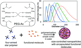
A simple method based on the vortex mixing of precursor chemicals in surfactant-free aqueous conditions has been developed for preparing nanoparticles that encapsulate functional molecules within a four-armed star polymer. Through investigation into the effects of altering such variables as the vortex intensity, chemical composition, temperature etc., it was found that the size of these nanoparticles could be controlled within a range of 20 to 200 nm. Furthermore, a variety of functional molecules could be encapsulated without changing the size of the nanoparticles, suggesting that this method has the potential to be used for a wide range of applications, such as medicine, industrial production and cosmetics.
Although many different methods have been developed for the preparation of nanoparticles containing functional molecules, there are still difficulties in preparing nanoparticles with not only a controlled size, but also a capability to encapsulate a variety of molecules (including proteins). This stems from the fact that common preparation methods15–20 such as free radical polymerization (emulsion polymerization), living polymerization, reversible addition–fragmentation chain transfer polymerization (RAFT), atom transfer radical polymerization (ATRP) and microwave polymerization utilize a surfactant and organic solvent that can denature proteins. Thus, there is a great need to develop a fast and simple procedure for the preparation of functional molecule-containing nanoparticles with a controlled size.
The authors have developed a method of preparing nanoparticles from a four-armed polyethylene glycol (PEG) star polymer, which can encapsulate a range of functional molecules such as proteins and siRNA by wrapping them in the mesh structure formed during polymerization.5,6,21–23 This polymerization under surfactant-free physiological conditions (room temperature with no organic modifier) is ideal for the preparation of nanoparticles containing fragile biomolecules. In this study, we developed a preparation procedure that can control the type and quantity of the molecule encapsulated within size-controlled nanoparticles through a greater understanding of the polymerization reaction of the star polymer used.
The effect of changing the encapsulated molecule was also examined using three different concentrations (0.01, 0.05, and 0.1 mg mL−1) of FLU, which was compared against using trypsin or nothing at all as the encapsulated molecule. As shown in Fig. 2a, the size distribution of the nanoparticles was essentially the same in all cases, with an average particle size of about 38 nm. In other words, the nature of the encapsulated molecule has no bearing on the size of the nanoparticles, which means that the effect of different molecules on the properties of the nanoparticle can be easily ascertained without worrying about size effects.
![]() | ||
Fig. 2 DLS analyses of nanoparticles prepared using different (a) encapsulated molecules and concentrations, and (b) pH solutions. |
To examine the effect of the pH, nanoparticles were produced under five different pH conditions (3–11) using a Britton–Robinson buffer. However, as shown in Fig. 2b, this had no discernible effect on the particle size distribution. This is a significant finding, as it means that the solution pH can be tailored to optimize the solubility and charge of the functional molecule without the fear of adversely affecting the size of the nanoparticles.
Encapsulation of the functional molecule within the nanoparticles was confirmed by using different concentrations of FLU, which was readily quantified within the nanoparticles by fluorescence analysis due to the PEG polymer of the nanoparticle exhibiting no fluorescence. Fig. 3 shows the relationship between the FLU concentration of the prepared solution and the fluorescence intensity of the dispersed solution, in which there is clearly a correlation between the two. This means that the quantity of functional molecule encapsulated can be easily controlled by simply changing its concentration in the initial solution.
![]() | ||
Fig. 3 Fluorescence analysis of FLU-encapsulated nanoparticles prepared from different concentrations of FLU solution. |
The polymer and APS concentration, as well as the ionic strength of the preparation solution, were examined to better understand the formation mechanism of the nanoparticles and how it could be used for size control. Three different concentrations (20, 25, and 30 mg mL−1) were used for this, and in Fig. 4a, we see that a polymer concentration of 20 mg mL−1 produced a particle size of 50 nm. This increased to 60 and 100 nm with polymer concentrations of 25 and 30 mg mL−1, respectively, meaning that the polymer concentration had a large influence on the nanoparticle's size. In the case of APS concentration (Fig. 4b), the formation of nanoparticles was not observed by DLS analysis at low APS concentrations (10 mg mL−1), but the particle size increased when the APS concentration was raised from 15 to 30 mg mL−1, indicating that although APS is required for nanoparticle formation, a large quantity results in large nanoparticles. This differs from the results seen with nanoparticles prepared using radical polymerization, where the size becomes smaller with increasing APS concentration.17,19 This opposite tendency is thought to derive from the large molecular size of the PEG star monomer, or possibly the four reaction groups within each monomer. With the ionic strength (Fig. 4c), values of 0.01, 0.1, and 1 M produced nanoparticle sizes of 130, 100 and 50 nm, respectively. Thus, the effect of ionic strength on size clearly becomes much more pronounced as the ionic strength is increased.
![]() | ||
Fig. 4 DLS analyses of nanoparticles prepared using different (a) concentrations of the star polymer, (b) concentrations of APS, (c) ionic strengths, (d) holding temperatures, and (e) holding times. |
Varying the temperature at which the mixture was left at prior to adding water (Fig. 4d) revealed that holding it at less than 10 °C for 15 min resulted in there being no evidence of nanoparticle formation by DLS analysis; however, when held at ∼20 or 30 °C, 50 nm diameter nanoparticles were confirmed. When the temperature was further increased to 40 and 50 °C the particle size distribution became much wider and the average size increased. This means that room temperature actually represents ideal conditions for achieving a homogeneous size distribution of nanoparticles. The effect of the time spent at room temperature was also examined, and as shown in Fig. 4e, this is clearly an important part of the process given that no nanoparticles are present immediately following vortex. Increasing the holding to 20, 30 and 60 min produced nanoparticles that were 30, 50, and 80 nm in size, respectively, clearly indicating that this represents the period of nanoparticle growth. In other words, increasing the time or temperature here increases the amount of polymerization that occurs. This process, however, is halted once water is added due to the concentration of unreacted star polymer becoming dilute to continue the reaction.
The inverse correlation between particle size and vortex intensity (Fig. 1d) can be explained by the fact that the quantity of APS consumed through reaction with oxygen in the reaction tube is increased with increasing vortex intensity, thereby reducing the effective quantity of APS that can be used for polymerization of the star polymer. This reduced quantity of APS results in the small nanoparticles seen in Fig. 4b. To confirm this hypothesis, nanoparticles were prepared under conditions in which all of the oxygen in the reaction tube was first removed by sonication and substituted by N2, resulting in an almost consistent nanoparticle size (ESI Fig. 1†). By varying the preparation conditions, it proved possible to produce nanoparticles with a size of anywhere between 20 and 200 nm (Fig. 4f). Finally, the stabilities of the encapsulated molecule and the nanoparticles themselves were examined, and the effect of the formation process on the encapsulated molecule was evaluated using protein (trypsin)-containing photodegradable nanoparticles prepared from a star PEG monomer with a photocleavable nitrobenzyl group. The activity of the trypsin released upon light irradiation was correlated with the amount of trypsin used for the nanoparticle preparation (ESI Fig. 2†). As no change was observed by DLS when the dispersed nanoparticle solution was stored at 4 °C for 2 weeks (ESI Fig. 3†), it can be concluded that any degradation of the encapsulated molecule during nanoparticle formation or of the nanoparticle during storage are negligible.
Footnote |
† Electronic supplementary information (ESI) available. See DOI: 10.1039/c5ra12205h |
This journal is © The Royal Society of Chemistry 2015 |