DOI:
10.1039/C5RA11908A
(Paper)
RSC Adv., 2015,
5, 60373-60379
Interlocked dimerization of C3-Symmetrical boron difluoride complex: designing non-cooperative supramolecular materials for luminescent thin films†
Received
20th June 2015
, Accepted 6th July 2015
First published on 6th July 2015
Abstract
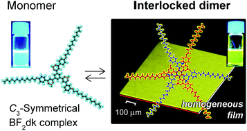
A lipophilic complex with radially-connected three β-diketonate boron difluoride (BF2dk) units to a central benzene ring was newly developed. The C3-symmetrical BF2dk complex (1) formed a self-complementary interlocked dimer (1)2 with increasing concentration in CHCl3 as revealed by NMR spectroscopy and quantum chemical calculations. A remarkable luminescence color change from blue to yellow was observed in response to the formation of interlocked dimers. Electrostatic interactions, hydrogen bonding between negative convexes (BF2 moiety) and positive concaves (three protons aligned on each arm) principally contribute to the dimerization, whereas the formation of interlocked dimers was accompanied by conformational changes of constituent molecules which interrupted further association. Consequently, casting of the chloroform solution of interlocked dimers on solid supports gave uniform thin films without uneven crystallization. It provides a new perspective for designing anti-cooperative systems for homogeneous molecular coatings.
Introduction
Self-assembly of photofunctional molecules has been attracting much interest as a means to create nanoarchitectures and their cooperative functions that are not accessible from the constituent molecules.1 Amphiphilic molecular design is widely adopted to achieve self-assembly of functional molecules in aqueous, organic and ionic media.2 In general, an increase in the concentration of the component molecules causes extended aggregation into developed structures,1 which are indispensable to achieve stand-alone supramolecular functions in given solvent media. In particular, formation of ordered aggregates with crystalline order has been preferred to achieve photofunctionalities such as long-range transfer of photon energy or charge carriers. On the other hand, in nanotechnology fields such as photovoltaics, photochemical events at the interface are of crucial importance. There exist strong demands for photofunctional molecular systems that evenly and densely spread on surfaces as homogeneous functional molecular coatings. Such molecular systems preferably show adaptive nature to varied surfaces, which needs to be designed from a different perspective from the widely-received highly-ordered self-assembly under thermodynamic equilibrium. That is, there are two issues to be solved. First, uniform thin films need to be facilely obtained by casting or spin coating method, which require inhibition of crystal nucleation in the kinetically controlled solvent evaporation processes, i.e., under far-from-equilibrium conditions. Second, to meet the above requirement, a molecular design principle is required to accumulate photofunctional units in high density while avoiding their extended aggregation in solution.
To avoid the formation of crystalline aggregates in solution, the simplest approach is to design suitably designed molecules that preferentially form dimers which do not further assemble to extended aggregates.
This is the case that the dimerization constant K is much larger than that for subsequent aggregation, which is referred to as anti-cooperative process.1 Although this process has received much less attention, it can be realized for example in aggregation of structurally flexible π-systems with peripheral alkyl chains, where dimerization induces changes in the conformation and orientation of alkyl chains to turn away to the other side of π-faces. Such dimerization-induced steric interaction between aliphatic side chains may consequently lead to the interruption of further association.
With this view in mind, we have designed a C3-symmetrical boron difluoride β-diketonate (BF2dk) complex in expectation of the formation of dimers which do not further aggregate in solution. Among varied molecular architectures developed so far, radial integration of functional units provides a means to introduce multiple photofunctional groups in a unit molecule, which is advantageous to enhance the density of these functionalities even in the condensed state. For example, molecules with C3-symmetrical axis have been widely studied in liquid crystals,3 supramolecular polymers4 and biomolecule-conjugates.5 On the other hand, the family of BF2dk complexes has been attracting much interest due to their unique properties as exemplified by large molar absorption coefficients,6a large ground dipole moment (μ = 6.7 Debye for BF2 dibenzoylmethane complex),6b high emission quantum yields,6c,d two-photon absorption,6a efficient electron transport6e and mechanochromic luminescence.6f The BF2dk complexes have been also introduced in molecular assemblies such as organogels,7 liquid crystals,6c,8 nanoparticles9 and self-assembled monolayers.10 In these systems, however, the design of BF2dk complexes that form discrete dimers with anti-cooperative nature has been unprecedented.
The design of a novel C3-symmetrical molecular framework for BF2dk complex 1 is shown in Scheme 1. Three BF2dk units are radially integrated in a central phenyl ring with long alkyl chains to ensure solubility in common organic solvents. The dodecyloxy groups are introduced at the para-position of dibenzoylmethane groups, in expectation for enhancing luminescent quantum yields.6c The boomerang-shaped mono-BF2dk complex (2)6d,8b and linearly-connected di-BF2dk complexes (3, 4)10b were synthesized as references in order to gain insight into the effect of molecular geometry on their intermolecular interactions, solubility and luminescence properties. Self-assembly of these complexes was examined in chloroform, and their accumulation on solid substrate was also investigated in terms of the development of homogeneous molecular coatings.
 |
| Scheme 1 Molecular structure of lipophilic BF2dk complexes 1–4. | |
Results and discussion
2.1. Spectroscopic characterization of BF2dk complexes in dilute solution
The effect of molecular structures on the spectroscopic property of BF2dk complexes (1–4) was measured in dilute solutions. Fig. S2 (ESI†) shows UV-vis absorption spectra of the complexes 1–4 and the corresponding ligands 1L–4L in chloroform (10 μM, 20 °C). Chloroform was chosen as solvent because of its superior property to dissolve a wide range of molecules. Absorption peaks of the complexes are observed around at 410–430 nm, which are red-shifted from the corresponding ligands (λmax at 365–390 nm) due to coordination of β-diketonate groups to BF2.6b In luminescence spectra, the complexes 1–3 showed blue luminescence at 440–450 nm whereas complex 4 gave cyan-colored luminescence at 480 nm (Fig. S3 and Table S1, ESI†).
All the BF2dk complexes showed high emission quantum yields (ΦE) above 88%, which are consistent with those reported for BF2dk derivatives possessing electron donating groups in the para positions.6c 1H-NMR spectra were obtained for the BF2 complexes and ligands, whose chemical shifts for the aromatic and enolic protons were summarized in Tables S2 and S3 (ESI†). The ligands 1L–4L in dilute CDCl3 solution showed a singlet signal at ca. 17.0 ppm, which are assignable to enolic protons of β-diketonate groups. The keto methylene protons were not observed, indicating that the enol tautomer is the dominant form in chloroform. Meanwhile, in BF2dk complexes the enolic proton signals disappeared completely even for diluted solutions (10 μM), reflecting the stability of BF2 complexes. The chemical shifts of the aromatic protons in BF2dk complexes showed remarkable downfield shifts compared to those observed for the corresponding ligands 1L–4L, reflecting electron withdrawal by the BF2 units. It is also to note that the trigonal complex 1 showed aromatic protons most downfield shifted at among all compounds tested in this study. This is ascribable to formation of intermolecular B–F⋯H hydrogen bonding in the C3-symmetrical complex 1 in organic media; the aromatic protons serve as hydrogen bond donors and BF2 units as acceptors, as discussed below.
2.2. Self-assembly characteristics of BF2dk complexes
The solubility of BF2dk complexes in chloroform is amenable to change depending on the chemical structure. The serially-concatenated complexes 3 and 4 show high crystallinity and were poorly soluble in chloroform. Micrometer-sized precipitates were formed upon standing the heat-dissolved 1.0 mM solutions at ambient temperature. In contrast, compounds 1 and 2 showed superior solubility and were homogeneously dissolved in chloroform. The formation of aggregates was characterized by a dynamic light scattering (DLS). The concentration dependence of the DLS count rate for the complex 1 showed apparent deviation from the linear relationship above ca. 1.0 mM, which is consistent with the formation of the molecular assembly (Fig. S4, ESI†). Correspondingly, 1H-NMR spectra of the complex 1 showed considerable upfield shifts in the aromatic protons of Hb, Hc, and Hd above the concentration of ca. 1.0 mM (Fig. 1a and c). The observed gradual upfield shifts in aromatic protons indicate that the C3-symmetric complex 1 forms parallel stacked aggregates which are in equilibrium with monomeric species on the 1H-NMR time scale. As shown in Fig. 1 inset, upon increasing the concentration of 1, the DLS diameter gradually increased from 1.66 ± 0.24 nm (3 mM) to 2.42 ± 0.31 nm (24 mM). These values indicate that the equilibrium consists of monomeric and small discrete aggregates and do not involve the formation of extended aggregates in solution. The VPO measurement showed that the complex 1 existed as monomers below the concentration of 1.0 mM. The number average molecular weight of 1 showed increase with the increase of concentration, which is explicable by the formation of dimeric aggregates (Fig. S6, ESI†). The complexation of ligand 1L with BF2 unit promotes the formation of dimers, as the trigonal ligand 1L alone showed upfield shifts only above the high concentration of ∼10 mM (Fig. S5a, ESI†). In the case of 2, however the spectral shifts were observed above the higher concentration of 50 mM (Fig. S5b, ESI†), and thus the mono-BF2dk complex 2 has lower tendency to form aggregates in solution.
 |
| Fig. 1 Concentration dependence of proton and fluorine signals in NMR measurements in CDCl3 at 20 °C. (a) 1H- and (b) 19F-NMR spectra of the complex 1 at varied concentrations. Changes in chemical shifts of (c) aromatic protons (Ha, Hb, Hc and Hd) and (d) fluorine in the complex 1 as a function of the concentration. The circles indicate the experimental values and the solid lines indicate the calculated profile according to eqn (1). Inset, DLS size distribution curves of complex 1 in chloroform. | |
The contribution of B–F⋯H hydrogen bonding in the self-assembly of complex 1 was then investigated by 19F-NMR spectroscopy. The fluorine signal of 1 showed presence of a satellite peak component derived from the naturally-abundant 19.58% 10B (I = 3) isotope (Fig. 1b).11 A broad peak was observed for BF2dk complex, which would be ascribed to the fast relaxation influenced by the quadrupolar boron nucleus.12 As can be seen in Fig. 1d, the fluorine signal of the complex 1 is concentration-dependent and showed downfield shift above the concentration of ca. 1.0 mM. This concentration threshold coincides with that observed for the upfield shifts of aromatic protons in 1H-NMR measurements, and these observations are indicative of the formation of intermolecular B–F⋯H hydrogen bonding in the aggregates formed.13 Pseudo-association constants K2 were then determined for 1 and 1L based on the concentration dependence of the 1H- and 19F-NMR chemical shifts. The dimer model was adopted since the formation of dimer as unit assembly is supported by vapor pressure osmometry (VPO) as described above and the quantum chemical calculation. The nonlinear least-squares fitting to eqn (1) (Experimental section)14 was carried out to determine K2 values in CDCl3 at 20 °C and 30 °C (solid line in Fig. 1c and d). The averaged K2 value calculated from 1H- and 19F-NMR data are 27.0 M−1 at 20 °C and 25.0 M−1 at 30 °C, respectively.
A K2 value of 45.2 M−1 was obtained by VPO at 30 °C, which is basically consistent with that obtained from NMR (K2 = 25.0 M−1 at 30 °C) where the observed variance in two different measurements is inherent in weak aggregation systems.2c Meanwhile, the association constant for 1L was determined to be less than 1.0 M−1 at 20 °C (Table S4, ESI†). From these results, it is apparent that the radially-connected three BF2 units play an essential role for the formation of dimeric self-assembly in chloroform.
2.3. Molecular geometry and electric potential surface of the C3-symmetrical core unit
Quantum chemical calculations were further conducted to elucidate the intermolecular interactions in dimers of 1 and the role of core moiety. The details of the calculations are described in ESI.† In the calculations, the dodecyloxy substituents in the complex 1 were omitted for simplification. The optimized structure of the monomer of 1 is shown in Fig. 2a. The radial orientation of BF2dk complexes with C3-symmetrical axis is shown to be the most stable conformation as compared to the other structural isomer by 5.18 kcal mol−1, indicating the complex 1 adopt nearly a planar structure in solution (Fig. S7, ESI†). The electric potential surface of 1 (Fig. 2b) shows positive concaves and negative convexes, which are formed on each arm of the molecule. The negative convex is due to the BF2 moiety, which serves as hydrogen bond acceptors and the positive concave is given by the series of Ha–Hb–Hd atoms. Interestingly, the electrostatic interactions and formation of H-bonds formed interlocked dimer (1)2 (Fig. 2c and d) with an interaction energy of 24.49 kcal mol−1. This energy is much larger than that of regular π–π stacking, which is generally in order of 1.5–2.5 kcal mol−1. While the parallel alignment of the central rings in the dimer allows π–π stacking, the arms of the molecules are rotated to alleviate steric hindrance and to maximize Coulombic interactions and H-bonds.
 |
| Fig. 2 (a) Geometry, (b) electric potential surface of the core unit, and (c and d) geometry of the interlocked dimer formed between two core units of the model compound from top and side views. | |
The average distances between Ha, Hb, Hd atoms and a fluorine atom in BF2dk complex are shown in Table S5 (ESI†): B–F⋯Ha = 2.484 Å, B–F⋯Hb = 2.248 Å and B–F⋯Hd = 2.978 Å. The angle between B–F and F⋯H bonds is approximately 120° (120.4° (Ha), 118.0° (Hb) and 125.8° (Hd), Table S6, ESI†). In this dimer geometry, the strongest interaction is expected between the F atom and Hb, which is in line with the observed 1H-NMR shifts as discussed in the previous section. The dimerization requires conformational changes in 1, that is, rotation of the BF2 groups toward the neighbouring molecule. It makes one F atom of the BF2 to adopt an “equatorial position” and the other one pointing toward the donor groups of the neighbouring molecules (Fig. 2d). The intermolecular distance in this dimer geometry is around 4.0 Å, which is a general figure since the aromatic rings are not perfectly orientated parallel (Fig. S8, ESI†). This aggregation-induced conformational change of the core units would render alkyl chains on the periphery of this interlocked dimer turning a way towards non-aggregated faces of the dimeric core, and thus encumbered aggregation into further extended structures. Although the whole self-assembly is determined by contribution of each intermolecular force and solvophobic interactions to the total Gibbs free energy, the formation of this interlocked dimers is promoted by both the B–F⋯H hydrogen bonding and Columbic interactions, rather than the π–π stacking.
2.4. Photoluminescence properties of interlocked dimers in solution and in cast films
The photoluminescence characteristics associated with the formation of interlocked dimers were then investigated. Fig. 3a shows the change in UV-vis absorption spectra of the complex 1 as a function of the concentration in chloroform. As described before, the dilute 10 μM solution gave a monomeric absorption peak at 424 nm (Fig. S2a, ESI†), and similar spectrum was also observed for a 1.0 mM solution. Meanwhile, upon increasing the concentration, the spectral maximum showed a blue shift to 408 nm (concentration; 24 mM). Such a blue-shift in absorption peak caused by self-assembly is generally ascribed to the excitonic interactions among transition dipole moments aligned in parallel geometry (H-aggregates).7a,15 This parallel chromophore orientation is basically consistent with the structure of interlocked dimers (1)2 with some rotational displacement as shown in Fig. 2.
 |
| Fig. 3 Concentration dependences of (a) absorption and (b) emission spectra of the complex 1 in chloroform (λex = 365 nm, 20 °C). (c) Dependence of relative luminescence intensity at 450 and 560 nm (I560/I450) on the concentration of 1. Inset shows photographs of the solutions (concentrations; 1 and 24 mM) under illumination by a UV lamp (λex = 365 nm). | |
In luminescence spectra, blue emission with a peak at 450 nm was observed for monomeric species (concentration; below 1.0 mM). On the other hand, with increasing the concentration of 1 beyond 1.0 mM, a broad peak appeared around at 560 nm. Fig. 3c shows a dependence of I560/I450, the relative luminescence intensity at 450 and 560 nm, on the concentration of 1. The red-shifted broad emission at 560 nm became dominant above the concentration of ∼24 mM. A full width at half maximum (FWHM) of 122 nm and a large Stokes shift of 152 nm are obtained from the emission spectrum recorded at 46 mM. Observation of such remarkably red-shifted emission spectra are often discussed in terms of excimers formed in the photorelaxation process.16 Excimers are usually formed when an electronically excited molecule and a ground state molecule collide and come into specific and close contact each other, allowing excitation exchange interactions.17 The photon emission from excimers is usually followed by molecular dissociation of the ground state molecules due to the π-orbital repulsion. In the case of 1, the red-shifted yellow emission is observed above the critical aggregation concentration of 1.0 mM, and it is reasonably assigned to emission from the interlocked dimers existing in solution and not from typical excimers formed by diffusion and collision of chromophores. The presence of exciton interactions, i.e., delocalization of excitation energy in interlocked dimers (1)2 was inferred from the blue-shifted UV-vis spectra (Fig. 3a). The interlocked dimers (1)2 are formed in its ground state with the face-to-face separation distance of ∼4 Å (Fig. 2d and S8, ESI†), which is close to the equilibrium separation of excimers (∼4 Å).17 It is also possible that the structural distortion of chromophores caused by the formation of inter-locked dimers (Fig. 2) may also contribute to the observed fluorescence characteristics. Absolute photoluminescence quantum yield determined for the monomeric complex 1 was 90% (1.0 mM), whereas it gradually decreased to 46% in response to the formation of interlocked dimers (46 mM, Fig. S9, ESI†). Such a decrease in luminescence quantum yield is often observed for π-stacked aromatic chromophores.18
To investigate if the interlocked dimers (1)2 show anti-cooperative feature and do not show detectable crystallization even in the non-equilibrium solvent casting process, the morphology and luminescence of BF2dk complexes (1–4) were observed for cast films by using confocal laser scanning microscopy (CLSM). The samples were prepared by drop casting of chloroform solutions (1.0 mM) on quartz plates. Fig. 4a–d show CLSM images of the complexes 1–4 after removal of the solvent. In the case of the complexes 3 and 4, micrometer-sized needle- or rod-like aggregates were already formed in chloroform and they were deposited on the substrates (Fig. 4c and d). The boomerang-shaped mono-BF2dk complex 2 also formed crystalline domains on the substrate after evaporation of the solvent (Fig. 4b). On the other hand, the complex 1 did not show such crystallization on the surface and gave a very uniform and homogeneous film (Fig. 4a). In X-ray diffraction (XRD) measurement, no apparent peaks were detected, indicating amorphous nature of the film (Fig. S10, ESI†). These results clearly indicate that the dimer complex 1 does not readily form crystalline order aggregates on the substrate during the solvent evaporation process. As described before, the radially attached solvophilic alkyl chains in the interlocked dimers (1)2 tend to enclose the aromatic core and it must have effectively suppressed the formation of crystalline aggregates. The cast film of 1 showed an absorption peak at 398 nm and exhibited yellow emission centred at 560 nm (Fig. 4e) with a quantum yield of 40%. The observed absorption peak is slightly blue shifted as compared to those observed for the inter-locked dimers in chloroform at higher concentrations (408 nm at 24 mM, Fig. 3a), the emission maximum of the film is essentially same as that observed for the interlocked dimer (1)2 in solution. These observations confirm the validity of our approach to develop homogeneous photofunctional supramolecular coatings at the interface by taking advantage of the accumulation of interlocked dimers with anti-cooperative self-assembling characteristics. Although the crystallization of interlocked dimers is suppressed under the normal casting condition, powdery sample with lower crystallinity was obtained when the chloroform solution of 1 (1 mM, 1 liter) was slowly evaporated. It exhibited weak and broad diffraction peaks in the powder XRD measurement (Fig. S11, ESI†). Thus, formation of crystalline aggregates is almost suppressed for interlocked dimers but not completely prohibited depending on the kinetic solvent removal conditions. According to the perspective under conventional thermodynamic equilibrium, the observed suppression of crystalline aggregate in solution could be classified into a specific monomer-dimer model.1b However from the view of developing homogeneous supramolecular coatings in the course of solution to solid phase changes, i.e., under non-equilibrium conditions, it would be helpful to classify the present system into the category of anti-cooperative self-assembly. The formation of interlocked lipophilic dimers (1)2 is accompanied by conformational changes of the constituents which inhibit formation of extended crystalline aggregates in solution and even in the solvent-evaporation process. This anti-cooperative methodology with interlocked dimerization of C3-symmetrical functional units will find applications where homogeneous molecular coating with condensed functional groups are required.
 |
| Fig. 4 CLSM images of the solid state BF2dk complexes drop casted from a 1 mM chloroform solution of (a) 1, (b) 2, (c) 3, and (d) 4. The fluorescence images were obtained by excitation at 405 nm with a long-pass filter (420 nm). (e) Corresponding photoluminescence spectra of the complex 1–4 excited at 365 nm. | |
Experimental section
Materials
All solvents used in this study are of analytical grade, and they are used as received. Sample solutions were prepared by dissolving the compounds in chloroform by heating to ca. 60 °C and subsequent ultra-sonication. The films were prepared on quartz plates by drop casting from the chloroform solution (1.0 mM).
Characterization
1H- and 19F-NMR spectra were measured on an AVANCE 300 M Type 300 MHz NMR (Bruker BioSpin Co., Ltd) using CDCl3 as solvent. As external standards, tetramethylsilane (TMS) and 2,2,2-trifluoroethanol (−76.76 ppm) were used for 1H-NMR and 19F-NMR measurements, respectively.19 UV-vis absorption spectra were recorded on a JASCO V-670 spectrometer. High concentration samples were measured with thin liquid films sandwiched by two quartz plates (liquid crystal cells, 6.8 μm spacing). The excitation light was provided perpendicularly to the liquid films placed on a stage of optical microscope (Nikon ECLIPSE 80i) and luminescence spectra were obtained by using a photonic multichannel analyzer (Hamamatsu Photonics PMA-12) attached to the optical microscope at room temperature. Confocal laser scanning microscopy (CLSM) images were obtained by using a Carl Zeiss LSM510META excited with a blue diode laser at 405 nm. Absolute photoluminescence quantum yields were determined by absolute PL quantum yield measurement system (Hamamatsu Photonics, C9920-02) equipped with an integrating sphere instrument. The validity of the value was confirmed by using two standard solutions of acridine yellow in absolute ethanol (ΦE = 47%) and methanol (ΦE = 57%), respectively.20 Dynamic light scattering (DLS) were measured by using the Malvern Zeta sizer Nano-ZS. Plot of mean count rate was obtained as the average values for 10 times measurements. Vapor pressure osmometry (VPO) was carried out on an OSMOMAT 070 (GONOTEC GmbH Co., Ltd) using benzil (MW: 210.23) as a standard compound in chloroform at 30 °C. X-ray diffraction (XRD) measurement was performed on a Rigaku SmartLab diffractometer with Cu Kα radiation (λ = 1.5406 Å).
Determination of association constant by 1H and 19F-NMR spectroscopies
The pseudo-association constant K2 was calculated based on a dimer model described by Martin (eqn (1)), where δ is the observed chemical shift, δmonomer is the chemical shift of monomer, Δδ is the difference in chemical shift between monomer (δmonomer) and dimer (δdimer). K2 is the association constant for dimerization in M−1, C is the concentration of the sample, and L = K2C.14 The chemical shifts versus concentration data were analyzed by nonlinear least squares fitting using Microsoft Excel 2010 Solver Add-In. |
 | (1) |
Quantum chemical calculation
The calculations were carried out with B3LYP method and 6-311G(d) basis set using Gaussian 09 program (full citation is in the ESI†). Default convergence criteria were used for the SCF calculations and the geometry optimizations. The stability of the geometries and the possibility of alternative structures were checked with frequency calculations, as well as re-optimizing the geometry after random distortions along selected distortion angles, respectively.
Conclusions
A lipophilic C3-symmetric BF2dk complex (1) that form interlocked dimers in organic media was developed. NMR spectroscopies and quantum chemical calculations revealed that the interlocked dimers are formed by electrostatic interactions between positive concaves and negative convexes of neighboring molecules, where H-bonds between the fluorine and aromatic protons play important roles. Both of the absorption and photoluminescence property of the complex 1 drastically changed in response to the formation of dimers (1)2. Homogeneous films were obtained by drop casting from the solution on solid surface, in which the interlocked dimers (1)2 are densely accumulated. These unique self-assembly features of the trigonal complex 1 are distinct from the conventional BF2dk complexes (2–4) that gave crystalline aggregates in CHCl3 and on the substrates. The present approach provides a new perspective in designing adaptive molecular coatings, which spread on surfaces without crystallization while maintaining high density of the functional groups. We presume that the present approach would be applicable to the design of functional interfaces such as light emitting layer for electroluminescence devices and sensory interfaces, where homogeneity and high density of functional groups are both required. Studies toward these applications are currently underway in these laboratories and will be reported in due course.
Acknowledgements
This work was supported by a Grant-in-Aid for the Global COE Program, “Science for Future Molecular Systems” from the Ministry of Education, Culture, Sports, Science and Technology of Japan, a Grants-in-Aid for Scientific Research (S) (25220805) and by JST, CREST.
Notes and references
-
(a) T. F. A. de Greef, M. M. J. Smulders, M. Wolffs, A. P. H. J. Schenning, R. P. Sijbesma and E. W. Meijer, Chem. Rev., 2009, 109, 5687 CrossRef CAS PubMed;
(b) Z. Chen, A. Lohr, C. R. Saha-Möller and F. Würthner, Chem. Soc. Rev., 2009, 38, 564 RSC;
(c) C. Kulkarni, S. Balasubramanian and S. J. George, ChemPhysChem, 2013, 14, 661 CrossRef CAS PubMed;
(d) C. Rest, R. Kandanelli and G. Fernández, Chem. Soc. Rev., 2015, 44, 2543 RSC.
-
(a) T. Kunitake, Angew. Chem., Int. Ed., 1992, 31, 709 CrossRef PubMed;
(b) T. Kawasaki, M. Tokuhiro, N. Kimizuka and T. Kunitake, J. Am. Chem. Soc., 2001, 123, 6792 CrossRef CAS PubMed;
(c) Y. Tobe, N. Utsumi, K. Kawabata, A. Nagano, K. Adachi, S. Araki, M. Sonoda, K. Hirose and K. Naemura, J. Am. Chem. Soc., 2002, 124, 5350 CrossRef CAS PubMed;
(d) T. Nakashima and N. Kimizuka, Polym. J., 2012, 44, 665 CrossRef CAS PubMed.
-
(a) A. R. A. Palmans, J. A. J. M. Vekemans, H. Fischer, R. A. Hikmet and E. W. Meijer, Chem.–Eur. J., 1997, 3, 300 CrossRef CAS PubMed;
(b) G. Hennrich, E. Cavero, J. Barberá, B. Gómez-Lor, R. E. Hanes, M. Talarico, A. Golemme and J. L. Serrano, Chem. Mater., 2007, 19, 6068 CrossRef CAS;
(c) M. Lehmann and M. Jahr, Chem. Mater., 2008, 20, 5453 CrossRef CAS;
(d) M. H. C. J. van Houtem, R. M-Rapún, J. A. J. M. Vekemans and E. W. Meijer, Chem.–Eur. J., 2010, 16, 2258 CrossRef CAS PubMed;
(e) A. Timme, R. Kress, R. Q. Albuquerque and H.-W. Schmidt, Chem.–Eur. J., 2012, 18, 8329 CrossRef CAS PubMed.
-
(a) K. P. van den Hout, R. M-Rapún, J. A. J. M. Vekemans and E. W. Meijer, Chem.–Eur. J., 2007, 13, 8111 CrossRef CAS PubMed;
(b) M. M. J. Smulders, A. P. H. J. Schenning and E. W. Meijer, J. Am. Chem. Soc., 2008, 130, 606 CrossRef CAS PubMed;
(c) F. García, J. Buendía and L. J. Sánchez, J. Org. Chem., 2011, 76, 6271 CrossRef PubMed;
(d) F. Aparicio, F. García and L. Sánchez, Chem.–Eur. J., 2013, 19, 3239 CrossRef CAS PubMed.
-
(a) Y. Nishida, T. Tsurumi, K. Sasaki, K. Watanabe, H. Dohi and K. Kobayashi, Org. Lett., 2003, 5, 3775 CrossRef PubMed;
(b) M. K. Müller and L. Brunsveld, Angew. Chem., Int. Ed., 2009, 48, 2921 CrossRef PubMed;
(c) K. B. Joshi, K. V. Krishna and S. Verma, J. Org. Chem., 2010, 75, 4280 CrossRef CAS PubMed;
(d) K. Matsuura, K. Watanabe, T. Matsuzaki, K. Sakurai and N. Kimizuka, Angew. Chem., Int. Ed., 2010, 49, 9662 CrossRef CAS PubMed.
-
(a) E. Congné-Laage, J.-F. Allemand, O. Ruel, J.-B. Baudin, V. Croquette, M. Blanchard-Desec and L. Jullien, Chem.–Eur. J., 2004, 10, 1445 CrossRef PubMed;
(b) N. M. D. Brown and P. Blandon, J. Chem. Soc. A, 1969, 526 RSC;
(c) I. Sánchez, M. J. Mayoral, P. Ovejero, J. A. Campo, J. V. Heras, M. Cano and C. Lodeiro, New J. Chem., 2010, 34, 2937 RSC;
(d) M. J. Mayoral, P. Ovejero, M. Cano and G. Orellana, Dalton Trans., 2011, 377 RSC;
(e) B. Domercq, C. Grasso, J.-L. Maldonado, M. Halik, S. Barlow, S. R. Marder and B. Kippelen, J. Phys. Chem. B, 2004, 108, 8647 CrossRef CAS;
(f) G. Zhang, J. Lu, M. Sabat and C.-L. Fraser, J. Am. Chem. Soc., 2010, 132, 2160 CrossRef CAS PubMed.
-
(a) X. Zhang, R. Lu, J. Jia, X. Liu, P. Xue, D. Xu and H. Zhou, Chem. Commun., 2010, 46, 8419 RSC;
(b) H. Maeda, Y. Haketa and T. Nakanishi, J. Am. Chem. Soc., 2007, 129, 13661 CrossRef CAS PubMed;
(c) Y. Haketa, S. Sakamoto, K. Chigusa, T. Nakanishi and H. Maeda, J. Org. Chem., 2011, 76, 5177 CrossRef CAS PubMed;
(d) C. Qian, M. Liu, G. Hong, P. Xue, P. Gong and R. Lu, Org. Biomol. Chem., 2015, 13, 2986 RSC.
-
(a) I. Sánchex, J. A. Campo, J. V. Heras, M. Cano and E. Oliveira, Inorg. Chim. Acta, 2012, 381, 124 CrossRef PubMed;
(b) O. A. Turanova, A. N. Turanov, D. V. Lapaev, O. I. Gnezdilov, S. V. Lobkov and Y. G. Galyametdinov, Russ. J. Gen. Chem., 2006, 76, 730 CrossRef CAS.
- G. Zhang, G. M. Palmer, M. W. Dewhirst and C.-L. Fraser, Nat. Mater., 2009, 8, 747 CrossRef CAS PubMed.
-
(a) D. Rohde, C.-J. Yan, H.-J. Yan and L.-J. Wan, Angew. Chem., Int. Ed., 2006, 45, 3996 CrossRef CAS PubMed;
(b) D. Rohde, C.-J. Yan and L.-J. Wan, Langmuir, 2006, 22, 4750 CrossRef CAS PubMed.
- L. Ronconi and P. J. Sadler, Coord. Chem. Rev., 2008, 252, 2239 CrossRef CAS PubMed.
- K. Ono, A. Nakashima, Y. Tsuji, T. Kinoshita, M. Tomura, J.-i. Nishida and Y. Yamashita, Chem.–Eur. J., 2010, 16, 13539 CrossRef CAS PubMed.
- S. Madhu, M. R. Rao, M. S. Shaikh and M. Ravikanth, Inorg. Chem., 2011, 50, 4392 CrossRef CAS PubMed.
- R. B. Martin, Chem. Rev., 1996, 96, 3043 CrossRef CAS PubMed.
- M. Kasha, H. R. Rawls and M. A. El-Bayoumi, Pure Appl. Chem., 1965, 11, 371 CrossRef CAS.
-
(a) F. M. Winnik, Chem. Rev., 1993, 93, 587 CrossRef CAS;
(b) P. K. Lekha and E. Prasad, Chem.–Eur. J., 2011, 17, 8609 CrossRef CAS PubMed.
- N. J. Turro. Modern Molecular Photochemistry, University Science Books, Sausalito, California, 1978 Search PubMed.
- W. West and S. Pearce, J. Phys. Chem., 1965, 69, 1894 CrossRef CAS.
- A. Foris, Magn. Reson. Chem., 2004, 42, 534 CrossRef CAS PubMed.
- J. Olmsted III, J. Phys. Chem., 1979, 83, 2581 CrossRef.
Footnote |
† Electronic supplementary information (ESI) available. See DOI: 10.1039/c5ra11908a |
|
This journal is © The Royal Society of Chemistry 2015 |
Click here to see how this site uses Cookies. View our privacy policy here.