DOI:
10.1039/C5RA11768B
(Paper)
RSC Adv., 2015,
5, 60441-60452
Potential antiviral activities of camel, bovine, and human lactoperoxidases against hepatitis C virus genotype 4
Received
18th June 2015
, Accepted 6th July 2015
First published on 6th July 2015
Abstract
Illnesses caused by hepatitis C virus (HCV) represent a major threat in Egypt and worldwide since there are still no efficient protective vaccines against the HCV infection. A large number of patients use camel milk as an alternative medicine to control the HCV infection because of the high cost of the available standard therapy. Milk contains several proteins (including lactoperoxidase, LPO) with a broad spectrum of activity against pathogenic microorganisms and viruses. In this study, the effectiveness of human, camel, and bovine LPOs against HCV genotype 4 was assayed using the RT-nested PCR and real time PCR techniques. Pre-incubation of the HepG2 cells with human, camel, or bovine LPOs which were then infected with HCV failed to block the receptors of the virus on the cell surface. However, direct interaction of HCV with LPO at concentrations of 1.5 mg ml−1 led to a complete neutralization of the viral particles and prevented the HCV entry to the cells. LPOs were also able to inhibit virus amplification in infected HepG2 cells at concentrations of 1.25 and 1.5 mg ml−1 with a relative activity of 100%. The highest anti-infectivity was demonstrated by the camel LPO, which showed a neutralizing activity on HCV particles and inhibition of virus amplification at a concentration of 1.0 mg ml−1 with the relative activity of 100%. This is first report that evaluates the effectiveness of LPO in controlling HCV infectivity.
Introduction
Milk is known to possess antimicrobial properties attributed to several proteinaceous systems.1,2 Based on their solubility at moderately acidic pH, milk proteins are traditionally grouped into two major classes, caseins (insoluble) and whey proteins (soluble). Caseins are intrinsically disordered proteins3–44 accounting for about 80% (w/w) of the total milk protein content. They can easily be isolated from skimmed milk by isoelectric precipitation or rennet-driven coagulation that results in the formation of curd (caseins) releasing whey (serum) proteins that remain in solution at pH 4.6, whereas caseins coagulate. In contrast to caseins, whey proteins are typical globular proteins that do not contain phosphorus and are characterized by amino acid compositions typical of the soluble globular proteins.45 The major constituents of whey are α-lactalbumin (α-LA), β-lactoglobulin (β-LG), immunoglobulins (IGs), lactoferrin (LF), and lactoperoxidase (LPO). Furthermore, β2-microglobulin, insulin-like growth factor-I (IGF-I), transthyretin, transforming growth factor-beta2 (TGF-β2), angiogenin, heart fatty acid-binding protein, and several other proteins are found in the whey as minor components.46
Early on, the antibacterial property of milk was shown to be associated with the presence of oxidizing enzymes.47 Subsequent in vitro studies revealed that the purified LPO preparations from milk can inhibit the Streptococcus cremoris growth and that the hydrogen peroxide produced by the microorganism was essential for this inhibition.48 Data accumulated over the years clearly showed that the range of the LPO antimicrobial potential is very broad, with this protein being active against a wide variety of milk spoilage and pathogenic microorganisms, such as various bacteria, molds, yeasts, mycoplasma, and protozoa.49–53 In fact, the raw milk of different species contains activated LPO that is able to inhibit other bacterial strains such as Staphylococcus aureus, Streptococcus faecalis, Escherichia coli and some similar pathogens.54–58 Besides LPO, the milk defense system contains other compounds that have antimicrobial effect especially antiviral effect such as lactoferrin and immunoglobulin.59–66 In addition, milk casein has anti-cancer effect67 and other protective activities.3
LPO is a heme containing glycoprotein with a molecular weight of ∼78 kDa that has a single chain of 612 residues and about 10% of carbohydrate content.68 LPO plays an important role in protecting the lactating mammary gland and the intestinal tract of newborn infants against pathogenic microorganisms.69–71 Although LPO is predominantly secreted into milk, this protein is found in many other secretory fluids in various parts of the body such as tears and saliva, where it is also involved in the antimicrobial activities. The oxidation of halides and pseudohalides by LPO in the presence of hydrogen peroxide lead to formation products with a broad spectrum activity against pathogenic microorganisms.70,72,73
The antibacterial activity of raw milk and the ability of LPO to inhibit the growth of lactic acid streptococci were shown to be dependent on the presence of the thiocyanate ions (SCN−) and hydrogen peroxide (H2O2).74,75 Subsequent studies showed that LPO mediates its antibacterial action through a specialized inhibitory system (LPO system) consisting of LPO, SCN− and H2O2, where LPO and SCN− are naturally present in milk, whereas H2O2 is generated by bacteria.57,76,77 Curiously, LPO was also shown to contribute to the bactericidal activity of saliva.54
It is recognized now that the antibacterial LPO effects could range from oxidative killing to blockage of glycolytic pathways, to interference with the cytopathic effects.78 Furthermore, the LPO-system might play an important role in the innate immunity since the activity of this system is not limited by the antimicrobial potential, but the LPO-system plays an active role in the aflatoxin degradation.79
Besides its profound bactericidal potential, milk is known to contain proteins that are effective against several pathogenic viruses, such as herpes virus, cytomegalovirus, hepatitis B virus (HBV), hepatitis C virus (HCV), and some others. In fact, human, camel, bovine, and sheep LFs were shown to prevent the HCV entry into HepG2 cells by direct interaction with the virus.62 Furthermore, although various LFs inhibited virus amplification in HCV infected HepG2 cells, the highest anti-infectivity was described for camel protein.62 Much less is currently known about the anti-viral activity of LPO. To fill this gap, the current study was dedicated to screening and evaluation of the anti-infectivity potentials of camel, bovine, and human LPOs against HCV genotype 4 in HepG2.
Results
Cytotoxic effects of human, camel, and bovine LPOs
To avoid any possibility that the elimination of the HCV was caused by a reduction in the viability of the cells, we tested the cytotoxic effects of the human, camel, and bovine LPOs on Vero and HepG2 cell lines.
The Vero cells (1.0 × 10⁵) and the HepG2 cells (1.0 × 10⁵) were treated with human, camel or bovine LPO at concentrations of 0.25, 0.5, 0.75, 1.0, 1.25 and 1.5 mg ml−1 for 4 days. The cell viability was observed in comparison with the untreated Vero and HepG2 cells as control cells. The result of this analysis revealed that after four days of incubation, human, camel, and bovine LPOs at concentration of 0.25, 0.5 and 0.75 mg ml−1 caused a slight reduction in the viability of the Vero cells to ∼94% as shown in Table 1. While, the LPO concentrations 1.0, 1.25 and 1.5 mg ml−1 showed a slightly higher reduction was ranged ∼86–92% in both cell lines as shown in Tables 1 and 2.
Table 1 Cytotoxic effect of human, camel and bovine lactoperoxidase on the viability of Vero cells by MTT method
|
Vero cells viability% |
0.25 mg ml−1 |
0.50 mg ml−1 |
0.75 mg ml−1 |
1.00 mg ml−1 |
1.25 mg ml−1 |
1.50 mg ml−1 |
HLPO, CLPO and BLPO correspond to human, bovine and camel LPOs. |
Control |
100 ± 0.01 |
100 ± 0.01 |
100 ± 0.02 |
100 ± 0.01 |
100 ± 0.01 |
100 ± 0.01 |
HLPOa |
99 ± 0.03 |
98 ± 0.05 |
98 ± 0.02 |
96 ± 0.03 |
95 ± 0.04 |
93 ± 0.05 |
CLPO |
97 ± 0.02 |
97 ± 0.01 |
97 ± 0.02 |
96 ± 0.06 |
94 ± 0.03 |
93 ± 0.02 |
BLPO |
98 ± 0.01 |
96 ± 0.0 |
95 ± 0.03 |
95 ± 0.02 |
95 ± 0.01 |
92 ± 0.03 |
Table 2 Cytotoxic effect of human, camel and bovine lactoperoxidase on the viability of HepG2 cells by MTT method
|
HepG2 cells viability% |
0.25 mg ml−1 |
0.50 mg ml−1 |
0.75 mg ml−1 |
1.00 mg ml−1 |
1.25 mg ml−1 |
1.50 mg ml−1 |
HLPO, CLPO and BLPO correspond to human, bovine and camel LPOs. |
Control |
100 ± 0.01 |
100 ± 0.01 |
100 ± 0.02 |
100 ± 0.01 |
100 ± 0.01 |
100 ± 0.01 |
HLPOa |
96 ± 0.02 |
94 ± 0.02 |
92 ± 0.03 |
91 ± 0.05 |
89 ± 0.05 |
87 ± 0.03 |
CLPO |
95 ± 0.03 |
94 ± 0.02 |
91 ± 0.02 |
90 ± 0.02 |
88 ± 0.03 |
86 ± 0.05 |
BLPO |
96 ± 0.03 |
95 ± 0.04 |
92 ± 0.03 |
90 ± 0.02 |
89 ± 0.04 |
87 ± 0.04 |
Table 2 shows that all LPOs at concentrations of 0.25 and 0.5 mg ml−1 exhibited similar cytotoxic effects on the HepG2 cells, the viability of which was ∼95% after four days of incubation. In addition, these proteins being added at concentrations of 0.75 and 1.0 mg ml−1 reduced the viability of the HepG2 cells to ∼90% after 4 days of incubation (see Table 2).
Finally, the viability of HepG2 cells was reduced to ∼88% when these cells were co-incubated with human, camel, and bovine LPOs at concentrations of 1.25 and 1.5 mg ml−1 (Table 2).
Protection effects of LPOs against the HCV particles
HepG2 cells were cultured, in duplicate, as described in the Materials and Methods section. The cultures were treated with human, camel or bovine LPO for 90 min, and then the cells were washed three times with the PBS buffer or fresh medium and infected with HCV for 90 min. The inoculated cells were cultured for seven days.
Fig. 1 represents the results of this analysis and shows that all tested proteins failed to protect cells from the HCV entry at all tested concentrations. This is evidenced by the presence of the specific 174 bp band corresponding to the HCV in samples of the HepG2 cells pre-treated with different concentrations of human, camel, and bovine lactoperoxidases. Under the identical conditions, the HepG2 cells pre-treated with camel lactoferrin were efficiently blocked from the virus entry (note the lack of the aforementioned 174 bp band in the corresponding samples).
 |
| Fig. 1 Human, camel and bovine lactoperoxidase were tested for their ability to block the HCV virus entry into HepG2. HepG2 cells were treated with purified proteins prior to infection with HCV at concentration of 0.5 mg ml−1 (A) and concentrations of 1.0 and 1.5 mg ml−1 (B). Lane 1 DNA ladder; lane 2, negative control; lane 3, positive control, camel lactoferrin was tested as a positive inhibitor against HCV (lane 7A) as indicated under the gel graph. Rluc served as internal control. HepG2 cells served as negative control, and infected HepG2 cells with HCV served as positive control. Amplified products were resolved in 3% agarose gel/ethidium bromide staining. | |
Neutralization effects of LPOs on HCV particles
Human, camel or bovine LPO were incubated with HCV-infected serum at concentrations of 0.5, 1.0 and 1.5 mg ml−1 for 90 min. Then, the HepG2 cells were infected with these mixtures.
After incubation for 90 min, the cell cultures were washed three times with the PBS buffer or fresh media, and the inoculated cells were cultured for seven days.
Human and bovine LPO were able to completely neutralize the HCV particles and inhibit the entry of the virus into HepG2 cells at concentration of 1.5 mg ml−1, while at concentrations of 0.5 and 1.0 mg ml−1 these proteins failed to block the HCV entry into HepG2 cells (Fig. 2). Camel LPO was able to neutralize of HCV particles completely and inhibit the entry of the virus into the HepG2 cells at concentrations of 1.0 and 1.5 mg ml−1 but it failed to block the HCV entry into HepG2 cells at concentration of 0.5 mg ml−1 (Fig. 2). These results were confirmed by using camel LF at concentration of 0.5 mg ml−1 which has a strong activity against HCV G4 (see Fig. 2).
 |
| Fig. 2 Activity of human lactoperoxidase (A), camel lactoperoxidase (B) and bovine lactoperoxidase (C) against HCV entry into HepG2 cells. Lactoperoxidases were tested for their ability to neutralize the HCV virus entry into HepG2 cells through direct interaction between the HCV particles with human, camel or bovine lactoperoxidases (lanes 4, 5 and 6). DNA ladder (lane1), negative control (lane 2), and amplified 174 bp of HCV from used positive control (lane 3). Camel lactoferrin was tested as a positive inhibitor against HCV (lane 7) as indicated under the gel graph. Rluc served as internal control. HepG2 cells served as negative control, and infected HepG2 cells with HCV served as positive control. | |
Evaluation of the effects of human, camel, and bovine LPOs on the intracellular replication of HCV
Human, camel, and bovine LPOs at concentrations of 0.25, 0.5, 0.75, 1.0, 1.25 and 1.5 mg ml−1 were investigated for their in vitro ability to inhibit the viral replication inside the HCV-infected HepG2 cells. In these experiments, camel LF at concentration of 0.5 mg ml−1 was used as a positive control since this protein is known to have a strong activity against HCV G4.
Fig. 3 shows that both human and bovine LPOs were able to inhibit HCV replication inside the infected HepG2 only at high concentrations of 1.25 and 1.5 mg ml−1 after 4, 7 and 10 days of incubation. However, these two LPOs at lower concentrations (0.25, 0.5, 0.75 and 1.0 mg ml−1) failed to inhibit HCV replication inside the infected HepG2 even after 4, 7 and 10 days of incubation.
 |
| Fig. 3 Intracellular inhibition of HCV replication in HepG2 cells by human lactoperoxidase (A), camel lactoperoxidase (B) and bovine lactoperoxidase (C). HCV infected HepG2 cells were treated with bovine lactoperoxidase for 4 days. RT nested PCR was performed to amplify viral RNA segments. Lane 1, DNA ladder; lane 2, negative control; lane 3, positive control; lane 4–9, lactoperoxidase at concentrations of 0.25–1.5 mg ml−1, respectively. Camel lactoferrin was tested as a positive inhibitor against HCV (lane 10) as indicated under the gel graph. Rluc served as internal control. HepG2 cells served as negative control, and infected HepG2 cells with HCV served as positive control. | |
The HCV replication inside infected HepG2 was inhibited by somewhat lower concentrations of the camel LPO (1.0, 1.25 and 1.5 mg ml−1) after 4, 7 and 10 days of incubation, whereas at the concentrations of 0.25, 0.5 and 0.75 mg ml−1 this protein also failed to inhibit HCV replication inside the infected HepG2 cells even after 4, 7 and 10 days of incubation. We did not observe noticeable differences between the results obtained for different incubation times. Therefore, we report here only data for the four days of incubation (Fig. 3).
Evaluation of the antiviral activity of human, camel, and bovine LPOs against HCV using real time PCR
Based on the HCV copy number calculations, we revealed that human, bovine, and camel LPOs at concentration of 1.5 mg ml−1 were able to completely inhibit the HCV particles entry into HepG2 cells with the relative activity of 100%. In addition, camel protein at concentration of 1.0 mg ml−1 completely inhibited the viral particles entry into the cells. On the other hand, these proteins at concentration of 1.0 mg ml−1 failed to protect the HepG2 cells against HCV entry completely and their relative activity was ∼20% only (Table 3).
Table 3 Detection of HCV RNA in infected HepG2 cellsa
Protein |
Type of experiment |
Protein concentration (mg ml−1) |
Calculated concentration (IU ml−1) |
Relative activity (%) |
Real time PCR data for human, camel and bovine lactoperoxidase activity against HCV in comparison with camel lactoferrin at concentration of 0.5 mg ml−1. HepG2 cells served as negative control, and infected HepG2 cells with HCV served as positive control. |
Control |
Control |
Positive |
101 000 |
0.00 ± 0.01 |
Control |
Negative |
0.0 |
100 ± 0.0 |
HLPO |
Neutralization |
0.5 |
82 000 |
18.81 ± 0.02 |
1.0 |
40 500 |
59.90 ± 0.01 |
1.5 |
0.0 |
100 ± 0.0 |
CLPO |
Neutralization |
0.5 |
60 200 |
40.39 ± 0.02 |
1.0 |
978 |
99.030 ± 0.006 |
1.5 |
0.0 |
100 ± 0.0 |
BLPO |
Neutralization |
0.5 |
85 300 |
15.54 ± 0.01 |
1.0 |
52 800 |
47.72 ± 0.02 |
1.5 |
0.0 |
100 ± 0.0 |
HLPO |
Protection |
1.0 |
80 600 |
20.19 ± 0.02 |
CLPO |
Protection |
1.0 |
79 650 |
21.13 ± 0.02 |
BLPO |
Protection |
1.0 |
80 100 |
20.69 ± 0.01 |
CLF |
Neutralization |
0.5 |
0.0 |
100 ± 0.0 |
CLF |
Protection |
0.5 |
65 000 |
35.64 ± 0.01 |
Furthermore Table 4 shows that the ability of LPOs to prevent intracellular HCV replication was steadily increased with increase in the protein concentration from 0.25 to 1.5 mg ml−1. The activity of human LPO against HCV replication at concentrations of 0.25, 0.5, 0.75 and 1.0 mg ml−1 was approaching about 28%, 50%, 72% and 84%, respectively, whereas the activity became 100% at concentrations of 1.25 and 1.5 mg ml−1. Camel LPO was more efficient and reduced the viral particles from replication inside infected cells at lower concentrations, showing the relative activity of 28%, 60% and 82% at concentrations of 025, 0.5 and 0.75 mg ml−1, respectively. This protein reached a 100% relative activity at concentrations of 1.0, 1.25 and 1.5 mg ml−1.
Table 4 Detection of HCV RNA in infected HepG2 cellsa
Protein |
Protein concentration (mg ml−1) |
Calculated concentration (IU ml−1) |
Relative activity (%) |
Real time PCR data for in vitro human, camel and bovine lactoperoxidase activity treatment against HCV-infected HepG2 cells in comparison with camel lactoferrin at concentration of 0.5 mg ml−1. HepG2 cells served as negative control, and infected HepG2 cells with HCV served as positive control. |
Control |
Positive |
140 500 |
0.0 ± 0.01 |
Negative |
0.0 |
100 ± 0.0 |
HLPO |
0.25 |
100 700 |
28.32 ± 0.02 |
0.5 |
69 800 |
50.32 ± 0.01 |
0.75 |
39 000 |
72.24 ± 0.03 |
1.0 |
22 250 |
84.16 ± 0.01 |
1.25 |
0.0 |
100 ± 0.0 |
1.5 |
0.0 |
100 ± 0.0 |
CLPO |
0.25 |
100 350 |
28.57 ± 0.01 |
0.5 |
55 200 |
60.71 ± 0.02 |
0.75 |
24 900 |
82.27 ± 0.02 |
1.0 |
0.0 |
100 ± 0.0 |
1.25 |
0.0 |
100 ± 0.0 |
1.5 |
0.0 |
100 ± 0.0 |
BLPO |
0.25 |
103 400 |
26.4 ± 0.02 |
0.5 |
71 090 |
49.4 ± 0.02 |
0.75 |
43 000 |
69.39 ± 0.02 |
1.0 |
20 180 |
85.63 ± 0.01 |
1.25 |
0.0 |
100 ± 0.0 |
1.5 |
0.0 |
100 ± 0.0 |
CLF |
0.5 |
0.0 |
100 ± 0.0 |
The behavior of bovine LPO was similar to that of human protein, and the activity of bovine LPO against intracellular HCV replication increased from 26% at 0.25 mg ml−1, to 49% at 0.5 mg ml−1, 69% at 0.75 mg ml−1, 85% at 1.0 mg ml−1, to become 100% at concentrations of 1.25 and 1.5 mg ml−1 after four days of treatment. The control experiments where camel LF was used at concentration of 0.5 mg ml−1 revealed that this protein showed relative activity of 100% against HCV replication at concentration of 0.5 mg ml−1 as shown in Tables 3 and 4.
Discussion
Milk contains a variety of compounds that protect the neonate as well as the milk itself from spoiling by deleterious microorganisms. One of those compounds is the enzyme lactoperoxidase (LPO).80 LPO has potent activities against both bacteria and fungi in the presence of hydrogen peroxide.81 The available studies on camel milk show that the LPO activation may induce a longer-lasting bacteriostatic effect than the effect of bovine milk due to the presence of higher levels of other indigenous antimicrobial components,82 although the quantity of bovine LPO in milk is higher than in other sources.83
In the present study, we investigated the potential activity of human, camel, and bovine milk LPOs against hepatitis C virus genotype 4 infectivity in HepG2 cells. Our analysis revealed that these proteins decrease the viability of Vero cells to ∼95% for all concentrations, whereas in the case of HepG2, these LPOs decrease the viability to ∼90%. These results match the results of El-Fakharany et al.62 who revealed that the cytotoxicity of human, camel, sheep, or bovine lactoferrins on HepG2 cells was around 90% while those proteins did not possess any cytotoxicity for peripheral blood mononuclear cells (PBMCs).
To analyze the anti-HCV activity of human, camel, and bovine LPOs, we conducted experiments where different potential mechanisms of infection inhibition (protection or neutralization) and treatment were analyzed. To check for the protection and neutralization activities, we analyzed the ability of LPOs to prevent virus interactions with the cells, when either the target cells (protection activity) or the viral particles (neutralization activity) were incubated with LPOs prior the infection. The treatment potential of LPOs was investigated by adding proteins to the cultures of already infected cells.
The present in vitro study showed that LPOs of various origin displayed direct neutralization effects on the HCV particles, instead of being involved in interaction with the HCV receptors on the surface of the target cells to prevent HCV access to the cells. We showed that camel LPO has the ability to neutralize HCV particles at concentrations of 1.0 and 1.5 mg ml−1 with relative efficiency of about 100%, whereas both human and bovine proteins neutralize HCV particles at concentration of 1.5 mg ml−1. Similarly, the inhibition of viral replication inside the HCV-infected cells in vitro is more efficient for the camel LPO than for the human and bovine proteins. Camel LPO has antiviral effect with treatment for 4 days at concentrations of 1.0–1.5 mg ml−1 with relative efficiency of about 100%, but at concentrations of 0.25–0.75 mg ml−1 failed to achieve a similar effect. On the other hand, both human and bovine LPOs achieved 100% inhibition efficiency of the amplification of HCV RNA sequences at higher concentrations of 1.25 and 1.5 mg ml−1. The ability of LPOs to suppress the intracellular RNA replication can also be related to the intracellular virus neutralization. The alternative mechanism related to the ability of LPO to treat the viral infection can be based on the action of the LPO-based oxidative system. Here, being internalized by the cell, LPO might interact with its substrates and catalyze the production of short-lived reactive oxidative species (ROS), such as OSCN−/HOSCN or HIO−. These ROS may inhibit synthesis or assembly of the viral nucleic acids and proteins inside the cell-line (as will be explained at the next pages). However, there is still unanswered important question on the difference between the extra- and intracellular efficiencies of the camel LPO that neutralizes the extracellular HCV particles at 1.5 mg ml−1, being active intracellularly at 1 mg ml−1. This difference might be due to the higher availability of the LPO substrates inside the cells than their abundance outside the cells. Importantly, the results on the intracellular HCV neutralization clearly show that even at 0.75 mg ml−1, camel LPO was more efficient neutralizer that bovine and human LPOs.
The novelty of this study is in the use of LPOs isolated from human, bovine, and camel milk for the comparative analysis of the inhibitory effects of these proteins on the HCV entry and amplification in HepG2 cells. This inhibitory activity seems to be agreement with the results of previous study, where camel LF was used to inhibit HCV (genotype 4) entry into the human PBMCs.59 Also El-Fakharany et al.60,62 and Redwan et al.64,65 revealed that camel LF has a noticeable ability to inhibit HCV replication. In addition, the results Laio et al.61 and El-Fakharany et al.62 indicated that the natural and recombinant forms of camel LF have the ability to inhibit HCV more efficiently than human, sheep or bovine LFs. This finding agrees with the current results showing that the camel LPO has a stronger potential to inhibit HCV than human and bovine LPOs. Also this our study complements the results of previous examinations of the anti-HCV potentials of four camel milk proteins, such as amylase, immunoglobulin G (IgG), casein, and α-lactalbumin, which revealed that this anti-HCV activity can range from undetectable as in cases of amylase, casein, and α-lactalbumin to intermediate as in case of IgG.60,67,84
The potential activity of the LPO in milk, whey and synthetic media has been earlier demonstrated against a wide range of microorganisms, including bacteria, HIV-1 virus, fungi, yeasts, mycoplasma, and protozoa.53,85–90 Shin et al.91 reported the potential of oral administration of LPO to attenuate pneumonia in influenza-virus-infected mice through the suppression of infiltration of inflammatory cells in the lung. Also, the same study showed that LPO and glucose oxidase can be virucidal to HIV-1 in the presence of sodium iodide, as assessed by the loss of viral replication in a syncytium-forming assay or by the inhibition of cytopathic effects on infected cells.92 These in vitro findings supported the important notion that the LPO might provide potent virucidal activity against HIV-1.
It is likely that antibacterial and antiviral activities in the upper and lower respiratory mucosa are driven by a multicomponent system that includes the dual oxidase protein LPO which catalyzes the H2O2-dependent oxidation of the psudohalide and SCN−, and the epithelial ion transporters that mediate the secretion of SCN− into airway surface fluid to eventually produce the antimicrobial OSCN−. In addition to the ability of LPO to catalyze the oxidation of SCN− to OSCN−, this reductase also oxidizes I− to produces hypoiodous acid HIO−. Both, OSCN− and HIO− are short-lived highly reactive intermediates with pronounced bactericidal and virucidal potentials.66
Fisher et al. postulated that the LPO-based oxidative host defense system can be active against the respiratory viruses.93 Curiously, hypothiocyanite (OSCN−) did not revealed any activity against adenovirus or respiratory syncytial virus (RSV), but a significant activity against both viruses was achieved when the SCN− was substitute with alternative LPO substrate (I−).93 Therefore, hypoiodous acid (HIO−), but not bactericidal hypothiocyanite (OSCN−), was needed to inactivate RSV. The administration of a single dose of 130 mg of oral potassium iodide to human subjects increased the serum I− concentration and resulted in the accumulation of I− in the upper airway secretion, leading to the enhanced respiratory mucosal antiviral defenses.93 These findings clearly indicated that the LPO/I−/H2O2 but not LPO/SCN−/H2O2 system can augment the airway antiviral innate immunity.93 In agreement with these observations, Derscheild et al. reported that the newborn or 3 week-old lambs given potassium iodide (KI) show a significant improvement in their defenses against RSV and also document the lack of direct RSV killing by individual KI, LPO or H2O2.94 Also, Gerson et al. reported that in vivo inhibition of the airway LPO in sheep is accompanied by a significant decrease in the bacterial clearance from the airways.95 Furthermore, the LPO antibiotic system was shown to effectively function in human airway tissue and secretions, since airway secretions showed LPO-dependent activity against Pseudomonas aeruginosa, Burkholderia cepacia, and Haemophilus influenza.96
The LPO-based antiviral activity may be directly dependent on the LPO protein interactions with some viral molecules and/or LPO may possess an indirect effect by catalyzing the production of short-lived reactive intermediate products, such as OSCN−/HOSCN or HIO−. These oxidative products probably interfere with the viral surface proteins preventing them from binding to the host cells or inhibiting virus cellular entry.97 Curiously, different viruses can be differently affected by these oxidative products. For example, the RSV inactivation required hypoiodous acid (HIO−), whereas bactericidal hypothiocyanite (OSCN−) has noticeable virucidal activity in vitro against the A/H1N1/2009 pandemic influenza virus.97
These oxidative species may inhibit the synthesis or assembly of the viral nucleic acids and proteins, thereby hindering the release of the virus particles from the infected cells. For example, the OSCN− might affect the structures and conformational properties of the viral surface/envelope proteins by oxidizing their thiol groups.98,99 Such scenario is indirectly supported by the fact that human upper airway and eye mucosal secretions lack the LPS-based defense mechanism,100–105 which probably contributes to the reported strong survival capability of influenza virus in the nasal mucus.106 Furthermore, viral shedding has been detected in nasal secretions.107 Viruses are shed in high numbers, and shedding may occur before symptom onset and continue for several days or weeks after symptoms have ceased.108
Wright and Tramer,109 Korhonen,85 and Barrett et al.110 reported that the main compounds responsible for the antimicrobial properties of the LPO system are oxidation products, hypohalides, because of their propensity to oxidize sulphydryl (SH) groups of microbial proteins. Therefore, the anti-viral activity of LPOs might be dependent on their ability to catalyze production of some short-lived highly reactive intermediate products, such as OSCN−/HOSCN or HIO−. The aforementioned oxidative products may compete and inhibit the synthesis or assembly of the viral proteins and nucleic acids, thereby blocking the releases of the virus particles from the infected cells. It is unclear at the moment what the levels of the different oxidative species within the treated infected cells are, and this important point definitely requires further experimental analysis.
Alternatively/concomitantly, these products may interfere with the viral surface proteins, eventually preventing the binding of viral particles to their corresponding cellular receptors.97 For example, the HOSCN was shown to change structures of the viral surface/envelope proteins being able to react selectively with the sulfhydryl groups leading to the oxidation of proteins and thio-based antioxidants.98,99,111–113
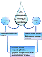
Skaff et al.114 revealed that the HOSCN may react with the neucleophilic selenols such as selenocysteine, and Hawkins111 suggested that HOSCN has a potential to react with nitrogen atom. Also, these intermediate active products may be implicated in the host antiviral defense based on the regulation of pendrin by IFN-γ.115 These and other potential mechanisms of LPO action against the microbes are summarized in Fig. 4.
 |
| Fig. 4 Suggested mechanisms of the LPO action against the microbes. | |
In the current study we demonstrated that lactoperoxidase has the ability to bind viral particles, as well as the ability to prevent viral replication inside the infected HepG2 cells. All results were compared with the antiviral effects of camel lactoferrin at concentration of 0.5 mg ml−1, which has the well-established abilities to inhibit entry of HCV particles into the target cells and to inhibit the intracellular replication of HCV as reported by Redwan et al.64 The anti-HCV activity of these milk proteins may be due to their direct interaction with viruses, but not with the infected cells. In addition, since LPO is a calcium- and iron-containing glycoprotein89,116 it might have some other mechanisms contributing to its antiviral activity, since regulation of iron homeostasis might represent an important factor to prevent the infection of the hepatic cells with HCV. In previous studies, LPO was shown to demonstrate a significant activity against both DNA and RNA viruses such as HIV, herpes simplex, respiratory syncytial, and echovirus type 11 viruses.117–120 Lenander-Lumikari121 and Popper and Knorr122 reported that LPO has potential activity against filamentous fungi and yeast (Candida albicans), as well as against bacteria.
Our analysis revealed that the anti-HCV activity of camel LPO is superior in comparison with those of human and bovine LPOs. Mode of action of LPOs against viruses may depend on the direct interaction of these proteins (based on their cationicity and/or hydrophobicity) with the viral molecules. Human LPO is moderately cationic, with a pI of 7.5, but bovine and camel LPOs are more cationic, being characterized by the pIs of 7.9 and 8.63, respectively. Both camel and bovine LPOs have four glycosylation sites, are able to covalently bind a ferric heme and show strong binding of a calcium ion.89,116,123 The cationicity of LPOs (and other mammalian peroxidases) is one of the major intrinsic structural characteristics (in addition to hydrophobicity, cationicity, and helical propensity), serving as important determinants of their antimicrobial potency.64,65
The virucidal activities of LPO may depend on these properties too. To shed more light on the potential mechanisms defining the noticeable differences in the anti-HCV activities of camel, human and bovine LPOs, we analyzed amino acid sequences of these proteins. Fig. 5A shows that these three LPOs are characterized by relatively high sequence identity.
 |
| Fig. 5 Sequence alignments of camel, human, and bovine LPOs (UniProt IDs: Q9GJW6, P22079, and P80025, respectively). (A) Results of multiple sequence alignment by the ClustalW2 algorithm (http://www.ebi.ac.uk/Tools/msa/clustalw2/). (B) Disorder PONDR® VSL2 profiles of various LPOs analyzed in this study, where results for camel, human, and bovine LPOs are shown by thick green, red, and black lines, respectively. A disorder threshold is indicated as a thin black line (at score of 0.5) to show a boundary between disorder (>0.5) and order (<0.5). Data for the alternatively spliced isoform of HLPO is included for the comparison. | |
In fact, camel LPO is 84.67 and 83.97% identical to human and bovine proteins, respectively, and there is 83.01% identity between the sequences of human and bovine proteins. This suggests that the overall numerical comparison of the amino acid sequences does not provide grounds for functional differentiation.
On the other hand, Fig. 5A shows that differences are not evenly spread throughout the sequences of these three LPOs, with their N-terminal domains (residues 1–170) being the most diversified parts. This conclusion is further illustrated by Fig. 5B representing the results of the evaluation of intrinsic disorder propensities of human, bovine and camel LPOs by PONDR® VSL2, which is one of the more accurate stand-alone disorder predictors.124–126 In agreement with our previous study,66 Fig. 5B shows that the most disordered parts are the N-terminal regions of the LPO preproproteins containing signal peptide (residues 1–26) and propeptide (residues 27–100). On the other hand, the peculiarities of the disorder distribution within the LPO catalytic domains are remarkably similar. Since even in the mature proteins (residues 101–712) the most diversified regions are their N-terminal domains, it is tempting to hypothesize that the described in this work difference in the anti-HCV activities of camel, human and bovine LPOs can be at least in part attributed to the differences in the disorder propensities of their N-terminal tails.
Although we show in this study that LPO has some promises as an antiviral agent, it is not clear at the moment if this protein can be used in the clinic and how it can be delivered to the infected individuals in vivo to have a curative effect. Although these are very important questions, it seems that they are a bit premature. In fact, our report is the first study evaluating the LPO activity against HCV infectivity. We believe that more information should be accumulated on these and related topics before making suggestions on potential clinical applications of LPO, which clearly should be a subject of subsequent studies.
Materials and methods
Milk processing
Human milk was harvested from the breast-feeding volunteer at the Gynecology Department of the Kasr Al Ainy School of Medicine, Cairo University (Cairo, Egypt). The informed consent was obtained from this volunteer that her milk will be used in the research study, and that her name will not be mentioned in the resulting published work. Pooled human, camel, and bovine milk was produced, collected and transferred to the laboratory in frozen aliquots. To prevent microbial growth, 0.02% sodium azide was added to the milk before processing. Milk was defatted by centrifugation at 4500 rpm for 30 min and caseins were precipitated by decreasing the pH to 4.2 with 1 M HCl.84,127 After centrifugation at 9500 rpm and 4 °C for 20 min, the supernatant was dialyzed overnight against 50 mM Tris–HCl buffer, pH 7.0 and centrifuged at 9500 rpm and 4 °C for 30 min. The resulting samples were used for purification of human, camel, and bovine LPOs according to the established protocols.
Assaying cytotoxic effect of human, camel, and bovine LPOs
Cytotoxic effect of LPO on the viability of the cells was assayed by (MTT) thiazolyl blue tetrazolium bromide method as previously described by Almahdy et al.67 In brief, Vero and HepG2 cells (10 × 103) were cultured in a 96-well plate and incubated at 37 °C, 5% CO2 and 88% humidity before LPO treatment, then the medium was refreshed with new DEMEM supplemented medium containing 0.25, 0.5, 0.75, 1.0, 1.25 and 1.5 mg ml−1 of human, camel or bovine LPO. The cells were maintained with 200 μl of fresh medium for 4 days at 37 °C, 5% CO2 and 88% humidity. After incubation, the cells were washed from debris and dead cells trice with the PBS buffer or fresh media. 20 μl MTT solution (5 mg of Thiazolyl Blue Tetrazolium Bromide (MTT) per 1 ml PBS buffer) was added to each well and the plate was placed on a shaking table at 150 rpm for 5 min to thoroughly mix the MTT into the media. The cells were incubated at 37 °C, 5% CO2 and 88% humidity for 5 hours to allow the MTT to metabolize. 200 μl dimethylsulfoxide (DMSO) was added to each well and the plate was shook again on the shaking table at 150 rpm for 5 min and then the optical density was measured at 630 nm by ELISA reader. The viability of the cells was calculated by comparison with control cells.
Potential activity of human, camel, and bovine LPOs against HCV genotype 4
To examine the protection effect of human, camel, and bovine LPOs for cells, HepG2 cells (1.0 × 10⁵) were plated in three 24-well microtiter plates. The purified human, camel, or bovine LPO was added to HepG2 cells (in 50 ml of DEMEM supplemented medium) to a final concentration of 0.5 and 1.0 mg ml−1 then incubated for 90 min at 37 °C. Free human, camel, and bovine LPOs were removed by washing three times with 1 ml of PBS or fresh media. After addition 1 ml of medium containing 2% HCV-infected serum (6.5 million copies ml−1, RNA G4), the cells were incubated for 90 min at 37 °C. The cells were washed three times with PBS and cultured for seven days at 37 °C, 5% CO2 and 88% humidity. To examine the neutralization effect of human, camel, and bovine LPOs with HCV, 1 ml of infected serum and human, camel or bovine LPO at concentrations of 0.5 and 1.0 mg ml−1 was pre-incubated in 50 ml of medium for 90 min at 24 °C, and then the mixture of HCV and human, camel or bovine LPO was added to the HepG2 cells cultured as described above, and incubated for 90 min at 37 °C, 5% CO2 and 88% humidity. The cells were washed three times with 1 ml of PBS and further incubated for 7 days at 37 °C, 5% CO2 and 88% humidity. Positive HepG2 (1.0 × 10⁵) control cells were infected with HCV and negative HepG2 (1.0 × 10⁵) control cells without viral infection cultures were included. Also camel LF at concentration of 0.5 mg ml−1 was used as positive indication for inhibition potential activity against HCV.62,64 The cells were washed three times from debris and dead cells by using RPMI-1640 supplemented media, followed by total RNA extraction.128
Evaluation of the effect of the human, camel, and bovine LPOs on the replication of hepatitis C virus in the HCV-infected HepG2 cells
After suspending HepG2 cells to 2 × 10⁵ cells ml−1 in the supplemented DEMEM culture media, the cells were left to adhere on two sets of 12-well plates for 24 h at 37 °C, 5% CO2 and 88% humidity, then infected with HCV-infected serum (6.5 million copies ml−1, RNA G4) and incubated for 48 h at 37 °C, 5% CO2 and 88% humidity. Then, the purified human, camel or bovine LPO was added to the final concentrations of 0.25, 0.5, 0.75, 1.0, 1.25, or 1.5 mg ml−1. In the positive cultures, the HepG2 cells (2.0 × 10⁵) were infected with HCV and then treated with LPO, whereas in the control cultures, the HepG2 cells (2.0 × 10⁵) were treated with LPO only, without infection. The HCV infected and the non-infected HepG2 cells were incubated with LPO for 4, 7, and 10 days at 37 °C, 5% CO2 and 88% humidity. In addition, camel LF at concentration of 0.5 mg ml−1 was used as positive indicator of HCV inhibition.62,64 At the end of incubation period, the cells were washed three times to clear from debris and dead cells using RPMI-1640 supplemented media. Then, the cells were tested by RT-PCR for the presence of HCV RNA.59,60,129
RNA extraction from the HepG2 cells
RNA was isolated from HepG2 cells as described previously by El-Fakharany et al.60 Briefly, cells were precipitated by centrifugation at 1500 rpm for 5 min at 4 °C and washed three times with PBS or basal media to remove adherent viral particles before lysis in 4 mol l−1 guanidinium isothiocyanate containing 25 mM sodium citrate, 0.5% sarcosyl and 100 mM β-mercaptoethanol and 100 μl sodium acetate. The lysed cells were centrifuged (Microcentrifuge, Heraeus Sepatech, Germany) at 12
000 rpm for 10 min at 4 °C. The aqueous layer was collected and mixed with equal volume of isopropanol. After incubation at −20 °C overnight, RNA was precipitated by centrifugation at 12
000 rpm for 30 min at 4 °C and the precipitate RNA was washed twice with 70% ethanol.
Synthesized RNA of internal control
RNA for internal control was synthesized as described by Ikeda et al.128 Briefly, RNA encoding Renilla luciferase (Rluc) was used as an internal control to monitor the efficiency of RT-nested PCR. The pRL-TK plasmid vector encoding Rluc was linearized by cutting at the Xba I site and then used as a template for in vitro transcription with T7 RNA polymerase. The synthesized RNA was treated with DNase and purified using an RNaeasy Mini kit.
RT-nested-PCR for HCV and internally controlled RNA
Reverse transcription-nested PCR was performed as described previously by Liao et al.61 and El-Fakharany et al.62 The complimentary DNA (cDNA) and the first PCR reaction of the nested PCR detection system for the HCV and Rluc RNA was performed in a 50 μl volume single-step reaction using the Ready-To-Go RT-PCR beads, 400 ng of total RNA, 10 μM of the reverse primer 1CH (for plus strand), 10 μM of the forward primer 2CH (for minus strand) and 10 μM of reverse primer P2. The second PCR reaction was done similar to the first, except for use of the nested reverse primer D2 and forward primer F2 at 10 μM each. A fragment of 174 bp was identified in positive samples. To avoid the reduction of the efficiency of HCV amplification reaction, cDNA was amplified with 5
:
1 HCV-to-Rluc primer concentrations in the first and second rounds of PCR. Amplified DNA (174 bp for HCV and 376 bp for Rluc) were detected by staining with ethidium bromide after separation on a 3% agarose gel electrophoresis.
Real time PCR
Briefly, HCV RNA was extracted from HepG2 cells as described above. Amplification of HCV RNA in samples and standards was measured by SYBR Green kit with two-step PCR, the RNA is first reverse-transcribed into cDNA using 1CH, 2CH and P2 primers, then the second step takes place with D2 and F2 primers. An aliquot of the reverse transcription reaction is then used for analysis of viral load using the Rotor-Gene real time PCR machine and the report was generated by Rotor-Gene Q Series Software 1.7 (Build 94) Copyright© 2008 Corbett Life Science, a QIAGEN. As described previously by El-Fakharany et al.62 and Redwan et al.64 the relative activity (%) was calculated as [(A) count of positive control − (B) count of tested protein]/(A) count of positive control × 100%.
Sequence analyses
Amino acid sequences of camel, human, and bovine LPOs (UniProt IDs: Q9GJW6, P22079, and P80025, respectively) were taken from UniProt (http://www.uniprot.org/uniprot/). Sequences were aligned using the multiple sequence alignment tool, ClustalW2 (http://www.ebi.ac.uk/Tools/msa/clustalw2/).
The intrinsic disorder propensities of camel, human, and bovine LPOs were evaluated by the PONDR® VSL2 algorithm, which is one of the more accurate stand-alone disorder predictors available at the moment.124–126
Conclusions
In conclusion, this study is the first attempt to examine the HCV inhibitory effects of the native lactoperoxidases purified from camel, human, or bovine milk. Results obtained support the efficacy of these proteins against virus activity and suggest that the use of LPOs might represent a promising approach for the HCV therapy.
Acknowledgements
This work was supported by grant from KACST and in part by a grant from the King Abdulaziz University (56-130-35-HiCi).
Notes and references
- L. A. Hanson and J. Winberg, Arch. Dis. Child., 1972, 47, 845–848 CrossRef CAS.
- E. Seifu, in New Issues in Food Policy, Control and Research, ed. A. P. Riley, Nova Science Publishers, Inc., Hauppauge, NY, USA, 2007, pp. 1–43 Search PubMed.
- E. M. Redwan, B. Xue, H. A. Almehdar and V. N. Uversky, Curr. Protein Pept. Sci., 2015, 16, 228–242 CrossRef CAS.
- T. L. McMeekin, J. Food Prot., 1952, 15, 57–63 Search PubMed.
- M. Halwer, Arch. Biochem. Biophys., 1954, 51, 79–87 CrossRef CAS.
- G. C. Kresheck, Acta Chem. Scand., 1965, 19, 375–382 CrossRef CAS PubMed.
- C. Holt and L. Sawyer, J. Chem. Soc., Faraday Trans., 1993, 89, 2683–2692 RSC.
- C. Holt, J. A. Carver, H. Ecroyd and D. C. Thorn, J. Dairy Sci., 2013, 96, 6127–6146 CrossRef CAS PubMed.
- J. Garnier, Biochim. Biophys. Acta, 1963, 66, 366–377 CrossRef CAS.
- J. L. Mangan, Br. J. Nutr., 1972, 27, 261–283 CrossRef CAS.
- P. F. Fox and J. Guiney, J. Dairy Res., 1973, 40, 229–234 CrossRef CAS.
- G. A. Broderick, Br. J. Nutr., 1987, 58, 463–475 CrossRef CAS.
- S. A. Gibson and G. T. Macfarlane, J. Gen. Microbiol., 1988, 134, 19–27 CAS.
- H. E. Swaisgood, J. Dairy Sci., 1993, 76, 3054–3061 CrossRef CAS.
- D. E. Johnson, B. Xue, M. D. Sickmeier, J. Meng, M. S. Cortese, C. J. Oldfield, T. Le Gall, A. K. Dunker and V. N. Uversky, J. Struct. Biol., 2012, 180, 201–215 CrossRef CAS PubMed.
- L. K. Creamer and T. Richardson, Arch. Biochem. Biophys., 1984, 234, 476–486 CrossRef CAS.
- L. C. Chaplin, D. C. Clark and L. J. Smith, Biochim. Biophys. Acta, 1988, 956, 162–172 CrossRef CAS.
- O. G. Issinger, C. Brockel, B. Boldyreff and J. T. Pelton, Biochemistry, 1992, 31, 6098–6103 CrossRef CAS.
- J. E. Plowman, L. K. Creamer, M. J. Liddell and J. J. Cross, J. Dairy Res., 1997, 64, 377–397 CrossRef CAS.
- M. Lecouvey, C. Frochot, L. Miclo, P. Orlewski, A. Driou, G. Linden, J. L. Gaillard, M. Marraud, M. T. Cung and R. Vanderesse, Eur. J. Biochem., 1997, 248, 872–878 CAS.
- M. H. Alaimo, E. D. Wickham and H. M. Farrell Jr, Biochim. Biophys. Acta, 1999, 1431, 395–409 CrossRef CAS.
- A. Chakraborty and S. Basak, J. Photochem. Photobiol., B, 2007, 87, 191–199 CrossRef CAS PubMed.
- A. Chakraborty and S. Basak, J. Photochem. Photobiol., B, 2008, 93, 36–43 CrossRef CAS PubMed.
- T. F. Kumosinski, H. Pessen, S. J. Prestrelski and H. M. Farrell Jr, Arch. Biochem. Biophys., 1987, 257, 259–268 CrossRef CAS.
- H. S. Rollema and J. A. Brinkhuis, J. Dairy Res., 1989, 56, 417–425 CrossRef CAS.
- E. L. Malin, M. H. Alaimo, E. M. Brown, J. M. Aramini, M. W. Germann, H. M. Farrell Jr, P. L. McSweeney and P. F. Fox, J. Protein Chem., 2001, 20, 391–404 CrossRef CAS.
- M. H. Smith, P. J. Edwards, K. P. Palmano and L. K. Creamer, J. Dairy Res., 2002, 69, 85–94 CrossRef CAS.
- R. W. Sleigh, A. G. Mackinlay and J. M. Pope, Biochim. Biophys. Acta, 1983, 742, 175–183 CrossRef CAS.
- T. L. James, CRC Crit. Rev. Biochem., 1985, 18, 1–30 CrossRef CAS.
- L. T. Kakalis, T. F. Kumosinski and H. M. Farrell Jr, Biophys. Chem., 1990, 38, 87–98 CrossRef CAS.
- N. M. Wahlgren, J. Leonil, P. Dejmek and T. Drakenberg, Biochim. Biophys. Acta, 1993, 1202, 121–128 CrossRef CAS.
- N. M. Wahlgren, P. Dejmek and T. Drakenberg, J. Dairy Res., 1994, 61, 495–506 CrossRef CAS.
- J. K. Thomsen, H. J. Jakobsen, N. C. Nielsen, T. E. Petersen and L. K. Rasmussen, Eur. J. Biochem., 1995, 230, 454–459 CrossRef CAS PubMed.
- E. Smyth, C. D. Syme, E. W. Blanch, L. Hecht, M. Vasak and L. D. Barron, Biopolymers, 2001, 58, 138–151 CrossRef CAS.
- C. D. Syme, E. W. Blanch, C. Holt, R. Jakes, M. Goedert, L. Hecht and L. D. Barron, Eur. J. Biochem., 2002, 269, 148–156 CrossRef CAS.
- L. Ashton, C. Johannessen and R. Goodacre, Anal. Chem., 2011, 83, 7978–7983 CrossRef CAS PubMed.
- A. N. Batista, J. M. Batista Jr, V. S. Bolzani, M. Furlan and E. W. Blanch, Phys. Chem. Chem. Phys., 2013, 15, 20147–20152 RSC.
- N. C. Maiti, M. M. Apetri, M. G. Zagorski, P. R. Carey and V. E. Anderson, J. Am. Chem. Soc., 2004, 126, 2399–2408 CrossRef CAS PubMed.
- H. M. Farrell Jr, P. X. Qi, E. D. Wickham and J. J. Unruh, J. Protein Chem., 2002, 21, 307–321 CrossRef CAS.
- P. X. Qi, E. D. Wickham and H. M. Farrell Jr, Protein J., 2004, 23, 389–402 CrossRef CAS.
- S. Jindal and A. Naeem, J. Fluoresc., 2013, 23, 367–374 CrossRef CAS PubMed.
- C. Holt, Curr. Opin. Struct. Biol., 2013, 23, 420–425 CrossRef CAS PubMed.
- D. C. Thorn, H. Ecroyd, J. A. Carver and C. Holt, Int. Dairy J., 2015, 46, 2–11 CrossRef CAS PubMed.
- M. A. Naqvi, S. Rauscher, R. Pomès and D. Rousseau, Biochemistry, 2014, 53, 6402–6408 CrossRef CAS PubMed.
- E. W. Evans, in Developments in Food Proteins, ed. B. J. F. Hudson, Applied Science Publishers, London, 1982, pp. 131–169 Search PubMed.
- W. Ben Ounis, S. F. Gauthier, S. L. Turgeon, S. Roufik and Y. Pouliot, Int. Dairy J., 2008, 18, 1043–1050 CrossRef CAS PubMed.
- F. S. Hanssen, Br. J. Exp. Pathol., 1924, 5, 271–280 CAS.
- G. R. Jago and M. Morrison, Proc. Soc. Exp. Biol. Med., 1962, 111, 585–588 CrossRef CAS.
- S. Sharma, A. K. Singh, S. Kaushik, M. Sinha, R. P. Singh, P. Sharma, H. Sirohi, P. Kaur and T. P. Singh, Int. J. Biochem. Mol. Biol., 2013, 4, 108–128 CAS.
- F. Bafort, O. Parisi, J. P. Perraudin and M. H. Jijakli, Enzyme Res., 2014, 2014, 517164 CAS.
- R. Floris, I. Recio, B. Berkhout and S. Visser, Curr. Pharm. Des., 2003, 9, 1257–1275 CrossRef CAS.
- J. Tenovuo, Oral Dis., 2002, 8, 23–29 CrossRef CAS.
- A. C. van Hooijdonk, K. D. Kussendrager and J. M. Steijns, Br. J. Nutr., 2000, 84(suppl. 1), S127–S134 CAS.
- S. J. Klebanoff and R. G. Luebke, Proc. Soc. Exp. Biol. Med., 1965, 118, 483–486 CrossRef CAS.
- L. Bjorck, C. Rosen, V. Marshall and B. Reiter, Appl. Microbiol., 1975, 30, 199–204 CAS.
- L. Bjorck, J. Dairy Res., 1978, 45, 109–118 CrossRef CAS.
- B. Reiter, V. M. Marshall, L. Bjorck and C. G. Rosen, Infect. Immun., 1976, 13, 800–807 CAS.
- D. R. Purdy and A. L. Koch, J. Bacteriol., 1976, 127, 1188–1196 CAS.
- R. M. Redwan el and A. Tabll, J. Immunoassay Immunochem., 2007, 28, 267–277 CrossRef PubMed.
- E. M. El-Fakharany, A. Tabll, A. A. El-Wahab, B. M. Haroun and E. M. Redwan, Hepat. Mon., 2008, 8, 101–109 Search PubMed.
- Y. Liao, E. El-Fakkarany, B. Lonnerdal and E. M. Redwan, J. Med. Microbiol., 2012, 61, 375–383 CrossRef CAS PubMed.
- E. M. El-Fakharany, L. Sanchez, H. A. Al-Mehdar and E. M. Redwan, Virol. J., 2013, 10, 199 CrossRef CAS PubMed.
- A. H. Albar, H. A. Almehdar, V. N. Uversky and E. M. Redwan, Curr. Protein Pept. Sci., 2014, 15, 778–797 CrossRef CAS.
- E. M. Redwan, E. M. El-Fakharany, V. N. Uversky and M. H. Linjawi, BMC Complementary Altern. Med., 2014, 14, 219 CrossRef PubMed.
- E. M. Redwan, V. N. Uversky, E. M. El-Fakharany and H. Al-Mehdar, C. R. Biol., 2014, 337, 581–595 CrossRef PubMed.
- H. A. Almehdar, E. M. El-Fakharany, V. N. Uversky and E. M. Redwan, Curr. Protein Pept. Sci., 2015, 16, 352–365 CrossRef CAS.
- O. Almahdy, E. M. El-Fakharany, E. El-Dabaa, T. B. Ng and E. M. Redwan, Hepat. Mon., 2011, 11, 724–730 CrossRef.
- S. M. Wolf, R. P. Ferrari, S. Traversa and K. Biemann, J. Mass Spectrom., 2000, 35, 210–217 CrossRef CAS.
- L. Gothefors and S. Marklund, Infect. Immun., 1975, 11, 1210–1215 CAS.
- D. A. Dionysius, P. A. Grieve and A. C. Vos, J. Appl. Bacteriol., 1992, 72, 146–153 CrossRef CAS PubMed.
- R. Kumar, K. L. Bhatia, Z. Dauter, C. Betzel and T. P. Singh, Acta Crystallogr., Sect. D: Biol. Crystallogr., 1995, 51, 1094–1096 CrossRef CAS PubMed.
- T. Soukka, M. Lumikari and J. Tenovuo, Scand. J. Dent. Res., 1991, 99, 390–396 CAS.
- M. J. Benoy, A. K. Essy, B. Sreekumar and M. Haridas, Life Sci., 2000, 66, 2433–2439 CrossRef CAS.
- B. Reiter, J. D. Oram, G. S. Pope and A. Pickerin, J. Gen. Microbiol., 1963, 33, 12 Search PubMed.
- B. Reiter, A. Pickering and J. D. Oram, 4th International Symposium on Food Microbiology, Gothenburg, Sweden, 1964 Search PubMed.
- B. Reiter, Ciba Found. Symp., 1978, 285–294 CAS.
- B. Reiter, V. M. Marshall and S. M. Philips, Res. Vet. Sci., 1980, 28, 116–122 CAS.
- A. S. Naidu, in Natural food antimicrobial systems, ed. A. S. Naidu, CRC Press, Boca Raton, FL, 2000, pp. 103–132 Search PubMed.
- M. P. Doyle and E. H. Marth, Z. Lebensm.-Unters. Forsch., 1978, 166, 271–273 CrossRef CAS.
- J. W. Boots and R. Floris, Int. Dairy J., 2006, 16, 1272–1276 CrossRef CAS PubMed.
- A. Welk, C. Meller, R. Schubert, C. Schwahn, A. Kramer and H. Below, BMC Microbiol., 2009, 9, 134 CrossRef CAS PubMed.
- J. P. Ramet, The technology of making cheese from camel milk (Camelus dromedarius), Food and Agriculture Organization (FAO), Rome, Italy, 2001 Search PubMed.
- M. Sisecioglu, M. Cankaya, I. Gulcin and H. Ozdemir, Protein Pept. Lett., 2009, 16, 46–49 CrossRef CAS.
- E. M. El-Fakharany, E. A. Serour, A. M. Abdelrahman, B. M. Haroun and R. M. Redwan el, Prep. Biochem. Biotechnol., 2009, 39, 105–123 CrossRef CAS PubMed.
- H. Korhonen, World Anim. Rev., 1980, 35, 23–29 Search PubMed.
- IDF, Bull. Int. Dairy Fed., 1991, 264, 2–19 Search PubMed.
- L. M. Wolfson and S. S. Sumner, J. Food Prot., 1993, 56, 887–892 CAS.
- J. Stadhouders and R. R. Beumer, The IDF Seminar on Indigenous Antimicrobial Agents of Milk - Recent Developments, Uppsala, Sweden, 1994 Search PubMed.
- J. N. de Wit and A. C. M. van Hooydonk, Neth. Milk Dairy J., 1996, 50, 227–244 CAS.
- E. Seifu, E. M. Buys and E. F. Donkin, Trends Food Sci. Technol., 2005, 16, 137–154 CrossRef CAS PubMed.
- K. Shin, H. Wakabayashi, K. Yamauchi, S. Teraguchi, Y. Tamura, M. Kurokawa and K. Shiraki, J. Med. Microbiol., 2005, 54, 717–723 CrossRef CAS PubMed.
- E. Seifu, E. M. Buys and E. F. Donkin, Trends Food Sci. Technol., 2005, 16, 137–154 CrossRef CAS PubMed.
- A. J. Fischer, N. J. Lennemann, S. Krishnamurthy, P. Pocza, L. Durairaj, J. L. Launspach, B. A. Rhein, C. Wohlford-Lenane, D. Lorentzen, B. Banfi and P. B. McCray Jr, Am. J. Respir. Cell Mol. Biol., 2011, 45, 874–881 CrossRef CAS PubMed.
- R. J. Derscheid, A. van Geelen, A. R. Berkebile, J. M. Gallup, S. J. Hostetter, B. Banfi, P. B. McCray Jr and M. R. Ackermann, Am. J. Respir. Cell Mol. Biol., 2014, 50, 389–397 Search PubMed.
- C. Gerson, J. Sabater, M. Scuri, A. Torbati, R. Coffey, J. W. Abraham, I. Lauredo, R. Forteza, A. Wanner, M. Salathe, W. M. Abraham and G. E. Conner, Am. J. Respir. Cell Mol. Biol., 2000, 22, 665–671 CrossRef CAS PubMed.
- C. Wijkstrom-Frei, S. El-Chemaly, R. Ali-Rachedi, C. Gerson, M. A. Cobas, R. Forteza, M. Salathe and G. E. Conner, Am. J. Respir. Cell Mol. Biol., 2003, 29, 206–212 CrossRef CAS PubMed.
- L. Cegolon, C. Salata, E. Piccoli, V. Juarez, G. Palu, G. Mastrangelo and A. Calistri, Int. J. Hyg. Environ. Health, 2014, 217, 17–22 CrossRef CAS PubMed.
- S. J. Klebanoff, J. Exp. Med., 1967, 126, 1063–1078 CrossRef CAS.
- M. Morrison and G. S. Bayse, Biochemistry, 1970, 9, 2995–3000 CrossRef CAS.
- J. Tenovuo, O. P. Lehtonen, A. S. Aaltonen, P. Vilja and P. Tuohimaa, Infect. Immun., 1986, 51, 49–53 CAS.
- J. Tenovuo, E. Grahn, O. P. Lehtonen, T. Hyyppa, L. Karhuvaara and P. Vilja, J. Dent. Res., 1987, 66, 475–479 CrossRef CAS PubMed.
- T. Hyyppa, L. Karhuvaara, J. Tenovuo, M. Lumikari and P. Vilja, Pediatr. Dent. J., 1989, 11, 30–36 CAS.
- G. Marcozzi, V. Liberati, F. Madia, A. Pizzinga and G. de Feo, Maturitas, 2003, 45, 225–229 CrossRef CAS.
- G. Mastrangelo, R. Zanibellato, U. Fedeli, E. Fadda and J. H. Lange, Int. J. Environ. Health Res., 2005, 15, 313–317 CrossRef CAS PubMed.
- G. Mastrangelo, R. Zanibellato, E. Fadda, J. H. Lange, L. Scoizzato and R. Rylander, Ann. Occup. Hyg., 2009, 53, 161–165 CrossRef CAS PubMed.
- F. L. Schaffer, M. E. Soergel and D. C. Straube, Arch. Virol., 1976, 51, 263–273 CrossRef CAS.
- R. B. Couch, in Fields Virology, ed. B. N. Fields, D. M. Knipe, R. M. Chanock, M. S. Hirsch, J. L. Melnick, T. P. Monath and B. Roizman, Raven Press, New York, 1990, pp. 607–629 Search PubMed.
- J. Barker, D. Stevens and S. F. Bloomfield, J. Appl. Microbiol., 2001, 91, 7–21 CrossRef CAS.
- R. C. Wright and J. Tramer, J. Dairy Res., 1958, 25, 104–118 CrossRef CAS.
- N. E. Barrett, A. S. Grandison and M. J. Lewis, J. Dairy Res., 1999, 66, 73–80 CrossRef CAS.
- C. L. Hawkins, Free Radical Res., 2009, 43, 1147–1158 CrossRef CAS PubMed.
- P. Nagy, G. N. Jameson and C. C. Winterbourn, Chem. Res. Toxicol., 2009, 22, 1833–1840 CrossRef CAS PubMed.
- J. D. Chandler and B. J. Day, Biochem. Pharmacol., 2012, 84, 1381–1387 CrossRef CAS PubMed.
- O. Skaff, D. I. Pattison, P. E. Morgan, R. Bachana, V. K. Jain, K. I. Priyadarsini and M. J. Davies, Biochem. J., 2012, 441, 305–316 CrossRef CAS PubMed.
- Y. Nakagami, S. Favoreto Jr, G. Zhen, S. W. Park, L. T. Nguyenvu, D. A. Kuperman, G. M. Dolganov, X. Huang, H. A. Boushey, P. C. Avila and D. J. Erle, J. Immunol., 2008, 181, 2203–2210 CrossRef CAS.
- M. J. Davies, C. L. Hawkins, D. I. Pattison and M. D. Rees, Antioxid. Redox Signaling, 2008, 10, 1199–1234 CrossRef CAS PubMed.
- P. Courtois, D. van Beers, M. de Foor, I. M. Mandelbaum and M. Pourtois, J. Biol. Buccale, 1990, 18, 71–74 CAS.
- M. Pourtois, C. Binet, N. van Tieghem, P. Courtois, A. Vandenabbeele and L. Thiry, J. Biol. Buccale, 1990, 18, 251–253 CAS.
- Y. Yamaguchi, M. Semmel, L. Stanislawski, A. D. Strosberg and M. Stanislawski, Antimicrob. Agents Chemother., 1993, 37, 26–31 CrossRef CAS.
- H. Mikola, M. Waris and J. Tenovuo, Antiviral Res., 1995, 26, 161–171 CrossRef CAS.
- M. Lenander-Lumikari, Oral Microbiol. Immunol., 1992, 7, 315–320 CrossRef CAS PubMed.
- L. Popper and D. Knorr, Nahrung, 1997, 41, 29–33 CrossRef CAS PubMed.
- T. J. Dull, C. Uyeda, A. D. Strosberg, G. Nedwin and J. J. Seilhamer, DNA Cell Biol., 1990, 9, 499–509 CrossRef CAS.
- K. Peng, S. Vucetic, P. Radivojac, C. J. Brown, A. K. Dunker and Z. Obradovic, J. Bioinf. Comput. Biol., 2005, 3, 35–60 CrossRef CAS PubMed.
- Z. L. Peng and L. Kurgan, Curr. Protein Pept. Sci., 2012, 13, 6–18 CrossRef CAS.
- X. Fan and L. Kurgan, J. Biomol. Struct. Dyn., 2014, 32, 448–464 CAS.
- E. M. Redwan, N. L. Larsen and I. A. Wilson, Journal of the Egyptian German Society of Zoology, A: Comparative Physiology, 2003, 40, 251–260 Search PubMed.
- M. Ikeda, A. Nozaki, K. Sugiyama, T. Tanaka, A. Naganuma, K. Tanaka, H. Sekihara, K. Shimotohno, M. Saito and N. Kato, Virus Res., 2000, 66, 51–63 CrossRef CAS.
- E. M. El-Fakharany, B. M. Haroun, T. B. Ng and E. R. Redwan, Protein Pept. Lett., 2010, 17, 1031–1039 CrossRef CAS.
|
This journal is © The Royal Society of Chemistry 2015 |
Click here to see how this site uses Cookies. View our privacy policy here.