DOI:
10.1039/C5RA11765H
(Paper)
RSC Adv., 2015,
5, 83304-83313
Synthesis and characterization of new modified silica coated magnetite nanoparticles with bisaldehyde as selective adsorbents of Ag(I) from aqueous samples†
Received
18th June 2015
, Accepted 14th September 2015
First published on 17th September 2015
Abstract
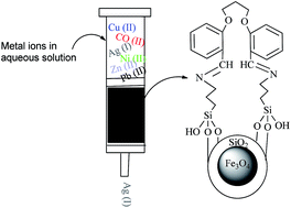
In this study, new silica-coated magnetic nanoparticles modified with bisaldehyde (BISA–APTSCMNPs) were synthesized using a normal method and a vice versa method. The crystal structure of the newly obtained nanoparticles was characterized by Fourier transform infrared spectroscopy (FT-IR), X-ray diffraction (XRD), Scanning Electron Microscopy (SEM), Transmission Electron Microscopy (TEM), Vibrating Sample Magnetometry (VSM), thermogravimetric analysis (TGA) and ultra-violet visible spectroscopy (UV-vis). The surface of the nanoparticles was modified with bisaldehyde (BISA–APTSCMNPs), and these were used for the highly selective removal of Ag(I) ion from an aqueous mixed metal ion solution containing Cu(II), Co(II), Ni(II), Zn(II) and Pb(II) using the syringe and batch techniques. Moreover, the silver ion desorption was most efficient in HCl.
1. Introduction
Nanotechnology is one of the most important branches of modern science in the preparation and usage of nanoparticles.1 In recent years, nanomaterials have attracted considerable interest in the research community due to their large specific surface areas and high reactivities.2 Magnetic nanoparticles are a class of nanoparticle which is commonly composed of magnetic elements such as iron, cobalt, nickel and their respective oxides.3,4 Among the various nanostructure materials, iron oxides have many important and diverse applications and play a major role in numerous areas of chemistry, physics and materials science. In particular, various derivatives of these nanoparticles, such as magnetite (Fe3O4), maghemite (γ-Fe2O3), and hematite (α-Fe2O3), have been investigated intensively for environmental and biological applications.5 In addition to their convenient magnetic properties and low toxicity and price, iron oxide nanoparticles show high surface to volume ratios, depending on their particle size and shape and disperse well in solvent.6 The size and shape of magnetite particles are generally controlled by their synthesis methods.7 To date, numerous chemical methods have been developed to produce magnetite nanoparticles such as microemulsion, chemical co-precipitation, hydrolysis, thermal decomposition and sol–gel techniques.8 Recently, several researchers have used Schiff base moieties for anchoring different catalytic species on magnetic nanoparticles.9–13
Magnetic solid phase extraction has developed rapidly in recent years due to its unique advantages such as easy operation, high extraction efficiency and reusability of sorbent.14 At present, magnetic nanoparticles (MNPs) have been studied as solid phase extraction (SPE) sorbents with highly magnetic characteristics for the separation and removal of chemical species such as metals, dyes and gases.15 In many cases, nanoparticles whose surfaces are functionalized with special functional ligands can improve the sorption capacity and efficiency of extractions.16,17 Recently, a number of functionalized Fe3O4 nanoparticles have been employed as solid phase extraction (SPE) adsorbents for the removal of metal ions from aqueous solutions. For example, silica-coated magnetic nanoparticles modified with imidazole and quercetin have been used for the removal of iodine and uranyl ions from aqueous solutions, respectively.18,19
In this study, we report the synthesis of silica-coated magnetic nanoparticles modified with bisaldehyde (BISA–APTSCMNPs) by a normal method and a vice versa method. The resulting nanoparticles were employed for the selective adsorption of Ag(I) ion from an aqueous solution in the presence of equal amounts of Cu(II), Co(II), Ag(I), Ni(II), Zn(II) and Pb(II) ions using the syringe and batch techniques.
2. Experimental
2.1. Chemicals and reagents
Ferric chloride hexahydrate (FeCl3·6H2O) with 98% purity, ferrous chloride tetrahydrate (FeCl2·4H2O) with 98% purity, absolute ethanol, glycerol with 99% purity and ammonia (NH3) with 25% purity were purchased from Merck, Germany. Tetraethyl orthosilicate (TEOS) with 99.8% purity, 3-aminopropyltrimethoxysilane (APTS) with 97% purity and 2-hydroxybenzaldehyde with 99.9% purity were purchased from Sigma-Aldrich, USA. Toluene was bought from Shanghai Experiment Reagent Co., Ltd (Shanghai, China). 1,3-Dibromopropane with 98% purity was purchased from Fluka Chemicals, Switzerland.
2.2. Instrumentation
FT-IR spectra (Shimadzu prestige-21) were used to determine the identity of the as prepared nanoparticles and to characterize the coated Fe3O4 nanoparticles. X-ray powder diffraction measurements were performed using an X-ray diffractometer (XRD) (Perkin Elmer) at ambient temperature. The surface morphology of the silica-supported ligands was identified with a scanning electron microscope (LECO SEM, Michigan, USA). TEM images of the nanoparticles were obtained with an H-912 transmission electron microscope. Magnetic measurements were performed by means of the vibrating sample magnetometery method, using a VSM 7407 magnetometer, at room temperature. Thermogravimetric analysis (TGA) was performed using a Perkin Elmer thermogravimetric analyzer. UV-visible spectra in the 200–1000 nm range were obtained in DMF solvent on a Perkin Elmer Lambda 45 spectrophotometer. The concentration of metal ions in the solution was measured using a flame atomic absorption spectrophotometer (FAAS, GBC 932 AA, Victoria, Australia). A Jenway model 4510 pH-meter was used for pH measurements by use of a combined electrode. An ultrasonication probe (Karl Deutsch, Germany) was used to disperse the nanoparticles in the solution.
2.3. Synthesis of 2,2′-(propane-1,3-diylbis(oxy))bisbenzaldehyde (BISA)
A solution of 1.22 g (0.01 mol) salicylaldehyde in hot ethanolic KOH (prepared by dissolving 0.56 g (0.01 mol) KOH in 100 mL of absolute ethanol) was stirred until a clear solution was obtained, which was then evaporated under vacuum. The residue was dissolved in DMF (25 mL) and 0.005 mol of 1,3-dibromopropane was added. The reaction mixture was refluxed for 5 h, during which KBr was separated out. The solvent was then removed in vacuo and the remaining solid was washed with water and crystallized using ethanol to obtain high quality crystals (mp 393 K) suitable for X-ray analysis in good yield (84%).20
2.4. Synthesis of modified magnetite nanoparticles
2.4.1. Synthesis of magnetite nanoparticles (MNPs). The magnetic nanoparticles (MNPs) were prepared according to ref. 14 with minor modifications. Briefly, FeCl3·6H2O (11.68 g) and FeCl2·4H2O (4.30 g) were dissolved in 200 mL deionized water under nitrogen gas with vigorous stirring at 85 °C. Then, 20 mL of 30% aqueous ammonia was added to the solution. The color of the bulk solution changed from orange to black immediately. The magnetic precipitates were washed twice with deionized water and once with 0.02 mol L−1 sodium chloride. The washed magnetite was stored in deionized water at a concentration of 40 g L−1.
2.4.2. Synthesis of silica-coated magnetic nanoparticles (SCMNPs). The silica-coated magnetic nanoparticles were synthesized according to previously reported methods with a minor modification.14 The magnetite suspension prepared above (20 mL) was placed in a 250 mL round-bottom flask and allowed to settle. The supernatant was removed, and an aqueous solution of tetraethylorthosilicate (TEOS, 10% (v/v), 80 mL) was added, followed by glycerol (60 mL). The pH of the suspension was adjusted to 4.6 using glacial acetic acid, and the mixture was then stirred and heated at 90 °C for 2 h under a nitrogen atmosphere. After cooling to room temperature, the suspension was washed sequentially with deionized water (3 × 500 mL), methanol (3 × 500 mL), and deionized water (5 × 500 mL). The silica magnetite composite was stored in deionized water at a concentration of 40 g L−1.
2.4.3. Synthesis of amine-modified silica magnetite (APTSCMNPs). These nanoparticles were synthesized according to previously reported methods with minor modifications.14 The silica coated magnetite nanoparticles were first dispersed by sonication in ethanol (100 mL); a solution of 3-aminopropyltrimethoxysilane (APTS, 10% in ethanol) was then added to the above mentioned mixture. After heating at 333 K for 6 h and stirring for 12 h at room temperature under a dry nitrogen atmosphere, the resulting solid was magnetically separated, washed with ethanol several times to remove the unreacted residue of silylating reagent and then vacuum dried at 353 K.
2.4.4. Synthesis of silica-coated magnetic nanoparticles modified with bisaldehyde (BISA–APTSCMNPs) using normal method. The amine-modified silica magnetite (APTSCMNPs) (3 g) was dispersed by sonication in 50 mL of absolute ethanol, and 2,2′-(propane-1,3-diylbis(oxy))bisbenzaldehyde (BISA) (1 g, 3.5 mmol) dissolved in 50 mL absolute ethanol was added. The resulting mixture was stirred for 24 h at room temperature and was refluxed for 12 h. Then, the formed magnetite nanoparticles were washed thoroughly with dichloromethane, methanol and chloroform and dried under vacuum at 60 °C.
2.4.5. Synthesis of BISA–AP with Schiff base reaction. BISA–AP ligand was synthesized by the reaction of 2,2′-(propane-1,3-diylbis(oxy))bisbenzaldehyde (BISA) (1 g, 3.5 mmol) in 30 mL dry toluene and 3-aminopropyltrimethoxysilane (APTS) (1.26 mL, 7.0 mmol) in 50 mL dry toluene in the presence of 3 to 4 drops formic acid as a catalyst. The mixture was stirred for 6 h under an N2-atmosphere at room temperature. The crystals were filtered by vacuum.
2.4.6. Synthesis of silica-coated magnetic nanoparticles modified with bisaldehyde (BISA–APTSCMNPs) using vice versa method. 1 g of the silica coated magnetite nanoparticles (SCMNPs) was first dispersed by sonication in water (100 mL) and BISA–AP ligand (2.13 g, 3.5 mmol) dispersed in water (100 mL) was added to the above mentioned mixture. After heating at 373 K for 2 h and stirring for 24 h at room temperature under dry nitrogen atmosphere, the modified magnetite nanoparticles were prepared by an “on water reaction”; furthermore, the resulting solid was magnetically separated, washed with hot ethanol several times to remove the unreacted ligands and then vacuum dried at 353 K.
2.5. Solid phase extraction techniques
2.5.1. Batch method. All adsorption experiments were carried out at room temperature in a gas bath shaker. To determine the sorption capacity, 20 mL of aqueous solution containing a 10−4 mol L−1 mixture of metal nitrate salts (Pb(NO3)2, Co(NO3)2, Zn(NO3)2, Cu(NO3)2, Ni(NO3)2 and Ag(NO3)) were added to 0.05 g sorbent (BISA–APTSCMNPs synthesized by the normal method and the vice versa method). The mixtures were shaken at 300 rpm and the extraction process was allowed to proceed for 1 h. Subsequently, an Nd–Fe–B strong magnet (5 cm × 4 cm × 2 cm, 1.4 T) was placed at the bottom of the beaker and BISA–APTSCMNPs were isolated from the solution. The upper aqueous phase was removed to measure the remaining metal ion concentration by AAS.To determine the adsorption capacity, the magnetic adsorbents were separated from the solution and washed with 10 mL deionized water. Then, 5 mL eluent solution containing 0.5 M HNO3, 0.5 M HCl and 0.5 M S2O32− (prepared in methanol) was added to the solution and agitated for 15 min by an Ultrasonic-50 Hz to separate the metal ions from the sorbent. In addition, the nanoparticles were isolated from the solution with an Nd–Fe–B strong magnet placed at the bottom of the beaker, and the upper aqueous phase was removed to measure the remaining metal ion concentration by AAS. The studies of metal ions (Pb(II), Co(II), Zn(II), Cu(II), Ni(II) and Ag(I)) were performed by varying the amount of BISA–APTSCMNP sorbents (0.01 g and 0.1 g), which were produced by the normal and vice versa methods. To check the reproducibility, all experiments were performed at least in triplicate.
2.5.2. Syringe method. According to the obtained results in the batch method, we designed a column which checked the rate of adsorption and sorption of Ag(I) ion solution by BISA–APTSCMNPs as the mobile phase. For the construction of home-made solid phase extraction syringes, some glass wool was placed at the bottom of a syringe. Afterwards, 0.05, 0.1 and 0.2 g of BISA–APTSCMNPs were weighed and placed into the syringe, and then finally another piece of glass wool was placed at the top of the syringe. The syringe, with a plastic stopper, was placed in the mouth of a Meyer flask so that it was fully coupled with the opening of the vacuum flask to prevent air infiltration. Then, a 50 mL burette was placed on top of the column so that the opening is placed on top of the column. The pump was turned on, and then the syringes were conditioned with 50 mL water before introducing the sample through the syringes. An aliquot of 50 mL of silver nitrate solution (10−4 mol L−1) was applied through the syringe with flow rates of 1, 3 and 5 mL min−1. After completion of the extraction process, the pump was turned off and the resulting solution was analyzed by AAS. The adsorption of Ag(I) on BISA–APTSCMNPs was performed using 5 mL HCl (0.5 M) with a flow rate of (1 mL min−1). After each complete cycle, the eluent was directly injected into the nebulizer of AAS.
3. Results and discussion
3.1. Preparation of the modified magnetite nanoparticles
In this study, BISA–APTSCMNPs nanoparticles were synthesized by a normal method and a vice versa method and were successfully used as a sorbent for the highly selective removal of Ag(I) ion from an aqueous mixed metal ion solution containing Cu(II), Co(II), Ni(II), Zn(II) and Pb(II) by the syringe and batch techniques. Compared with the normal method, the highest sorption percentage of metal ions was obtained with the nanoparticles synthesized using the vice versa method. In addition, the FT-IR peak of the organic compound was clearer in the spectrum of these nanoparticles. Therefore, it can be concluded that more organic compound attached onto the surface of these nanoparticles. The synthetic procedures of the modified magnetite nanoparticles are shown in Scheme 1. In the normal method, the external surface of magnetite nanoparticles (MNPs) was coated with a silica shell to obtain SCMNPs. Reaction of the silica-coated magnetic nanoparticles (SCMNPs) with 3-aminopropyltrimethoxysilane (APTS) afforded amine-modified silica magnetite (APTSCMNPs). The next step involves reaction of the APTSCMNPs amine groups with bisaldehyde (BISA) to yield the modified magnetite nanoparticles, BISA–APTSCMNPs. In the vice versa method, the BISA–AP ligands were synthesized from the reaction of bisaldehyde (BISA) and 3-aminopropyltrimethoxysilane (APTS). Then, the reaction of the resulting ligands with silica coated magnetite nanoparticles (SCMNPs) produced the nanomaterial BISA–APTSCMNPs. The crystal structures of the newly obtained nanoparticles were characterized by FT-IR, XRD, SEM, TEM, VSM, TGA and UV-vis.
 |
| Scheme 1 Synthesis of modified magnetite nanoparticles by a normal method and a vice versa method. | |
3.2. Characterization of the modified magnetite nanoparticles
3.2.1. FT-IR analysis. Fig. 1 shows the FT-IR spectra of all the nanoparticles prepared in this study. The presence of the magnetite core nanomaterial was confirmed by the observation of two broad bands at around 459 cm−1 and 573 cm−1, which correspond to the Fe–O stretching and Fe2O3 stretching, respectively (Fig. 1a). The IR spectrum of the silica coating of magnetite nanoparticles (SCMNPs) shows absorption peaks at 1103 and 949 cm−1, which are due to the Si–O–Si and Si–OH stretching vibrations, respectively (Fig. 1b). In addition, the presence of the anchored propyl groups was confirmed by the stretching vibrations, which appeared at about 2933 cm−1 in the FT-IR spectrum of 2,2′-(propane-1,3-diylbis(oxy))bisbenzaldehyde (BISA). The peak corresponding to the CH ring stretching vibration appeared at around 2933 cm−1.13 In addition, the absorption bands of the stretching vibrations of the C–O (ether groups) and C
N (imine) bands were observed at 1293 cm−1 and 1640 cm−1, respectively. The peak at 1599 cm−1 is a characteristic of the bending vibrations of the C
C aromatic ring. The peak at 3427 cm−1 belongs to the stretching vibration of O–H adsorbed on the surface of the Fe3O4 nanoparticles. The broad absorption bands at 3440 cm−1 and 1600 cm−1 were due to OH stretching vibrations, which corresponds to OH groups on the surface of the iron oxide; this band can be attributed to the adsorbed water molecules (Fig. 1d and e).
 |
| Fig. 1 FTIR spectra of the synthesized magnetite nanoparticles: (a) MNPS, (b) SCMNPs, (c) APTSCMNPs, (d) BISA–APTSCMNPs (normal method) and (e) BISA–APTSCMNPs (vice versa method). | |
3.2.2. XRD analysis. The crystallinity of the as-prepared magnetic nanoparticles was determined by X-ray diffraction (XRD) using a diffractometer with Cu Kα radiation (40 kV/30 mA). Fig. 2 shows the XRD profiles of the BISA–APTSCMNPs nanoparticles prepared in this study. As shown in Fig. 2, the presence of diffraction peaks located at 2θ = 30.2°, 35.4°, 37.1°, 43.1°, 53.3°, 57°, and 62.5° correspond to the (2 2 0), (3 1 1), (2 2 2), (4 0 0), (4 2 2), (5 1 1) and (4 4 0) planes, respectively. These results indicated that magnetite (Fe3O4) has a face-centered cubic (fcc) structure. Moreover, we did not observe other iron oxides, such as α-Fe2O3, β-Fe2O3 and γ-Fe2O3, which indicates that Fe3O4 was not oxidized to other forms in the synthetic steps. This pattern indicates that the organic species are well dispersed on the surface of SCMNPs and that the crystalline phase of the organic components is not present in the resulting nanomaterials.
 |
| Fig. 2 X-ray diffraction patterns of (a) BISA–APTSCMNPs (normal method) and (b) BISA–APTSCMNPs (vice versa method). | |
3.2.3. SEM analysis. A scanning electron microscope (SEM) is extremely useful for characterizing the morphological structure and size of magnetite nanoparticles. The SEM image of the prepared BISA–APTSCMNPs nanomaterials is shown in Fig. 3. SEM images of synthesized nanoparticles clearly show the BISA–APTSCMNPs nanoparticles are spheroidal in shape. As shown in Fig. 3a, the diameter of BISA–APTSCMNPs (prepared using the normal method) is about 30–45 nm, whereas the diameter of the synthesized BISA–APTSCMNPs using the vice versa method is about 35–50 nm, which is slightly larger than the average particle size obtained from the normal method (Fig. 3b). Moreover, the SEM images clearly indicate that the sizes of the particles changed very slightly after the addition of the organic compound; therefore, the effect of this modification on particle size was negligible.
 |
| Fig. 3 SEM images of magnetite nanoparticles: (a) BISA–APTSCMNPs (normal method) and (b) BISA–APTSCMNPs (vice versa method). | |
3.2.4. TEM analysis. To obtain more direct information on the size and morphology of the particles, transmission electron microscopy (TEM) micrographs of the obtained nanoparticles were obtained. Fig. 4a and b illustrates the TEM images of the synthesized BISA–APTSCMNPs. TEM analysis of the nanoparticles indicated that the both the BISA–APTSCMNPs (normal method) and BISA–APTSCMNPs (vice versa method) have spherical morphologies. According to the TEM images, the mean particle size is about 45 nm for BISA–APTSCMNPs (normal method) and 50 nm for BISA–APTSCMNPs (vice versa method), which indicates that the size of the particles obtained using the vice versa method is slightly larger than that of the particles obtained using the normal method.
 |
| Fig. 4 TEM images of magnetite nanoparticles: (a) BISA–APTSCMNPs (normal method) and (b) BISA–APTSCMNPs (vice versa method). | |
3.2.5. VSM analysis. To study the magnetic properties of the magnetite nanoparticles before and after silica coating, we investigated the hysteresis loops of magnetite and functionalized magnetite nanoparticles at room temperature using vibrating sample magnetometry (VSM). Fig. 5 shows the room temperature magnetic hysteresis loops of the prepared magnetite nanoparticles. The VSM results indicate that all the prepared magnetite nanoparticles possess superparamagnetism properties at room temperature, which is due to their small size. On the other hand, coating the surface of the magnetite nanoparticles with silica and bisaldehyde (BISA) compounds results in a decrease in the saturation magnetization (Ms), which may be due to the superparamagnetic properties and the screening effects of the silica layer, since the magnetic silica nanocomposites could be readily and stably dispersed in water and remained in the suspension in the absence of an external magnetic field. The saturation magnetization values for the Fe3O4 particles, SCMNPs and BISA–APTSCMNPs nanomaterials were 67, 59 and 18 emu g−1, respectively. The slow decrease in the saturation magnetization for the BISA–APTSCMNPs nanoparticles compared to the Fe3O4 microspheres can be attributed to the silica shell.
 |
| Fig. 5 Hysteresis loops of (a) MNPS, (b) SCMNPs, (c) APTSCMNPs, (d) BISA–APTSCMNPs (normal method) and (e) BISA–APTSCMNPs (vice versa method) nanoparticles at room temperature using VSM. | |
3.2.6. TGA analysis. A thermogravimetric analyzer (TGA) was used to confirm the coating formation and to estimate the binding efficacy of bisaldehyde (BISA) on the surface of the magnetite nanoparticles. The TGA measurements were carried out by weighing a powder sample of 5–10 mg and loading it into a platinum pan. The mass change in the temperature range from 30 to 800 °C at a heating rate of 20 °C min−1 under a nitrogen flow was monitored and recorded.Fig. 6 shows the result of TGA on the nanoparticles in the range of 30–800 °C. The TGA curves of the prepared nanoparticles gave two mass loss processes before 600 °C. The mass loss rates are about 1.5% and 10.7%. The first weight loss rate at about 1.5% with the temperature below 260 °C is due to the evaporation of water in the magnetite BISA–APTSCMNPs nanoparticles, and the second weight loss of about 10.7% between 260 and 450 °C is attributed to the thermal decomposition of the organic species, i.e. the aminopropyl groups and bisaldehyde in the nanomaterial. The TGA curve above 600 °C indicates that the organic species in the magnetic composite nanoparticles have been completely decomposed and that the Fe3O4 nanoparticles remain.
 |
| Fig. 6 TGA curves of magnetite nanoparticles: (a) BISA–APTSCMNPs (normal method) and (b) BISA–APTSCMNPs (vice versa method). | |
3.2.7. UV-vis analysis. The light absorption properties of all the synthesized nanomaterials were measured using a UV-vis spectrophotometer (Perkin Elmer Lambda 45) with a wavelength range of 200–700 nm. As shown in Fig. 7, the UV-vis absorption spectrum showed the absorption peak of bisaldehyde (BISA) in the range of 210–285 nm due to the π–π* transitions of the (C
C) aromatic ring and imine (C
N) group.
 |
| Fig. 7 The UV-vis spectra of MNPS, SCMNPs, APTSCMNPs and BISA–APTSCMNPs nanoparticles. | |
3.3. Extraction condition optimization
3.3.1. Effect of extraction time. The effect of the extraction time was also studied in the range of 5–60 min for the batch method and at different flow rates, varying from 1.25 mL min−1 to 10 mL min−1 for the syringe method in order to determine the optimum extraction time for maximum adsorption. Mixed standard solutions of Cu(II), Co(II), Ag(I), Ni(II), Zn(II) and Pb(II) (1 mg L−1) were used for all experiments. The adsorption of Ag(I) increased during the first 5 min and then leveled off as an equilibrium (above 95% sorption) was reached in 5 min. The optimum flow rates of all metal ions were found to be 3 mL min−1 for the syringe method. The rapid adsorption equilibrium indicates the strong affinity between the ligands and the metal ions. This behavior indicates that these ions have good accessibility through the chelating sites on the modified silica; the binding constant between the metal ions and the imine and hydroxyl groups immobilized on the silica surface is possibly high. According to these results, the sorbent is suitable for application in a syringe system wherein a shorter extraction time or faster adsorption is required.
3.3.2. Effect of sorbent amount. The amount of sorbent is a very important parameter that affects the recovery. Nanoparticles have been shown to be superior sorbents because they have higher surface areas than conventional sorbents. Therefore, smaller amounts of modified silica coated magnetite nanoparticles (nano-sorbents) can achieve satisfactory results. The optimum sorbent amount was determined using the batch and syringe methods. The influence of the sorbent amount for the quantitative extraction of Cu(II), Co(II), Ag(I), Ni(II), Zn(II) and Pb(II) from an aqueous sample was tested in the range of 5–50 mg for the batch method and 25–100 mg for the syringe method at pH 9. The effects of the adsorbent amount on the extraction of metal ions in the batch procedure are shown in Fig. 8 and 9. The results for the syringe procedure are shown in Table 1. As can be seen from Fig. 7 and 8, the best sorption percentage and the highest adsorption coefficient values were obtained for Ag(I) ion from water samples with BISA–APTSCMNPs (0.05 g) synthesized using the vice versa method. According to these results, the synthesized nanomaterial shows excellent silver(I) ion adsorption selectivity at pH = 9 in a mixed metal ion solution containing Cu(II), Co(II), Ni(II), Zn(II) and Pb(II). Furthermore, the experimental results indicated that 0.05 and 0.1 g of the nano-sorbent are enough for the preconcentration of Ag(I) using the batch and syringe methods, respectively. Therefore, 0.05 and 0.1 g of sorbent were used in the subsequent experiments.
 |
| Fig. 8 Removal and recovery percentage of metal ions at different amounts of adsorbent obtained with the normal method in various eluents. | |
 |
| Fig. 9 Removal and recovery percentage of metal ions at different amounts of adsorbent obtained with the vice versa method in various eluents. | |
Table 1 Doping of silver metal by nanoparticles modified with bisaldehyde (BISA–APTSCMNPs)a
Attraction Ag+ 10−4 M |
1 mL min−1 |
3 mL min−1 |
5 mL min−1 |
ND. not detected. |
BISA–APTSCMNPs (normal method) 0.05 g |
65 |
61 |
54 |
BISA–APTSCMNPs (normal method) 0.1 g |
97 |
93 |
74 |
BISA–APTSCMNPs (normal method) 0.2 g |
99 |
95 |
78 |
BISA–APTSCMNPs (vice versa method) 0.05 g |
71 |
67 |
55 |
BISA–APTSCMNPs (vice versa method) 0.1 g |
H |
98 |
80 |
BISA–APTSCMNPs (vice versa method) 0.2 g |
H |
H |
87 |
Cotton |
ND |
01 |
ND |
3.3.3. Effect of elution conditions on recovery. Various acids were used to identify the best eluent for the adsorbed metal ions in solid phase extraction (SPE) with BISA–APTSCMNPs produced by the normal and vice versa methods; the effects of these acids are shown in Fig. 8 and 9. To obtain a high enrichment factor, a suitable eluent should be used. Diluted hydrochloric acid (HCl), nitric acid (HNO3) and thiosulfate (S2O32−) as prepared in methanol solutions were used for the maximum elution of Cu(II), Co(II), Ag(I), Ni(II), Zn(II) and Pb(II) ions from the nanoparticle-supported complex, as previously described in the literature. An acid solution has been widely used for the elution of metal ions from a sorbent due to protonation at the chelating sites of the sorbent. The experimental results indicated that among the different eluents used, 0.5 M HCl provided higher recovery and reproducibility. Hydrochloric acid (HCl) is highly suitable for displacing metal ions from the binding sites; moreover, this acid does not interfere with the subsequent determination by AAS.
3.3.4. Reusability of sorbent. The reusability of a sorbent is very important in order to evaluate its potential for widespread usage in the laboratory. The stability and potential reusability of the adsorbent was investigated through several adsorption–elution cycles. In order to test the reusability, the sorbent was subjected to repeated adsorption and desorption operations as follows: 100 mg sorbent was placed in the syringe and washed with 0.5 M HCl and water to activate the sorbent. 10 mL of 1.0 mg L−1 Cu(II), Co(II), Ag(I), Ni(II), Zn(II) and Pb(II) mix standard solution was drawn at room temperature. The metal adsorbed ligands were washed with 10 mL 0.5 M HNO3 and the desorbed metal ions were detected with AAS. Finally, the eluted sorbent was treated with doubly distilled deionized water repeatedly until the supernatant reached neutrality. The absorbance and recovery of Cu(II), Co(II), Ag(I), Ni(II), Zn(II) and Pb(II) were calculated for the adsorption and desorption operations, respectively. The results agreed with 95% ± 5% recoveries for all metal ions within 0.1–5% relative error for the metal ions in five cycles, which fully demonstrates that the nanoparticle-supported ligands have desirable stability and reusability.
3.3.5. Effect of doping of silver ions by column filled with BISA modified nanoparticles. Ag(I) doping studies were performed by varying the concentration of the BISA–APTSCMNPs sorbents (0.05 g, 0.1 g and 0.2 g) produced by the normal and vice versa methods with flow rates of 1 mL min−1, 3 mL min−1 and 5 mL min−1. Data Tables 1 and 2 show the doping and adsorption of silver ions. As can be seen from the results, 0.1 g of synthesized BISA–APTSCMNPs using the vice versa method are sufficient for the preconcentration of Ag(I) using the syringe method with a flow rate of 3 mL min−1.
Table 2 Desorption of Ag(I) ion from the surface of nanoparticles modified with bisaldehyde (BISA–APTSCMNPs)
Pull Ag+ 10−4 M |
5 mL HCl 0.5 M |
Condensed |
BISA–APTSCMNPs (normal method) 0.1 g |
331 |
33.1 |
BISA–APTSCMNPs (vice versa method) 0.1 g |
356 |
35.6 |
4. Conclusion
In this study, aromatic bisaldehyde (BISA) was synthesized by coupling salicylaldehyde with 1,3-dibromopropane; then a nanocomposite material, BISA–APTSCMNPs, was synthesized using a normal method and a vice versa method. The newly synthesized nanoparticles were characterized by FT-IR, XRD, SEM, TEM, VSM, TGA and UV-vis. These nanoparticles were applied as a sorbent for the highly selective removal of Ag(I) ion from an aqueous mixed metal ion solution containing Cu(II), Co(II), Ni(II), Zn(II) and Pb(II) by the syringe and batch techniques. The adsorption mechanism of metal ions onto the surface of BISA–APTSCMNPs is more likely to be template effects or interactions between the metal ions and aromatic rings.21,22 The results indicated that the best sorption percentage and the highest adsorption coefficient values were obtained in regard to Ag(I) ion from water samples with BISA–APTSCMNPs synthesized using the vice versa method. According to these results, we can say the novel modified magnetite nanoparticles may be used as an alternative sorbent for the removal of Ag(I) from water samples. Moreover, it was observed that the silver ion desorption was most efficient in HCl. XRD results show that the prepared magnetite nanoparticles have good crystal structures with a face-centered (fcc) structure. Ag(I) ion doping studies showed that 0.1 g of BISA–APTSCMNPs synthesized using the vice versa method are sufficient for the preconcentration of Ag(I) using the syringe method at a flow rate of 3 mL min−1.
Acknowledgements
The authors acknowledge the Payame Noor University, Ardabil, Iran for providing facilities for this study.
References
- M. Faraji, Y. Yamini and M. Rezaee, J. Iran. Chem. Soc., 2010, 7, 1–37 CrossRef CAS.
- Q. Li, S. Mahendra, D. Y. Lyon, L. Brunet, M. V. Liga, D. Li and J. J. P. Alvarez, Water Res., 2008, 42, 4591–4602 Search PubMed.
- R. Kaur, A. Hasan, N. Iqbal, S. Alam, M. K. Saini and S. K. Raza, J. Sep. Sci., 2014, 37, 1805–1825 CrossRef CAS PubMed.
- T. K. Indira and P. K. Lakshmi, Int. J. Pharm. Sci. Nanotechnol., 2010, 3, 1035–1042 CAS.
- P. I. Girginova, A. L. Daniel-da-Silva, C. B. Lopes, P. Figueira, M. Otero, V. S. Amaral, E. Pereira and T. Trindade, J. Colloid Interface Sci., 2010, 345, 234–240 CrossRef CAS PubMed.
- N. Nuryono, N. M. Rosiati, B. Rusdiarso, S. C. W. Sakti and S. Tanaka, SpringerPlus, 2014, 3(515), 2–12 Search PubMed.
- L. Shen, Y. Qiao, Y. Guo, S. Meng, G. Yang, M. Wu and J. Zhao, Ceram. Int., 2013, 3, 1–6 Search PubMed.
- S. Laurent, D. Forge, M. Port, A. Roch, C. Robic, L. V. Elst and R. N. Muller, Chem. Rev., 2008, 108, 2064–2110 CrossRef CAS PubMed.
- H. Keypour, M. Balali, M. M. Haghdoost and M. Bagherzadeh, RSC Adv., 2015, 5, 53349–53356 RSC.
- M. Esmaeilpour, A. R. Sardarian and J. Javidi, Appl. Catal., A, 2012, 445–446, 359–367 CrossRef CAS PubMed.
- A. Ghorbani-Choghamarani, Z. Darvishnejad and M. Norouzi, Appl. Organomet. Chem., 2015, 29, 707–711 CrossRef CAS PubMed.
- A. D. Campo, T. Sena, J. P. Lellouche and I. J. Bruce, J. Magn. Magn. Mater., 2005, 293, 33–40 CrossRef PubMed.
- M. H. Ayob, J. Efendi, L. S. Yuan, S. Chandren, H. C. Siong and H. Nur, Jurnal Teknologi, 2014, 69, 21–23 CrossRef.
- C. Huang and B. Hu, Spectrochim. Acta, Part B, 2008, 63, 437–444 CrossRef PubMed.
- S. Shariati, M. Faraji, Y. Yamini and A. A. Rajabi, Desalination, 2011, 270, 160–165 CrossRef CAS PubMed.
- N. Neyaz, M. S. S. Zarger and W. A. Siddiqui, Indian J. Environ. Sci., 2014, 5, 260–269 CAS.
- M. Wierucka and M. Biziuk, Trends Anal. Chem., 2014, 59, 50–58 CrossRef PubMed.
- T. Madrakian, A. Afkhami, M. A. Zolfigol, M. Ahmadi and N. Koukabi, Nano-Micro Lett., 2012, 4(1), 57–63 CrossRef CAS.
- S. Sadeghi, H. Azhdari, H. Arabi and A. Zeraatkar Moghaddam, J. Hazard. Mater., 2012, 215–216, 208–216 CrossRef CAS PubMed.
- A. H. M. Elwahy and A. A. Abbas, Tetrahedron, 2000, 56, 885–895 CrossRef.
- V. K. Ahluwalia and R. Aggarwal, Organic Synthesis: Special Techniques, 2001, pp. 65–89, ISBN 1-84265-058-0 Search PubMed.
- Y. Habata, J. Kizaki, Y. Hosoi, M. Ikeda and S. Kuwahara, Dalton Trans., 2014, 44(3), 1170–1177 RSC.
Footnote |
† Electronic supplementary information (ESI) available: The FT-IR spectra were obtained on a Shimadzu Prestige-21 FT-IR spectrophotometer using KBr discs. These spectra were used to determine the identities of the as prepared nanoparticles and to characterize the coated Fe3O4 nanoparticles. The magnetic properties of the synthesized magnetite nanoparticles were performed by means of the vibrating sample magnetometery method using a VSM 7407 magnetometer at room temperature. The light absorption properties of all the synthesized nanomaterials were measured using a UV-vis spectrophotometer (Perkin Elmer Lambda 45) with in the wavelength range of 200–1000 nm. See DOI: 10.1039/c5ra11765h |
|
This journal is © The Royal Society of Chemistry 2015 |
Click here to see how this site uses Cookies. View our privacy policy here.