Performance of wastewater biological phosphorus removal under long-term exposure to CuNPs: adapting toxicity via microbial community structure adjustment

Hong Chena, Xiang Lia, Yinguang Chen*b, Yanan Liua, He Zhanga and Gang Xuea
aSchool of Environmental Science and Engineering, Donghua University, 2999 North Renmin Road, Songjiang District, Shanghai, 201620, China
bState Key Laboratory of Pollution Control and Resource Reuse, School of Environmental Science and Engineering, Tongji University, 1239 Siping Road, Shanghai 200092, China. E-mail: yg2chen@yahoo.com; Fax: +86-21-65986313; Tel: +86-21-65981263

Received 17th June 2015 , Accepted 9th July 2015

First published on 9th July 2015


Abstract

Copper nanoparticles (CuNPs) have been used in a wide range of applications, and the released CuNPs entering wastewater treatment plants (WWTP) might pose potential risks to the wastewater biological treatment process, such as phosphorus removal. Here we present the possible long-term effect of CuNPs on biological phosphorus removal, and simultaneously compare this with their acute impact. It was found that the terribly deteriorated phosphorus removal process under shock load of CuNPs returned to a normal level after long-term exposure to 50 mg L−1 of CuNPs; also, the inhibited transformations of intracellular metabolites, such as polyhydroxyalkanoates (PHA) and glycogen were gradually recovered. However, long-term exposure to 50 mg L−1 of CuNPs made both the bacterial diversity and the abundance of functional bacteria (polyphosphate accumulation organisms, PAO) in the EBPR system decrease, indicating that bacteria sensitive to CuNPs were washed out, and bacteria left via microbial community structure adjustment could undertake the task of phosphorus removal. Further mechanistic investigation revealed that the enhanced reactive oxygen species (ROS) and the decreased enzyme activity under the shock load of CuNPs returned to normal as well after long-term exposure to CuNPs.

1. Introduction

Nanoparticles (NPs) with at least one dimension less than 100 nm have been applied in various fields including personal care, commercial, pharmaceuticals, and the military due to their novel and unique physical–chemical properties.1 The increasing utilization of nanoparticle-containing products, however, has been observed to result in the release of NPs into the environment,2,3 and the released NPs were confirmed to induce adverse impacts on human and environment microorganisms. Therefore, more and more research has been conducted to investigate their potential toxicity in recent years.

Since the released NPs enter the WWTP via the civil sewage system, the possible impacts of NPs, such as SiO2, Al2O3, ZnO, TiO2 and Ag nanoparticles, on the performance of biological wastewater treatment have been studied intensively, such as the influences of NPs on activated sludge properties, the change of bacterial community structure, and the removals of chemical oxygen demand (COD), nitrogen and phosphorus. It was reported that AgNPs with concentration less than 0.5 mg L−1 had no obvious effect on COD and NH4+–N removal efficiencies,4–6 while the functional bacterial community changed remarkably.5 Also, the phosphorus removal efficiency had no significant variation under both the short-term and long-term exposure to AgNPs less than 5 mg L−1.7,8 As to ZnO NPs, 100 mg L−1 of ZnO NPs decreased the nitrogen and phosphorus removal efficiencies in the activated sludge treatment process,9 suppressed the methane production in the process of anaerobic granular sludge system,10 and 50 mg L−1 of ZnO NPs inhibited the microbial activities in the out layer of the biofilms.11 In addition, shock load of 50 mg L−1 of TiO2, Al2O3 and SiO2 nanoparticles would not inhibit the phosphorus and nitrogen removal, however their long-term exposure made the nitrogen removal decrease apparently, which resulted from the declined diversity of microbial community and the reduced abundance of functional bacteria.12–14 Therefore, different NPs showed different impacts on waste water biological treatment, and long-term nutrition removal deterioration mainly related to the declined diversity of microbial community and reduced abundance of functional bacteria.

Among various NPs, CuNPs are one of the most important engineered nanoparticles, which are used in a wide range of applications including supplements, cosmetics, paints, and electronics.15 It was reported that CuNPs could change the physical–chemical properties of activated sludge, and deteriorated the phosphorus removal efficiencies under the short-term exposure,16 however the released Cu2+ from CuNPs decreased the N2O production during activate sludge process.17 It is well known that controlling phosphorus discharge from WWTP is vital to keep water body from eutrophication, and wastewater biological phosphorus removal via EBPR is often adopted.18 Furthermore the chronic effect of NPs always showed different performance from the acute one, it is necessary to investigate the long-term influence of CuNPs on the performance of wastewater biological phosphorus removal. In addition, some key bacteria such as PAO plays vital role in biological phosphorus removal. Under the exposure of CuNPs, different bacteria might show different tolerance to the same toxicity, and the toxicity might perform the function of bacteria selection. Then the selected bacteria would have various contributions to phosphorus removal. Therefore, the corresponding microbial structure shift needs to investigate for explaining the long-term phosphorus removal performance change.

It was documented that intracellular ROS production induced by NPs was the main reason for the toxicity of NPs.7 When intracellular ROS was produced, the oxidative stress occurred,19 and the microbial would defense this kind of oxidative stress via eliminating the ROS. However, when the level of ROS production was too high that the microbial cannot clear them up, toxic effects damaging the components including protein, lipids and DNA would happen, therefore key intracellular enzyme relating to phosphorus removal might be influenced. During long-term exposure to CuNPs, with the shift of microbial structure, the capacity of ROS eliminating might be changed as well. Since the ROS production and key enzyme activity also contribute to the performance of phosphorus removal, they should be detected as well under both the conditions of short-term and long-term exposure to CuNPs.

Here, we present the potential long-term effect of CuNPs on the biological phosphorus removal, and simultaneously compared with their acute impact. Firstly, under the conditions of short-term and long-term exposure to CuNPs, biological phosphorus removal efficiencies and the corresponding variations of key intracellular metabolites transformations were detected and compared. Then, polymerase chain reaction-denatured gradient gel electrophoresis (PCR-DGGE) and fluorescence in situ hybridization (FISH) assays were applied to indicate the shift and adjustment of bacterial community structure after long-term exposure to CuNPs. Finally, key enzymes relating to phosphorus removal and the ROS production were measured to reveal the possible mechanisms of CuNPs long-term affecting biological phosphorus removal during the process of EBPR.

2. Material and methods

2.1 Copper nanoparticles

Commercially produced CuNPs (99.9% purity, 20–40 nm) was purchased from Alfa Aesar. The X-ray diffraction (XRD) analysis of CuNPs was conducted using a Rigaku D/Max-RB diffractometer equipped with a rotating anode and a Cu Kα radiation source, investigating the presence of CuO and Cu2O coating on the Cu core, and the result was shown in Fig. S1 (ESI). The stock suspension of CuNPs (200 mg L−1) was prepared by dispersing 0.2 g of NPs in 1 L of MilliQ-water, followed by 2 h of ultrasonication (25 °C, 500 W, 20 kHz). The average diameter of NPs in stock suspension was determined to be 128 nm by dynamic light scattering (DLS) analysis using a Malvern Autosizer 4700 (Malvern Instruments, UK).

2.2 Experiments of CuNPs affecting performance of biological phosphorus removal

Before the experiment was conducted, four sequence batch reactors (SBR #1–#4) of EBPR were operated using synthetic wastewater with working volume of 4 L according to our previous publication.16 The detailed wastewater composition and the set-up of parent SBR were described in the ESI. After around 100 days' acclimatization, the phosphorus anaerobic release and aerobic uptake as well as net phosphorus removal efficiency in the SBR reached relatively stable (98 ± 2%), implying that the activated sludges in these four reactors were cultured well. The total suspended solid (TSS) and volatile suspended solids (VSS) in these SBRs were 3200 ± 190 and 2380 ± 126 mg L−1. Then, experiments regarding short-term and long-term effects of CuNPs on the performance of phosphorus removal during EBPR process were carried out.

As to the batch experiment of short-term effect of CuNPs on phosphorus removal, 1600 mL of mixture withdrawn from the parent SBR #1 before the end of aerobic stage was centrifuged at 4000 rpm for 5 min, washed with 0.9% NaCl solution for 3 times, and resuspended in 400 mL of distilled water before being divided into 4 batch reactors (reactors #A–#D) which were covered by foil on the outside. Then 10, 60 and 100 mL of stock CuNPs solution (200 mg L−1) were added to reactors #A–#C. Reactor #D, with no CuNPs addition, was served as the control. The 100 mL of suspended sludge and 100 mL of stock synthetic wastewater (ESI) were supplemented into each reactor. Then, distilled water was added to make the final volume of the mixture in each reactor to be 400 mL, resulting in the initial concentrations of BOD 300 mg L−1 and soluble orthophosphorus (SOP) 15 mg L−1. The initial pH in each reactor was adjusted to 7.5 by adding 2 M NaOH or 2 M HCl. After being bubbled with nitrogen gas for 10 min, all batch reactors were sealed and anaerobically stirred for 2 h, and then aerobically stirred at DO of approximately 6 mg L−1 for 3 h. The batch experiment was replicated for 3 times, and the performance of biological phosphorus removal was detected during the EBPR cycle.

As to the experiment of long-term effect of CuNPs on biological phosphorus removal, the 100, 600 and 1000 mL of CuNPs stock suspension (200 mg L−1) were added to the SBR #2, #3 and #4 to make the CuNPs concentration of 5, 30 and 50 mg L−1 at the beginning of the anaerobic stage in one EBPR cycle. All the other operational conditions were the same as the parent SBRs. In order to keep the CuNPs concentration constant during the whole long-term process, certain amount of CuNPs stock solution was added to the reactors every day because of the CuNPs loss via removal during EBPR cycle and sludge discharge.

Taking the concentration of 5 mg L−1 CuNPs for example, the removal efficiency of 5 mg L−1 CuNPs was about 95% during one cycle of EBPR, indicating that 5% of CuNPs were lost via removal (determination of CuNPs removal efficiency can be seen in ESI). Thus, 4.3 mg L−1 CuNPs were left in the reactor and the amount of 2.9 mg CuNPs were lost after three cycles of one day. At the end of aerobic stage after the third cycle of one day, 400 mL activated sludge was wasted to keep the sludge age of 10 d, and another 1.7 mg CuNPs were lost. Therefore, 23 mL of CuNPs stock solution (200 mg L−1) was added at the beginning of anaerobic stage of the next cycle to supplement the loss (4.6 mg) via removal and sludge discharge. Every two days, the sludge mixture was digested, filtered through 0.22 μm mixed cellulose ester membrane, and determined by inductively coupled plasma optical emission spectrometry (ICP-OES, PerkinElmer Optima 2100 DV, USA) after acidified with 4% ultrahigh purity HNO3. Then appropriate amount of CuNPs solution was added to the SBR to keep the relatively constant CuNPs concentration. The whole procedure was illustrated as the flow-process diagram (Fig. S2) in ESI. SBR #3 and #4 with CuNPs concentrations of 30 and 50 mg L−1 followed the same procedures. The SBR #1 with no CuNPs addition was set as the control. After culturing for 62 d, the wastewater treatment performance in SBR #1–4 all reached relatively stable.

2.3 Determination of enzyme activities of exopolyphosphatase (PPX) and polyphosphate kinase (PPK)

Aliquots of activated sludge were washed for 3 times with 1.5 M NaCl buffer (containing 0.01 M EDTA and 1 mM NaF, pH 7.4).20 Then, the resuspended pellets were sonicated at 20 kHz and 4 °C for 5 min to break down the cell structure of bacteria in activated sludge. The debris was centrifuged at 12[thin space (1/6-em)]000g and 4 °C for 10 min and the crude extracts in supernatant were obtained for the enzyme activity measurement. All enzymes activities were based on protein content, which was determined by the method of Lowry et al. with bovine serum albumin as the standard.21

The determination of PPX activity was conducted according to the reference.22 The reaction was carried out at 30 °C after adding 50 μL crude extracts to the reaction mixture containing 0.5 M Tris–HCl buffer (pH 7.4), 5 mM MgCl2 and 2.5 mM p-nitrophenyl phosphate. After 45 min incubation, 2 mL of 0.5 M KOH was added to terminate the reaction, followed by measuring the absorbance at 405 nm. The specific PPX activity was defined as the production of μmol p-nitrophenol per min per mg protein.

The assay of polyphosphate (poly-P) utilization was used to determine the PPK activity.23 The reaction, in a final volume of 1 mL, contained 100 mM Tris–HCl (pH 7.4), 8 mM MgCl2, 200 mM D-glucose, 0.5 mM NADP, 150 μg of Sigma Type 45 poly-P, 1 unit of HK, 1 unit of G6P-DH, and 150 μL of crude extracts. The P1,P5-di(adenosine-5′) pentaphosphate (Ap5A, Sigma) was included in the assay to inhibit adenylate kinase. The reaction was started by adding the ADP resulting in a final concentration of 1 mM. The produced NADPH was measured spectrophotometrically at 340 nm.24 The specific PPK activity was determined as the production of μmol NADPH per min per mg protein.

2.4 Measurement of ROS production induced by CuNPs

Intracellular ROS production was determined using an established fluorescence assay.25 Activated sludge was centrifuged at 100g for 5 min and washed with 0.1 M phosphate buffer (pH 7.4) for 3 times. The pellets were resuspended in 0.1 M phosphate buffer containing 50 μM of dichlorodihydrofluorescein diacetate (H2DCF-DA, Molecular Probes, Invitrogen) and incubated at 21 ± 1 °C in dark. After 30 min of incubation, the phosphate buffer containing H2DCF-DA was removed by centrifugation. The pellets were resuspended in synthetic wastewater (pH 7.5, ESI) and plated into a 96-well plate. The generated fluorescein DCF was measured after 4.5 h using a microplate reader (BioTek, USA) with 485 nm excitation and 520 nm emission filter.

2.5 Analysis of microbial community diversity

PCR-DGGE was utilized to analyze the bacterial community diversity in the EBPR systems. Bacterial genomic DNA of activated sludge was extracted using the FastDNA Kit (BIO 101; Vista, CA, USA) according to the manufacturer's instructions. The 16S rDNA variable V3 region of the extracted DNA was amplified with primers 338f with a GC clamp (5′-CGCCCGCCGCGCGCGGCGGGCGGGGCGGGGGCACGGGGGGACTCCTACGGGAGGCAGCAG-3′) and 518r (5′-ATTACCGCGGCTGCTGG-3′) according to the literature.26 PCR amplification was carried out in a total volume of 25 μL containing 10 ng of template DNA, 1× Ex Taq reaction buffer, 1 U ExTaq polymerase, 1.5 mM MgCl2, 0.2 mM dNTPs and 0.5 μM primers (Takara Japan) using an Eppendorf Mastercycler Gradient thermocycler (Eppendorf, Germany). The amplification program consisted of an initial denaturation step at 94 °C for 5 min, 30 cycles of denaturation at 94 °C for 30 s, annealing at 58 °C for 30 s and extension at 72 °C for 30 s, followed by a final extension at 72 °C for 10 min. PCR products were electrophoresed on 8% polyacrylamide gel in 1× TAE buffer with gradients ranged from 30% to 60% denaturant (100% denaturant: 7 M urea and 40% (v/v) deionized formamide) at a constant voltage of 80 V for 15 h at 60 °C using a D-Code Universal Mutation Detection System (BioRad). The gel was stained with EB for 15 min and viewed with a BioRad Gel Documentation system (BioRad). Prominent bands were then excised from the gel, and after cleanup treatment the recovered DNA was reamplified (initial denaturation at 94 °C for 5 min, 30 cycles of denaturation at 94 °C for 30 s, annealing at 58 °C for 30 s and extension at 72 °C for 30 s, followed by a final extension at 72 °C for 10 min), purified, cloned to PRISM 3730 automated DNA sequencer (Applied Biosystems, USA). The sequences from this study have been submitted to the GenBank database under accession numbers KP126619–KP126627 and the closest matching sequences were searched using the BLAST program.

2.6 Microbial quantitative changes analyzed by FISH with 16S rRNA-targeted oligonucleotide probes

The quantifications of PAO, glycogen accumulation organisms (GAO) and total bacteria in activated sludge were conducted by FISH analysis. Activated sludge obtained from the SBRs was fixed with freshly prepared 4% paraformaldehyde for 8 h at 4 °C. After being rinsed with phosphate buffer (PBS, pH 7.2), 10 μL of samples were immobilized on gelatin coated glass slide, dehydrated in the ethanol serials (50%, 75%, 85% and 98%, 3 min per step), and finally dried in air. The following oligonucleotide probes, EUBMIX (containing EUB338, EUB338-II and EUB338-III, specific for most Bacteria), PAOMIX (containing PAO462, PAO651 and PAO846, specific for Accumulibacter) and GAOMIX (containing GAOQ431, GAOQ989 and GB_G2, TFO_DF218, TFO_DF618, specific for Candidatus Competibacter phosphatis), were used for hybridization and listed in Table S1 (ESI). These probes were commercially synthesized and labeled with FITC, AMCA, and Cy3 at the 5′ end, respectively. Hybridization on the slide glass was performed according to the method of Amann et al. with slight modification.27 Briefly, 20 μL of hybridization buffer (0.9 M NaCl, 20 mM Tris–HCl (pH 7.2), 0.01% sodium dodecyl sulfate, 35% deionized formamide and 0.2 ng probes) was hybridized with the fixed samples, and then the slides were incubated in a prewarmed Boekel InSlide Out Hybridization Oven (Boekel Scientific, USA) at 46 °C for 2 h, followed by a washing step at 48 °C for 20 min in a washing buffer (20 mM Tris–HCl (pH 7.2), 70 mM NaCl, 5 mM EDTA and 0.01% SDS). The washing buffer was removed by rinsing with sterile water and the slide was dried in air. FISH samples were finally observed using the Laser Scanning Confocal Microscope (Leica TCS SP2). Triplicated tests for each sludge sample were conducted and five fields were chosen to take image for every test, which meant that there were 15 images for each sludge sample used for FISH quantification. The abundance of Accumulibacter or Candidatus Competibacter phosphatis was determined as the ratio of the mean image area targeted by PAOMIX or GAOMIX to that targeted by EUBMIX using the image analyzing software (Image-Pro Plus, V6.0, Media Cybernetics).

2.7 Other analytical methods

The analyses of SOP, TSS and VSS were conducted according to standard methods.28 Protein, glycogen, acetic acid, and PHA (including polyhydroxybutyrate (PHB), ployhydroxyvalerate (PHV) and polyhydroxy-2-methylvalerate (PH2MV)) were assayed according to our previous publication.29

2.8 Statistical analysis

All tests were performed in triplicate and the results were expressed as mean ± standard deviation. An analysis of variance (ANOVA) was used to test the significance of results, and p < 0.05 was considered to be statistically significant.

3. Results and discussion

3.1 Effects of CuNPs on phosphorus removal

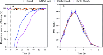
In our previous study,16 it was found that short-term exposure of CuNPs with concentrations of 30 and 50 mg L−1 would decrease the phosphorus removal efficiency significantly during the EBPR process, and both the anaerobic phosphorus release and aerobic phosphorus uptake were inhibited, while CuNPs with lower concentration (5 mg L−1) had no significant effect on removal efficiency (see ESI, Fig. S3). It is well known that wastewater biological treatment in WWTP is a progress of long-term run, and it is necessary and meaningful to further investigate the effect of long-term exposure to CuNPs on wastewater treatment progress, such as phosphorus removal. During the long-term culturing period with CuNPs addition, the performance of phosphorus removal was illustrated in Fig. 1. It was shown from Fig. 1A that the removal efficiency under the exposure to 5 mg L−1 of CuNPs during the whole culturing period was almost the same as control test, which meant that long-term exposure to low concentration of CuNPs still had no obvious effect on biological phosphorus removal. However, the performance of long-term exposure to higher concentrations of CuNPs (30 and 50 mg L−1) was significant difference from that of short-term. There was no net phosphorus removal occurred when 30 and 50 mg L−1 CuNPs were suddenly added (see ESI, Fig. S3). While with the extension of acclimation time, the phenomenon of net phosphorus removal reappeared, and the capability of phosphorus removal gradually recovered to the normal level, although the recovery time under the exposure to 50 mg L−1 of CuNPs took a longer time than that of 30 mg L−1. After the phosphorus removal efficiency reached stable, the transformations of SOP during one EBPR cycle were detected as Fig. 1B, which suggested that the anaerobic phosphorus release and aerobic phosphorus uptake returned to normal as well.
image file: c5ra11579e-f1.tif
Fig. 1 Effects of long-term exposure to CuNPs on performance of biological phosphorus removal. (A) The phosphorus removal efficiency, and −10–0 d represented the time before long-term exposure. The maximal standard deviations of triplicated measurements are less than 15%; (B) the transformation of SOP during one anaerobic and aerobic EBPR cycle. Error bars represent standard deviations of triplicate tests.

3.2 Effects of CuNPs on intracellular metabolites of activated sludge

It is well known that PAO will be enriched when the sequential anaerobic and aerobic phases were employed, and phosphorus removal in EBPR system is relevant to the anaerobic and aerobic transformations of polyphosphate, PHA and glycogen (Fig. S4, ESI). In anaerobic stage, PAO takes up short chain fatty acids and converts them to intracellular PHA, and the energy and reducing power requiring for PHA synthesis are provided mainly by the degradation of intracellular polyphosphate and glycogen, respectively. Under aerobic condition, the intracellular stored PHA are oxidized and used for microorganism growth, SOP uptake and glycogen replenishment. In this study, one EBPR cycle contained 8 h, including anaerobic stage for 2 h, aerobic stage for 3 h, and settling period for 3 h. Thus, the transformations of PHA and glycogen during one EBPR cycle under the condition of short-term and long-term exposure to CuNPs were unearthed as Fig. 2.
image file: c5ra11579e-f2.tif
Fig. 2 Transformations of PHA and glycogen during one EBPR cycle after short-term exposure (A) and long-term exposure (B) to CuNPs. Error bars represent the standard deviations of triplicate tests.

The PHA mainly contained PHB, PHV and PH2MV. When acetic acid is used as carbon source for phosphorus removal, the PHB was the major compound in the PHA. Under the shock loads of 30 and 50 mg L−1 of CuNPs, the anaerobic synthesis of PHB, PHV and PH2MV were all inhibited (Fig. S5, ESI), leading to the amounts of PHA decreased as 17.3% and 22.7%, respectively, when comparing to the control test (Fig. 2A). Since the phosphorus anaerobic release provides energy for PHA synthesis, the energy deficient induced by phosphorus release inhibition in anaerobic stage might be responsible for the declining of PHA synthesis. In addition, when smaller amount of PHA was synthesized, less reducing power was demanded, which was consistent with the decrease of glycogen anaerobic degradation as illustrated in Fig. 2A. In aerobic stage, the intracellular stored PHA are oxidized and used for microorganism growth, SOP uptake and glycogen replenishment. It was observed from Fig. 2A that the PHA aerobic degradation was suppressed significantly, resulting in less glycogen aerobic synthesis and no net phosphorus removal under the shock of 30 and 50 mg L−1 of CuNPs. It should be noted that the transformations of PHA and glycogen under the shock load of 5 mg L−1 CuNPs have no significant difference from the control test (p > 0.05, see Table S2, ESI, for statistical analysis). Therefore, short-term exposure to higher concentrations of CuNPs (30 and 50 mg L−1) had serious inhibition on transformations of intracellular metabolites, which were consistent with the deteriorated phosphorus removal efficiency.

However, after long-term culturing, as illustrated in Fig. 2B, the transformations of PHA and glycogen during one EBPR cycle returned to the normal level even under the exposure to higher concentrations of CuNPs, which matched the recovered phosphorus removal efficiency well. In EBPR system, some key bacteria such as PAO plays vital role in biological phosphorus removal. Under the exposure of CuNPs, different bacteria might show different tolerance to the same toxicity, and the toxicity might perform the function of bacteria selection. Thus, the selected microbial structure might be the reason for phosphorus removal recovery. In the following, the mechanism for this kind of adapting during long-term exposure was further dug out from the aspect of microbial structure shift.

3.3 Effects of CuNPs on microbial community structure

The efficient operation of WWTP depends on the stable microbial community structure and enough abundance of key functional microbial in the activated sludge.30 Some NPs, such as TiO2 NPs and Al2O3 NPs were reported to decrease the microbial diversity and the abundance of functional bacterial after long-term exposure, leading to the deterioration of nutrients removal for activated sludge.12,13 Taking biological phosphorus removal via EBPR for example, two main microbial groups-PAO and GAO exist in the system, and only PAO relates with biological phosphorus removal, therefore accumulating higher abundance of PAO is beneficial for higher phosphorus removal efficiency. Since the exposure to 30 mg L−1 of CuNPs showed similar biological phosphorus removal performance as that of 50 mg L−1, the bacterial community structure under long-term exposure to 30 mg L−1 of CuNPs was not considered.

In this study DGGE analysis was employed to determine the microbial diversity change in EBPR systems after long-term exposure to CuNPs. As shown in Fig. 3, the activated sludge exposed to 5 mg L−1 of CuNPs (L2) had similar bacterial diversity as the sludge of control (L1). However, bacteria diversity of the activated sludge exposed to 50 mg L−1 CuNPs (L3) decreased obviously when compared with the control sludge.


image file: c5ra11579e-f3.tif
Fig. 3 DGGE profiles of activated sludge after long-term exposure to 5 and 50 mg L−1 of CuNPs (L1, L2 and L3 represented control sludge, and sludge exposed to 5 and 50 mg L−1 CuNPs, respectively).

From the detailed bands information of DGGE profile (see Table 1), it was found that the Candidate division TM7, Candidatus Competibacter, Candidatus Accumulibacter, beta proteobacterium and Bacteroidetes appeared in sludge of control and sludge exposed to 5 mg L−1 of CuNPs. It is well known that Candidatus Accumulibacter is a kind of typical PAO, and can remove phosphorus by using short chain fatty acids. In addition, Candidate division TM7, beta proteobacterium and Bacteroidetes were found to make great contributions to the phosphorus removal in the literatures.31–33 After long-term exposure to 50 mg L−1 of CuNPs, the bands of 5, 6 and 7 in DGGE profile disappeared, which meant that Candidatus Accumulibacter and beta proteobacterium were gradually washed out. Although long-term exposure to high concentration of CuNPs made some bacteria disappear, the phosphorus removal efficiency returned to the level of control one, which inferred that Candidate division TM7 and Bacteroidetes were mainly responsible for phosphorus removal in the system.

Table 1 DGGE bands based on the V3 region of 16S rRNA gene and their closely related sequences
Band ID Accession no. Most closely related bacterial sequence Identity (%)
Species and strain Accession no.
1 KP126619 Uncultured Candidate division TM7 bacterium clone Skagen f60 DQ640706.1 100
2 KP126620 Uncultured bacterium clone SBRAC41 HQ158638.1 99
3 KP126621 Uncultured Candidatus Competibacter sp. clone 375 JQ726376.1 95
4 KP126622 Uncultured bacterium gene for 16S rRNA AB567959.1 92
5 KP126623 Uncultured Candidatus Accumulibacter sp. clone EMB clone_7 HM046420.1 98
6 KP126624 Uncultured beta proteobacterium clone MBfR_NSP-113 JN125267.1 96
7 KP126625 Uncultured bacterium clone ADK-MOh02-39 EF520623.1 96
8 KP126626 Uncultured Bacteroidetes bacterium clone BF2C11 JN820190.1 100
9 KP126627 Uncultured bacterium partial 16S rRNA gene HE646316.1 99


FISH was adopted to analyze the abundance of key bacteria (PAO and GAO) in this study according to the literature.34 The FISH images of sludge long-term exposed to 5 and 50 mg L−1 CuNPs and the control sludge were illustrated in Fig. 4. In control sludge, the abundances of PAO and GAO were 78 ± 5% and 20 ± 3%, respectively. After long-term exposure to 5 mg L−1 CuNPs, their abundances were similar as the control sludge, with PAO of 74 ± 4% and GAO of 21 ± 3%, respectively. However, the abundance of functional bacteria PAO decreased to 65 ± 5%, and the abundance of GAO increased to 30 ± 3% after long-term exposure to 50 mg L−1 of CuNPs.


image file: c5ra11579e-f4.tif
Fig. 4 Microscopes of activated sludge in the SBRs long-term exposed to CuNPs as visualized by FISH. The control sludge was shown in (A1), (B1) and (C1), sludge exposed to 5 mg L−1 CuNPs was shown in (A2), (B2) and (C2), and sludge exposed to 50 mg L−1 CuNPs was shown in (A3), (B3) and (C3). The PAO was hybridized with AMCA (blue, A1, A2 and A3), and GAO was hybridized with Cy3 (red, B1, B2 and B3). Probe EUBmix attained domain bacteria labelled with FITC (green, C1, C2 and C3).

It was obvious that long-term exposure to low concentration of CuNPs did not significantly induce the change of bacterial community structure in EBPR systems, and that the phosphorus removal efficiency kept stable during the entire culturing time was easily understood. Interestingly, the phosphorus removal efficiency returned to normal after long-term exposure to 50 mg L−1 CuNPs, even though the microbial diversity and the abundance of key functional bacteria (PAO) decreased. Thus, we speculated that the recovery process could be as follows: (1) shock load of CuNPs with higher concentration showed acute toxicity to the microbial in the EBPR system and the phosphorus removal efficiency was deteriorated; (2) during long-term culturing period, different bacteria performed various tolerant capacity to this kind of toxicity; (3) the bacteria which could not adapted the toxicity were washed out from the system, and the diversity of bacteria decreased; (4) the bacteria which could endure the toxicity of CuNPs were selected and left in the system; (5) the selected bacteria were gradually enriched to suitable abundance, and the microbial community structure was directionally adjusted to adapt the adverse environment; (6) the phosphorus removal efficiency and the transformation of intracellular metabolites returned to the normal level finally.

3.4 Intracellular ROS production and key enzyme activity under the exposure of CuNPs

Since the microbial structure has shifted during the long-term exposure to CuNPs, the changed bacteria might show different capacity of ROS eliminating, resulting in the activity of key enzyme various. It is reported that PPX and PPK are the key enzymes related to biological phosphorus removal.22,23 PPX is responsible for anaerobic phosphorus release, and the aerobic phosphorus uptake is related to the enzyme of PPK. Thus, ROS productions and the activities of some key enzymes (PPX and PPK) under the short and long-term exposure to CuNPs were explored in this study.

It was obvious to see from Fig. 5A that under the short-term exposure to different concentrations of CuNPs, the productions of ROS enhanced to 183%, 230% and 330% of control along with the increasing of CuNPs concentrations (5, 30 and 50 mg L−1). As to the enzyme activity, under the shock load of 30 and 50 mg L−1 of CuNPs, PPX decreased to 66.7 ± 6% and 44.4 ± 5% of control, and PPK decreased to 62.1 ± 6% and 37.9 ± 4% of control, which were consistent with the decreased anaerobic phosphorus release and the declined aerobic phosphorus uptake. However, shock load of 5 mg L−1 of CuNPs did not show obvious adverse impact on the activities of PPX and PPK (p > 0.05, statistic analysis, Table S3, ESI). While after long-term acclimation, the level of intracellular ROS production returned to the normal, and the enzyme activities had no significant variation when compared with control test (data not shown).


image file: c5ra11579e-f5.tif
Fig. 5 Intracellular ROS production under the short-term or long-term exposure to CuNPs (A), and the activities of enzyme PPX and PPK under the shock load of CuNPs (B). Error bars represent the standard deviations of triplicate tests.

When the system was exposed to CuNPs suddenly, the acute toxicity induced the oxidative stress, and the level of intracellular ROS increased as observed above. The produced ROS could be eliminated to some extent, and the function of microbial could perform normally, thus it could be reasonable that the enzyme activities and the phosphorus removal efficiency had no significant variation even the ROS level increased under the short term exposure to 5 mg L−1 of CuNPs. However, much higher production of ROS under short-term exposure to 30 and 50 mg L−1 of CuNPs could not be eliminated totally, inducing the decreasing of enzyme activities and the deterioration of phosphorus removal. After long-term culturing, the bacteria that could not adapt the toxicity of CuNPs were washed out from the system as observed in Fig. 3, and the adjusted bacteria could defense this kind of toxicity. Thus, the ROS production and the enzyme activity recovered to the normal level under the condition of long-term exposure, which was consistent with the phosphorus removal performance.

4. Conclusion

In summary, the chronic and acute impacts of CuNPs on the performances of biological phosphorus removal were compared. Results showed that terribly deteriorated phosphorus removal under the short-term exposure to CuNPs returned to normal level after long-term exposure to 30 and 50 mg L−1 of CuNPs, and the corresponding inhibited intracellular metabolites transformations were gradually recovered. Further investigation revealed that enhanced intracellular ROS and the decreased enzyme activities under the shock load of CuNPs returned to normal as well after long-term exposure to CuNPs. During the recovery period, some sensitive bacteria were washed out, and both the bacterial diversity and the abundance of functional bacteria (PAO) in EBPR system decreased. However, the left bacteria could adapt the toxicity induced by CuNPs and were responsible for phosphorus removal.

Acknowledgements

This work was financially supported by National Science Foundation of China (51425802), the Program of Shanghai Subject Chief Scientist (15XD1503400), and the Fundamental Research Funds for the Central Universities (2232015D3-23).

References

  1. G. Brumfiel, Nature, 2006, 440, 262 CrossRef CAS PubMed.
  2. S. K. Brar, M. Verma, R. D. Tyagi and R. Y. Surampalli, Waste Manage, 2010, 30, 504–520 CrossRef CAS PubMed.
  3. S. J. Klaine, P. J. J. Alvarez, G. E. Batley, T. F. Fernandes, R. D. Handy, D. Y. Lyon, S. Mahendra, M. J. McLaughlin and J. R. Lead, Environ. Toxicol. Chem., 2008, 27, 1825–1851 CrossRef CAS PubMed.
  4. C. Zhang, Z. Liang and Z. Hu, Water Res., 2014, 50, 350–358 CrossRef CAS PubMed.
  5. J. Chen, Y. Q. Tang, Y. Li, Y. Nie, L. L. Hou, X. Q. Li and X. L. Wu, Chemosphere, 2014, 104, 141–148 CrossRef CAS PubMed.
  6. L. Hou, K. Li, Y. Ding, Y. Li, J. Chen, X. Wu and X. Li, Chemosphere, 2012, 87, 248–252 CrossRef CAS PubMed.
  7. Y. Chen, H. Chen, X. Zheng and H. Mu, J. Hazard. Mater., 2012, 239–240, 88–94 CrossRef CAS PubMed.
  8. H. Chen, X. Zheng, Y. Chen and H. Mu, RSC Adv., 2013, 3, 9835–9842 RSC.
  9. X. Zheng, R. Wu and Y. Chen, Environ. Sci. Technol., 2011, 45, 2826–2832 CrossRef CAS PubMed.
  10. H. Mu, X. Zheng, Y. Chen, H. Chen and K. Liu, Environ. Sci. Technol., 2012, 46, 5997–6003 CrossRef CAS PubMed.
  11. J. Hou, L. Z. Miao, C. Wang, P. F. Wang, Y. H. Ao, J. Qian and S. S. Dai, J. Hazard. Mater., 2014, 276, 164–170 CrossRef CAS PubMed.
  12. X. Zheng, Y. Chen and R. Wu, Environ. Sci. Technol., 2011, 45, 7284–7290 CrossRef CAS PubMed.
  13. X. Zheng, Y. Su and Y. Chen, Environ. Sci. Technol., 2012, 46, 7182–7188 CrossRef CAS PubMed.
  14. Y. Chen, Y. L. Su, X. Zheng, H. Chen and H. Yang, Water Res., 2012, 46, 4379–4386 CrossRef CAS PubMed.
  15. PEN, Project on Emerging Nanotechnologies, 2011.
  16. H. Chen, X. Zheng, Y. Chen, M. Li, K. Liu and X. Li, PLoS One, 2014, 9, e92871 Search PubMed.
  17. Y. Chen, D. B. Wang, X. Y. Zhu, X. Zheng and L. Y. Feng, Environ. Sci. Technol., 2012, 46, 12452–12458 CrossRef CAS PubMed.
  18. D. B. Wang, X. M. Li, Q. Yang, W. Zheng, Y. Wu, T. J. Zeng and G. M. Zeng, Water Res., 2012, 46, 3868–3878 CrossRef CAS PubMed.
  19. F. Q. Schafer and G. R. Buettner, Free Radical Biol. Med., 2001, 30, 1191–1212 CrossRef CAS.
  20. J. W. McGrath and J. P. Quinn, Appl. Environ. Microbiol., 2000, 66, 4068–4073 CrossRef CAS.
  21. O. H. Lowry, N. J. Rosebrough, A. L. Farr and R. J. Randall, J. Biol. Chem., 1951, 193, 265–275 CAS.
  22. S. J. Lee, Y. S. Lee, Y. C. Lee and Y. L. Choi, J. Basic Microbiol., 2006, 46, 108–115 CrossRef CAS PubMed.
  23. J. W. van Groenestijn, M. M. Bentvelsen, M. H. Deinema and A. J. Zehnder, Appl. Environ. Microbiol., 1989, 55, 219–223 CAS.
  24. Z. Q. Zhang, J. Yu and R. C. Stanton, Anal. Biochem., 2000, 285, 163–167 CrossRef CAS PubMed.
  25. L. K. Limbach, P. Wick, P. Manser, R. N. Grass, A. Bruinink and W. J. Stark, Environ. Sci. Technol., 2007, 41, 4158–4163 CrossRef CAS.
  26. C. Yang, D. E. Crowley and J. A. Menge, FEMS Microbiol. Ecol., 2001, 35, 129–136 CrossRef CAS PubMed.
  27. R. I. Amann, W. Ludwig and K. H. Schleifer, Microbiol. Rev., 1995, 59, 143–169 CAS.
  28. APHA, American Public Health Association, Washington, DC, 1998 Search PubMed.
  29. Y. M. Jiang, Y. G. Chen and X. Zheng, Environ. Sci. Technol., 2009, 43, 7734–7741 CrossRef CAS PubMed.
  30. A. Oehmen, P. C. Lemos, G. Carvalho, Z. Yuan, J. Keller, L. L. Blackall and M. A. M. Reis, Water Res., 2007, 41, 2271–2300 CrossRef CAS PubMed.
  31. Y. H. Kong, Y. Xia, J. L. Nielsen and P. H. Nielsen, Microbiology-Sgm, 2007, 153, 4061–4073 CrossRef CAS PubMed.
  32. Y. H. Kong, M. Beer, G. N. Rees and R. J. Seviour, Microbiology, 2002, 148, 2299–2307 CAS.
  33. W.-D. Tian, W.-G. Li, K.-J. An, X.-R. Kang, C. Ma and Z.-L. Ran, J. Water Sustainability, 2011, 1, 269–278 CAS.
  34. G. R. Crocetti, P. Hugenholtz, P. L. Bond, A. Schuler, J. Keller, D. Jenkins and L. L. Blackall, Appl. Environ. Microbiol., 2000, 66, 1175–1182 CrossRef CAS.

Footnote

Electronic supplementary information (ESI) available: The composition of synthetic wastewater, the set-up and operation of parent sequencing batch reactors, the measurement of CuNPs removal efficiency, the XRD of CuNPs, the effect of short-term exposure to CuNPs on biological phosphorus removal, effect of shock load of CuNPs on transformations of PHB, PHV and PH2MV during one cycle of EBPR, and the statistical analysis of experimental data. See DOI: 10.1039/c5ra11579e

This journal is © The Royal Society of Chemistry 2015