DOI:
10.1039/C5RA11481K
(Paper)
RSC Adv., 2015,
5, 64604-64613
Synthesis and evaluation of a glucose attached pyrene, as a fluorescent molecular probe in sugar and non-sugar based micro-heterogeneous media†
Received
16th June 2015
, Accepted 20th July 2015
First published on 21st July 2015
Abstract
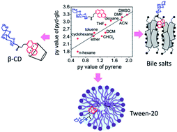
A new fluorescent pyrene–glucose conjugate (pyd-glc), 1-(4,6-O-butylidene-β-D-glucopyranosyl)-4-(1-pyrene)-butan-2-one, has been synthesized by attaching a pyrene molecule to acetal (butylidene) protected glucose via a butane-2-one linker. Detailed photo-physical studies of pyd-glc in homogeneous media have been carried out in both protic and aprotic solvents which lead to a py scale, a solvent polarity scale specific to this molecule, like that of pyrene. The molecule aggregates in water and partitions as a monomer in micro-heterogeneous media. The photo-physical response of pyd-glc has been studied in various sugar and non-sugar based micro-heterogeneous media. Similar to pyrene, this molecule offers multiple fluorescence parameters to investigate the organization of micro-heterogeneous media. The fluorescence parameters of pyd-glc faithfully reflect the state of organization of these anisotropic media.
Introduction
A leading trend in organic photochemistry is to attach standard fluorophores with bio-relevant organic molecules like, steroids,1 bile acids,2,3 lipids,4,5 sugar molecules,6,7 DNA fragments,8,9 amino acid residues10,11 etc. The major objective of synthesizing these derivatives is to probe bio-organized systems to get information about their bio-functionality.12,13 This kind of ‘system probe’, which causes less perturbation to the investigated systems,14 can be used to investigate a specific site of interest11 as they have a definite location in vitro.5 As a result, this class of probe is often considered as a better reporter of the medium. The vast volume of literature found for this trend implies its success. For example, H-aggregates of cholesterol attached coumarin have been employed to distinguish progressive association of di-hydroxy and tri-hydroxy bile salts.1 Fluorophore attached bile salts have been used to investigate aggregation behaviour, transport phenomenon, polarity etc.2,3,15–17 Long alkyl chain attached fluorophores are useful in lipid membrane studies, like configuration analysis, membrane fusion, phase transition, bilayer heterogeneity etc.4,5,14,18,19 Derivatization of sugar moieties, especially β-CD serves as sensor of different molecules.6,7,20,21 Molecular beacons are useful for the study of DNA conformations, DNA cleavage analysis etc.8,9,22 Unnatural fluorescent amino acids are useful tool for the analysis of protein conformations, distance between two sites, interior polarity etc.10,11,23,24 Here, a pyrene attached glucose analogue, 1-(4,6-O-butylidene-β-D-glucopyranosyl)-4-(1-pyrene)-butan-2-one (abbreviated as ‘pyd-glc’) (Fig. 1) has been synthesized and its probing ability in sugar and non-sugar based organized media has been investigated. For the past couple of decades it has been realized that the range of applications of fluorescence molecular probes can be enhanced by suitably conjugating the probe with different moieties which can specify the position of the probe in micro-heterogeneous media.5,11,14 The attachment of a glucose unit with the well known fluorescent probe pyrene, would enable a more efficient use of pyrene for carbohydrates and other sugar based media (e.g. β-CD, Tween 20). Another important aspect of attaching glucose moiety with pyrene is the possibility of probing the hydrophilic–hydrophobic interfaces, unlike pyrene which prefers pure non-polar media.25,26
 |
| Fig. 1 Molecular structure of 1-(4,6-O-butylidene-β-D-glucopyranosyl)-4-(1-pyrene)-butan-2-one (pyd-glc). | |
Here, acetal (butylidene) protected glucose has been attached with pyrene fluorophore via butane-2-one as a linker. Pyrene is a standard fluorophore because of the extensive applications of its various fluorescent parameters like, Ham effect, long fluorescence lifetime, excimer formation etc.25 Using all these parameters of pyrene moiety of pyd-glc, a progressive change of the micro-environment of certain organized media has been looked into. In fact, in recent years efforts have been made to introduce transition metal based complexes with high lifetime to overcome the background fluorescence of biological samples.27,28 From this point of view modification of pyrene with different conjugation is worth as it has a fairly long lifetime component which is also very sensitive towards the change of the micro-environment.
β-CD is a doughnut shaped cyclic oligosaccharide having cavity diameter 6.2 Å and length 8.0 Å.29,30 On account of the presence of hydrophilic –OH groups in its outer surface and hydrophobic inner core it serves as a micro-heterogeneous medium and can encapsulate a wide variety of molecules inside it.31 Inclusion complex forming ability of β-CD has been used occasionally in drug delivery, food industry, toxicity reduction etc.32,33 Here, encapsulation of the pyrene moiety of pyd-glc inside β-CD nano-cavity has been monitored by the above mentioned fluorescent parameters. Tween-20 is a sugar based non-ionic surfactant.34 Due to its non-toxicity it has its wide applications in drug, food, scientific research etc.35 It has its critical micellar concentration (CMC) ∼0.06 mM.35 Tween-20 has been used as another sugar based drug delivery system to show the probing ability of pyd-glc in sugar based media.
To emphasize the versatility of pyd-glc, it has also been employed in non-sugar micro-heterogeneous media like, bile salts, sodium cholate (NaC) and sodium deoxy cholate (NaDC). Bile salts are natural surfactants help in fat digestion.36 They possess unique structural features compared to the other conventional surfactants. Hydrophilic concave face containing –OH and –COOH groups and hydrophobic convex face containing methyl groups impart facial polarity. In aqueous medium, primary aggregation of bile salt is followed by the secondary aggregation as a function of concentration. Hydrophobic interaction of steroidal backbone leads to primary aggregation and hydrogen bonding of –OH groups bind primary aggregates together to form secondary aggregates.37
The main objectives of this work are (i) to synthesize a new glucose attached pyrene derivative, 1-(4,6-O-butylidene-β-D-glucopyranosyl)-4-(1-pyrene)-butan-2-one (pyd-glc), (ii) preliminary study of photo-physical parameters in homogeneous media and a comparison with the known behaviour of pyrene, and a detailed study of the photo-physics of pyd-glc in different micro-heterogeneous media like, (iii) sugar based organized media (e.g. β-CD, Tween-20) and (iv) non-sugar based organized media (e.g. bile salts) to establish it as a fluorescent molecular probe.
Materials and methods
Materials
β-CD was purchased from Sigma Chemical Co. (Bangalore, India). Sodium di-hydrogen phosphate, di-sodium hydrogen phosphate, Tween-20 were purchased from Merck Specialties Pvt. Ltd. (Mumbai). Bile salts, sodium cholate (NaC) and sodium deoxycholate (NaDC) were purchased from S. D. Fine Chemical Company, India. Triple distilled water used for all experiments was prepared by using KMnO4 and NaOH. All other solvents used were of spectroscopic grade. Fresh stock solutions were prepared for all the experiments. 1 mM acetonitrile solution of pyd-glc was used as a stock solution of probe. Final concentration of the fluorophore was maintained at 4 μM. 0 to 16 mM solutions of β-CD were prepared for this study from a primary stock solution of β-CD. A stock solution of 0.25 mM of Tween-20 was prepared in water. 0 to 0.17 mM of Tween-20 solutions were made by successive dilution for this study. Sodium cholate (NaC) and sodium deoxycholate (NaDC) concentrations were varied from 0–48 and 0–18 mM, respectively. pH of the solutions were kept at ∼7.4 with 50 mM phosphate buffer, similar to the physiological condition. ACN contamination was kept <1% in all the cases.
Photophysical studies
Steady state fluorescence measurements were carried out by using Fluoromax 4 (Horiba Jobin Yvon) spectrofluorimeter with 150 W Xenon lamp as the source of excitation. To obtain highly resolved vibronic spectra 1 second integration time, 0.1 nm increment and 1/1 nm slit width were maintained. This gives accurate value of II/IIII of the pyrene conjugate, pyd-glc.
Fluorescence lifetime measurements were done by Horiba Jobin Yvon TCSPC lifetime instrument in time-correlated single-photon counting arrangement. Nano-LED of 340 nm was used as excitation source. Forward mode of data acquisition was used with pulse repetition rate 25 kHz. Instrumental full width at half-maxima of the 340 nm LED, including the detector response was measured to be ∼800 ps. The instrument response function was collected by using scattered medium, LUDOX AS40 colloidal silica. IBH software was used for the decay analysis. Decays were fitted to get a symmetric distribution keeping χ2 value at 0.97 ≤ χ2 ≤ 1.34.
Average fluorescence life time was determined by using the following equation.38 Here, τi is the lifetime of a component having amplitude βi, i signifies number of components present.
All these experiments were carried out at room temperature.
Results and discussions
Synthesis of pyd-glc
α-,β-Unsaturated compound, 1, which upon reduction39 using Zn/NH4Cl in EtOH/H2O mixture gave the corresponding reduced chalcone derivative, pyd-glc in 82% yield (Scheme 1). Reduction was carried out under neutral condition with ethanol
:
H2O solvent mixture in the ratio of 9
:
1. However, the compound, 1 even after increasing the temperature to 50 °C failed to dissolve. But the solubility was obtained when two to three drops of DMSO was added to the reaction mixture, and resulted in good yield of the reduced product, pyd-glc. Remarkable chemoselective carbon–carbon double bond reduction was observed and the labile functionality like ketone was unaffected under these conditions. Structure of the synthesized sugar ketone derivative, pyd-glc was determined by (1H, 13C) NMR spectroscopy, DEPT-135, mass spectroscopy and elemental analysis.
 |
| Scheme 1 Synthesis of 1-(4,6-O-butylidene-β-D-glucopyranosyl)-4-(1-pyrene)-butan-2-one, pyd-glc. | |
The pyrene based sugar-chalcone derivative, 1 on reduction using Zn furnished the pyrene based sugar ketone derivative, pyd-glc. In 1H NMR spectrum of compound, pyd-glc the characteristic methylene peaks were appeared as two multiplets in the regions 3.1–3.0 and 2.8–2.6 ppm whereas two peaks at 45 ppm and 27 ppm corresponds to the methylene carbons in 13C NMR confirmed the formation of the reduced product. From the DEPT-135 experiment the appearance of six peaks which appeared on one side of the base line corresponds to the six methylene carbons confirms the formation of the sugar ketone derivative, pyd-glc.
Photophysical studies of pyd-glc in non-aqueous homogeneous media
D2h symmetry of pyrene imparts restriction in vibrational modes of motion.26 As a result, emission spectra of pyrene is highly structured with five distinct vibrational lines at 373 (I), 379 (II), 383 (III), 389 (IV) and 393 (V) nm. In polar solvents, symmetry forbidden transitions (e.g. 0–0 band, I) become favourable due to the reduction of local symmetry.26,40,41 As a result, vibrational band intensity ratio (II/IIII) of pyrene has been used as a faithful tool for the determination of solvent polarity. This is often referred as the ‘Ham effect’, which leads to a solvent polarity scale named the ‘py scale’.25,40 Like py scale of pyrene, intensity ratio (II/IIII), of pyd-glc has been calculated in solvents of different polarity. To obtain the vibrational spectral lines accurately, previously mentioned conditions were maintained. Absorbance and fluorescence spectra of pyd-glc in different solvents have been given in ESI (Fig. S1†). It shows absorbance band of pyd-glc (λabs ∼ 342 nm) is ∼10 nm red shifted as compared to pyrene (λabs ∼ 332 nm). Fig. 2 shows py value of pyd-glc with different solvent parameter scales using pyrene (literature) as a reference.
 |
| Fig. 2 py value (II/IIII) of pyd-glc with different solvent parameter scales, (a) ET(30) scale for aprotic solvents, (b) ET(30) scale for protic solvents, (c) Kosower Z scale for aprotic solvents and (d) Kosower Z scale for protic solvents, with respect to pyrene, at pyd-glc concentration 4 μM. | |
Fig. 2 shows py value of pyd-glc in different solvents is parallel with that of pyrene, except water. This exceptional behaviour has been discussed in the next section. Keeping py values of pyrene (literature) and pyd-glc in X-axis and Y-axis, respectively, linear plot has been obtained (Fig. 3). This implies that the py scale value obtained from pyd-glc has similar significance as pyrene and can be correlated by using the following equations.
|
py of pyd-glc = 0.89(±0.08) × py of pyrene + 1.54(±0.14) for aprotic solvents
| (1) |
|
py of pyd-glc = 1.0(±0.07) × py of pyrene + 1.36(±0.07) for protic solvents
| (2) |
 |
| Fig. 3 Plot of py values of pyrene (literature) vs. pyd-glc for (a) aprotic and (b) protic solvents. | |
This new scale has a slope similar to the py scale of pyrene but has a different intercept, reflecting the hydrophilicity introduced by the glucose conjugate.
Photophysical studies of pyd-glc in water
Deviation of II/IIII value of pyd-glc from linearity in Fig. 3b indicates the possibility of aggregate formation in water. Probable ground state association of this non-polar molecule pyd-glc in water has been analyzed by absorption spectra, fluorescence spectra and lifetime data. Ratio of the absorption peak and valley (Apeak/Avalley) of the most intense absorption band is higher (2.66) in case of solvents, like acetonitrile (dielectric constant, ε = 37.5) where this molecule is well soluble (Fig. 4a). In aqueous medium, spectral broadening occurs which decreases the Apeak/Avalley value (1.33) (Fig. 4b). This observation suggests ground state aggregate form in water (dielectric constant, ε = 80).42 The presence of aggregate in ground state is also reflected in the long tail in UV-visible absorption spectrum in water medium (Fig. 4b).
 |
| Fig. 4 Absorption spectra of pyd-glc in (a) acetonitrile and (b) water medium to determine Apeak/Avalley value at fluorophore concentration 4 μM. | |
Evidence for the presence of ground state association is also observable from fluorescence spectra. Binary solvent study, using a mixture of water and acetonitrile has been done for the above purpose (Fig. 5). Iso-emissive point at ∼460 nm shows two-state equilibrium. Moreover, the intensity of the aggregate form (at 480 nm) decreases with increasing proportion of solvating solvent, ACN (inset of Fig. 5).
 |
| Fig. 5 Fluorescence spectra of pyd-glc (4 μM) in different proportion of water and acetonitrile, at λex = 340 nm. | |
Change in the emission profile of pyd-glc in water with the change in the excitation wavelength (Fig. 6a) signifies the presence of more than one emitting state, unlike ACN (Fig. 6b). Excitation spectra of pyd-glc in water (ESI, Fig. S2†) with different emission wavelength are also different from each other.
 |
| Fig. 6 Fluorescence spectra of pyd-glc (4 μM) in (a) water and (b) acetonitrile with the variation of excitation wavelength. | |
The presence of pre-associated state is also evident from the excited state photo-physics. Table 1 summarizes fluorescence lifetime value of pyd-glc in different solvents. In all other solvents, except water, fluorescence decay shows mono-exponential behaviour. Only in case of water, it shows bi-exponential decay which points to the presence of aggregate form. As all these lifetime experiments have been done in non-degassed condition average fluorescence lifetimes have lower value.43 Fluorescence lifetime decay profiles of pyd-glc in solvents of different polarity have been given in ESI, Fig. S3.† Residue distribution plots for the same have been given in ESI, Fig. S4.†
Table 1 Fluorescence lifetime data of pyd-glc (λex = 340 nm, λem = 375 nm) in different solvents
Solvent |
τi(βi) |
χ2 |
Cyclohexane |
18.88 |
1.18 |
n-Hexane |
8.82 |
1.10 |
n-Heptane |
9.51 |
0.98 |
1,4-Dioxan |
43.64 |
1.16 |
THF |
19.30 |
1.14 |
CHCl3 |
19.65 |
1.21 |
Acetonitrile |
16.13 |
0.97 |
DMF |
36.15 |
1.21 |
Methanol |
18.18 |
1.19 |
Propanol |
27.06 |
1.04 |
Water |
6.27 (0.98), 43.43 (0.02) |
1.25 |
The above studies in homogeneous media suggest that for the evaluation of pyd-glc as a possible fluorescent molecular probe for aqueous solutions of β-cyclodextrin, Tween-20 and bile salts, three photo-physical parameters can be used: (i) II/IIII fluorescence intensity ratio, (ii) change in the average fluorescence lifetime value (τaveg) and (iii) efficiency of excimer formation.
Interaction of pyd-glc with β-CD
Fig. 7a shows the change in fluorescence intensity of pyd-glc with increasing concentration of β-CD. Increase in the monomeric emission intensity with the addition of β-CD is due to the increase in the population of the monomeric form. Longer wavelength emission peak at 480 nm is due to the excimer emission, like pyrene. An isoemissive point at ∼460 nm signifies two-state equilibrium between pyd-glc in aqueous solvent and inside β-CD nano-cavity. It is known that in presence of host molecule, guest molecule leaves its aqueous hydration sphere and goes into the hydrophobic cavity provided by the host molecule. Inset of Fig. 7a, shows the decrease of excimer intensity (excimer intensity has been taken after normalizing at 374 nm, peak I) followed by labelling effect, with increasing β-CD concentration. Fig. 7b is highly resolved spectra of the probe in β-CD medium taken in the above mentioned condition to get py value. Inset of Fig. 7b shows the ratio of II/IIII of pyd-glc with increasing concentration of β-CD. It shows an increase in the py value followed by the saturating effect. In case of pyrene, with increasing β-CD concentration, py value decreases with a same kind of saturating effect.44 py value obtained from pyrene (literature) and pyd-glc after saturation (10 mM β-CD) indicates almost same kind of polarity of β-CD cavity. py value indicates 30.20 and 31.67ET(30) value, respectively, for pyrene and pyd-glc (from eqn (1), ESI, Fig. S5a†). This signifies pyd-glc is as good as pyrene in indicating the polarity of β-CD cavity.44
 |
| Fig. 7 (a) Fluorescence spectra of pyd-glc (4 μM) with increasing concentration of β-CD, inset shows decrease of excimer fluorescence intensity, (b) fluorescence spectra of pyd-glc (4 μM) with increasing concentration of β-CD under high resolution, inset shows variation of II/IIII value; at λex = 340 nm. | |
Non-linear scattering method has been used to determine inclusion constant of pyd-glc into β-CD cavity.29,32 The following equation has been used for this purpose,
1/(R − R0) = 1/(rC) + 1/(rCKi[CD]) |
Here,
R0,
R = intensity ratio of
pyd-glc in absence and presence of β-CD,
r = constant,
C = concentration of the guest molecule, here
pyd-glc, 4 μM,
Ki = inclusion constant, [CD] = concentration of β-CD.
From the linear plot (Fig. 8a) of 1/[CD] vs. 1/(R − R0) inclusion constant (Ki) has been determined by taking intercept/slope value. Ki is obtained (550 ± 10) M−1 at room temperature from the above plot. This Ki value of pyd-glc is well comparable with sugar containing flavone, Rutin.33 With a length of 10.4 Å and width of 8.2 Å pyrene cannot enter into β-CD cavity fully, unlike γ-CD.29,30 The plausible scheme for the penetration of pyd-glc inside β-CD cavity has been shown in Fig. 8b, where only a part of the pyrene ring goes inside axially.
 |
| Fig. 8 (a) Determination of inclusion constant of pyd-glc in β-CD and (b) plausible scheme for the penetration of pyd-glc inside β-CD nano-cavity. | |
Change of the micro-environment around pyrene moiety of pyd-glc molecule is also evident from the excited state photo-physics. Lifetime data of pyd-glc in β-CD micro-environment has been summarized in ESI, Table S1.† Fig. 9 shows the fluorescence lifetime decay profiles of the probe in β-CD environment. From the inset of Fig. 9 it is evident that there is almost 10 fold increase in the average fluorescence lifetime (τaveg) value in presence of β-CD in the above mentioned concentration. Change in the location of pyrene in presence of β-CD decreases the non-radiative decay, as a result average fluorescence lifetime (τaveg) increases. Residue distribution plot for the same has been given in ESI, Fig. S6.†
 |
| Fig. 9 Fluorescence lifetime decay profiles of pyd-glc in β-CD media, at λex = 340 nm, λem = 375 nm. | |
Interaction of pyd-glc with Tween-20
Tween-20, also named as sorbitanmono-laurate is one of the surface active agent in Tween series.35 This type of carbohydrate based surfactants has one sugar moiety along with lipid chain; as a result they undergo micellization like other surfactants.34 The change in the micro-environment from pre-micellar state to post-micellar state has been monitored by the py value of pyd-glc. Fig. 10a shows the fluorescence spectra of pyd-glc with increasing concentration of Tween-20. Fig. 10b is the highly resolved fluorescence spectra of pyd-glc taken in the above mentioned condition, in presence of Tween-20. The onset of the py value has been found at the critical micellar concentration (0.06 mM) followed by the saturating effect (inset of Fig. 10b). Similar break in slope during micellization has also been found in pyrene.45 Spectra of Fig. 10a have been normalized at the peak I to get Fig. 10c. Along with the excimer emission at 480 nm there is also a prominent band at 450 nm. The structure of the excimer emission is due to the presence of different kind of static excimers.42,46,47 The 480 nm excimer band is arising from the normal excimer formed by the parallel overlap of two pyrene rings, whereas, 450 nm band arises due to the angular displaced overlap of the two pyrene rings. This kind of spectrum is commonly observed in films of fatty acids, neat liquids and also in single crystals.47,48 This kind of static excimer formation (at 450 nm) is also found in pre-micellar state, which signifies the sensitivity of pyd-glc towards Tween-20. The breaking of pre-associated aggregate also follows the micellization (inset of Fig. 10c). The py value obtained in 0.1 mM of Tween-20 for both pyrene (literature) and pyd-glc indicates similar kind of medium polarity. py value indicates 36.73 and 38.41ET(30) value, respectively, for pyrene and pyd-glc (from eqn (1), ESI, Fig. S5b†).45 This shows py scale of pyd-glc is well applicable as that of pyrene in this media.
 |
| Fig. 10 (a) Fluorescence spectra of pyd-glc (4 μM) with increasing concentration of Tween-20, (b) fluorescence spectra of pyd-glc (4 μM) with increasing concentration of Tween-20 under high resolution, inset shows variation of II/IIII value, (c) normalized spectra of pyd-glc (4 μM) at peak I with increasing concentration of Tween-20, inset shows decrease of excimer fluorescence intensity; at λex = 340 nm. | |
Lifetime data of pyd-glc in Tween-20 has been summarized in ESI, Table S2.† Almost 15 fold increase in the average fluorescence lifetime (τaveg) value has been found at the highest Tween-20 concentration used. Fig. 11 shows fluorescence lifetime decay profiles of the probe in Tween-20 miceller media. The onset of average fluorescence lifetime (τaveg) value also follows the cmc of Tween-20 (inset of Fig. 11). Residue distribution plots for the same have been given in ESI, Fig. S7.†
 |
| Fig. 11 Fluorescence lifetime decay profiles of pyd-glc in Tween-20 media, at λex = 340 nm, λem = 375 nm. | |
Interaction of pyd-glc with bile salts
From the above discussions it is evident that this newly synthesized fluorescent molecular probe has dependable probing ability in sugar based micro-heterogeneous media. At the same time applicability of this probe has also been demonstrated in non-sugar media, like bile salts. Fig. 12a shows the intensity enhancement of pyd-glc with increasing concentration of NaDC. Inset of Fig. 12a shows the decrease of excimer intensity (excimer intensity has been taken after normalizing at 374 nm, peak I), where the onset follows critical micellar concentration of NaDC (4–6 mM). Spectra of Fig. 12b has been collected under high resolution condition as mentioned before. Inset of Fig. 12b shows the py value of pyd-glc with increasing concentration of NaDC, where onset has been obtained at its CMC. Similar fluorescence response of pyd-glc has also been found during the CMC study of hydrophilic bile salt, NaC which has CMC ∼ 12–16 mM (Fig. S8a and b†).
 |
| Fig. 12 (a) Fluorescence spectra of pyd-glc (4 μM) with increasing concentration of NaDC, inset shows decrease of excimer fluorescence intensity and (b) fluorescence spectra of pyd-glc (4 μM) with increasing concentration of NaDC under high resolution, inset shows variation of II/IIII value; here λex = 340 nm. | |
py value obtained for both pyrene (literature) and pyd-glc after saturation indicates similar kind of polarity of bile salt micellar media. py value indicates 32.5 and 35.4ET(30) value, respectively, for pyrene and pyd-glc in NaDC media (from eqn (1), ESI, Fig. S9a†).49 Similarly, py value indicates 33.0 and 35.95ET(30) value, respectively, for pyrene and pyd-glc in NaC media (from eqn (1), ESI, Fig. S9b†).50
Excited state photo-physics of this probe also follows the progressive micellization of NaDC. Fig. 13 gives the fluorescence lifetime decay profiles of pyd-glc in NaDC media. ESI, Table S3† enlists the fluorescence lifetime data of pyd-glc in NaDC media. Almost 20 times increase in the average fluorescence lifetime (τaveg) value follows CMC of NaDC (inset of Fig. 13). Similar change in lifetime value has also been found in NaC media. In ESI, Fig. S8c† gives the decay profiles of pyd-glc in NaC media and Table S4† enlists the corresponding lifetime data. Residue distribution plots for the NaDC and NaC have been given in ESI, Fig. S10 and S11,† respectively. This shows the probing ability of pyd-glc in the bio-surfactant systems is like pyrene.49,50 This kind of surfactants are important from physiological point of view as they mimic bio-systems.
 |
| Fig. 13 Fluorescence lifetime decay profiles of pyd-glc in NaDC media, at λex = 340 nm, λem = 375 nm. | |
Conclusions
Photo-physical properties of the newly synthesized glucose attached pyrene derivative, 1-(4,6-O-butylidene-β-D-glucopyranosyl)-4-(1-pyrene)-butan-2-one, have been investigated in homogeneous solvents. This provides a py scale specific to pyd-glc that could be used to monitor the local polarity of the probe in various micro-heterogeneous media. In aqueous medium, pyd-glc undergoes aggregation due to the hydrophobicity of the protected glucose. With increase in medium micro-heterogeneity the aggregates break and the probe is incorporated in its monomeric form. The corresponding change of II/IIII ratio thus becomes a sensitive indicator of the nature of micro-heterogeneous media. Applicability of this new fluorescent glucose derivative has been explored in mainly sugar based organized media, like β-CD and Tween-20. It forms 1
:
1 complex with β-CD with inclusion constant value (550 ± 10) M−1 at room temperature. In Tween-20 media, pyd-glc forms different types of excimers and also shows specific interaction with Tween-20. Breaking of the excimer follows the CMC of Tween-20. Bile salt micellization study has been used to show this new probe's probing ability in non-sugar heterogeneous media also. py scale made for this probe (pyd-glc) indicates similar kind of polarity as that of pyrene (with <10% deviation) in various micro-heterogeneous media. From this study, this newly synthesized molecule has been emerged as a multi-parametric fluorescent molecular probe as pyrene.
Experimental section
General procedure for synthesis of sugar ketone derivative, pyd-glc
Sugar chalcone (1 mmol) in ethanol (50 ml) was added to water solution containing NH4Cl (20 mmol) at room temperature and stirred vigorously with Zn powder (3 mmol) added in three equal portions at intervals of 15 minutes. Two to three drops of DMSO was added for the solubility of the reaction mixture. Stirring was continued for 15 minutes by warming the reaction mixture. The completion of the reaction was monitored through TLC and the reaction mixture was filtered using celite bed to remove the unreacted Zn. The filtrate was then evaporated under reduced pressure and extracted using EtOAc–water mixture. The ethylacetate layer was dried over anhyd. Na2SO4 and concentrated to dryness. The product thus obtained was further purified by column chromatography.
Synthesis, physicochemical and spectral data of 1-(4,6-O-butylidene-β-D-glucopyranosyl)-4-(1-pyrene)-butan-2-one (pyd-glc)
Compound, pyd-glc was obtained as a colourless solid by the reduction of sugar-chalcone, 1 (0.49 g, 1 mmol) using Zn (0.20 g, 3 mmol) and NH4Cl (1.07 g, 20 mmol). (ESI, Fig. S12–15†).
Mp: 188–190 °C.
Yield: 0.40 g (82%).
1H NMR (300 MHz, CDCl3): δ 8.23–7.97 (m, 8H, Ar-H), 7.87 (d, J = 7.8 Hz, 1H, Ar-H), 4.33 (t, J = 5.1 Hz, 1H, Ace-H), 4.03 (d, J = 5.4 Hz, 1H, Sac-H), 3.84–3.77 (m, 1H, Sac-H), 3.65–3.59 (m, 3H, Sac-H), 3.28–3.18 (m, 3H, Sac-H), 3.06–2.95 (m, 3H, –CH2, Sac-H), 2.87 (dd, J = 3.9 Hz, J = 15.8 Hz, 1H, –CH2), 2.77–2.60 (m, 3H,–CH2), 1.60–1.54 (m, 2H, –CH2), 1.38 (q, J = 7.8 Hz, 2H, –CH2), 0.91 (t, J = 7.2 Hz, 3H, –CH3).
13C NMR (75 MHz, CDCl3): δ 208.0, 135.1, 131.4, 130.8, 130.1, 128.5, 127.6, 127.5, 127.2, 126.8, 125.9, 125.1, 125.0, 124.9, 122.9, 102.4, 80.3, 76.0, 75.2, 74.1, 70.5, 68.1, 45.5, 45.4, 36.2, 27.0, 17.4, 13.9.
ESI-MS Calc. for C30H32O6, 488; m/z found, 511 [M + Na]+.
Elemental analysis anal. Calc. for C30H32O6: C, 73.75; H, 6.60%. Found: C, 73.78; H, 6.62.
Acknowledgements
A. K. M. thanks DST, Government of India for financial assistance in the form of major project. T. M. acknowledge SERC-DST, New Delhi for financial support. T. M. thank DST, New Delhi for use of NMR facility under DST-FIST programme to the Department of Organic Chemistry, University of Madras, Guindy Campus, Chennai, India. I. S. and H. M. thank CSIR, New Delhi, India for their research fellowships.
References
- H. S. P. Rao, A. Desai, I. Sarkar, M. Mohapatra and A. K. Mishra, Phys. Chem. Chem. Phys., 2014, 16, 1247–1256 RSC.
- M. Gomez-Mendoza, M. L. Marin and M. A. Miranda, Org. Biomol. Chem., 2014, 12, 8499–8504 CAS.
- M. Gomez-Mendoza, M. L. Marin and M. A. Miranda, J. Phys. Chem. Lett., 2011, 2, 782–785 CrossRef CAS.
- H. Raghuraman, S. Shrivastava and A. Chattopadhyay, Biochim. Biophys. Acta, 2007, 1768, 1258–1267 CrossRef CAS PubMed.
- S. Haldar, M. Kombrabail, G. Krishnamoorthy and A. Chattopadhyay, J. Phys. Chem. Lett., 2012, 3, 2676–2681 CrossRef CAS.
- S. Nagarajan and T. Mohan Das, New J. Chem., 2009, 33, 2391–2396 RSC.
- M. Becuwe, D. Landy, F. Delattre, F. Cazier and S. Fourmentin, Sensors, 2008, 8, 3689–3705 CrossRef CAS PubMed.
- K. Matsumoto, Y. Shinohara, S. S. Bag, Y. Takeuchi, T. Morii, Y. Saito and I. Saito, Bioorg. Med. Chem. Lett., 2009, 19, 6392–6395 CrossRef CAS PubMed.
- J. J. Li, R. Geyer and W. Tan, Nucleic Acids Res., 2000, 28, 1–6 CrossRef PubMed.
- B. E. Cohen, T. B. McAnaney, E. S. Park, Y. N. Jan, S. G. Boxer and L. Y. Jan, Science, 2002, 296, 1700–1703 CrossRef CAS PubMed.
- G. Turcatti, K. Nemeth, M. D. Edgerton, U. Meseth, F. Talabot, M. Peitsch, J. Knowles, H. Vogel and A. Chollet, J. Biol. Chem., 1996, 271, 19991–19998 CrossRef CAS PubMed.
- S. Haldar, A. Chaudhuri and A. Chattopadhyay, J. Phys. Chem. B, 2011, 115, 5693–5706 CrossRef CAS PubMed.
- H. Yamaguchi, M. Okada, S. Akitaya, H. Ohara, T. Mikkaichi, H. Ishikawa, M. Sato, M. Matsuura, T. Saga, M. Unno, T. Abe, N. Mano, T. Hishinuma and J. Goto, J. Lipid Res., 2006, 47, 1196–1202 CrossRef CAS PubMed.
- P. Somerharju, Chem. Phys. Lipids, 2002, 116, 57–74 CrossRef CAS.
- J. M. Crawford, Y. J. Lin, B. A. Teicher, J. P. Narciso and J. L. Gollan, Biochim. Biophys. Acta, 1991, 1085, 223–234 CrossRef CAS.
- F. Holzinger, C. D. Schteingart, H. T. Ton-Nu, S. A. Eming, M. J. Monte, L. R. Hagey and A. F. Hofmann, Hepatology, 1997, 26, 1263–1271 CAS.
- U. Schramm, A. Dietrich, S. Schneider, H. P. Buscher, W. Gerok and G. Kurz, J. Lipid Res., 1991, 32, 1769–1779 CAS.
- C. S. Owen, J. Membr. Biol., 1980, 54, 13–20 CrossRef CAS.
- D. K. Struck, D. Hoekstra and R. E. Pagano, Biochemistry, 1981, 20, 4093–4099 CrossRef CAS.
- R. Corradini, A. Dossena, G. Galaverna, R. Marchelli, A. Panagia and G. Sartor, J. Org. Chem., 1997, 62, 6283–6289 CrossRef CAS.
- C. Huo, J. C. Chambron and M. Meyer, New J. Chem., 2008, 32, 1536–1542 RSC.
- Y. Saito, Y. Shinohara, S. S. Bag, Y. Takeuchi, K. Matsumoto and I. Saito, Tetrahedron, 2009, 65, 934–939 CrossRef CAS PubMed.
- J. Wang, J. Xie and P. G. Schultz, J. Am. Chem. Soc., 2006, 128, 8738–8739 CrossRef CAS PubMed.
- V. W. Cornish, D. R. Benson, C. A. Altenbach, K. Hideg, W. L. Hubbell and P. G. Schultz, Proc. Natl. Acad. Sci. U. S. A., 1994, 91, 2910–2914 CrossRef CAS.
- B. Valeur, Molecular Fluorescence: Principles and Applications, Wiley-VCH Verlag GmbH, 2001 Search PubMed.
- V. Glushko, M. S. R. Thaler and C. D. Karp, Arch. Biochem. Biophys., 1981, 210, 33–42 CrossRef CAS.
- K. Huang, C. Jiang and A. A. Marti, J. Phys. Chem. A, 2014, 118, 10353–10358 CrossRef CAS PubMed.
- N. P. Cook, V. Torres, D. Jain and A. A. Marti, J. Am. Chem. Soc., 2011, 133, 11121–11123 CrossRef CAS PubMed.
- A. M. Pena, T. Ndou, J. B. Zung and I. M. Warner, J. Phys. Chem., 1991, 95, 3330–3334 CrossRef.
- K. W. Street and W. E. Acree, Appl. Spectrosc., 1988, 42, 1315–1318 CrossRef CAS.
- G. C. Catena and F. V. Bright, Anal. Chem., 1989, 61, 905–909 CrossRef CAS.
- H. X. Zhang, X. Huang, P. Mei, K. H. Li and C. N. Yan, J. Fluoresc., 2006, 16, 287–294 CrossRef CAS PubMed.
- M. R. Guzzo, M. Uemi, P. M. Donate, S. Nikolaou, A. E. H. Machado and L. T. Okano, J. Phys. Chem. A, 2006, 110, 10545–10551 CrossRef CAS PubMed.
- H. Razafindralambo, C. Blecker and M. Paquot, Adv. Chem. Eng., 2012, 8, 215–229 Search PubMed.
- M. E. Mahmood and D. A. F. Al-Koofee, Glob. J. Sci. Front. Res. Chem., 2013, 13, 1–7 Search PubMed.
- G. Suglhara, K. Yamakawa, Y. Murata and M. Tanaka, J. Phys. Chem., 1982, 86, 2784–2788 CrossRef.
- S. Selvam and A. K. Mishra, Photochem. Photobiol. Sci., 2011, 10, 66–75 CAS.
- J. R. Lakowicz, Principles of Fluorescence Spectroscopy, Kluwer Academic, Plenum Publishers, New York, 1999 Search PubMed.
- J. P. Li, Y. X. Zhang and Y. Ji, J. Chin. Chem. Soc., 2008, 55, 390 CrossRef CAS PubMed.
- D. C. Dong and M. A. Winnik, Can. J. Chem., 1984, 62, 2560–2565 CrossRef CAS.
- K. Kalyanasundaram and J. K. Thomas, J. Am. Chem. Soc., 1977, 99, 2039–2044 CrossRef CAS.
- F. M. Winnik, Chem. Rev., 1993, 93, 587–614 CrossRef CAS.
- D. S. Karpovich and G. J. Blanchard, J. Phys. Chem., 1995, 99, 3951–3958 CrossRef CAS.
- F. Barrientos, G. Snieckute and A. Avram, Bachelor Project, Roskildensis University, 2013.
- ESI,† Chem. Commun., 2011, http://www.rsc.org/suppdata/cc/c1/c1cc10605h/c1cc10605h.pdf.
- R. Huenerbein and S. Grimme, Chem. Phys., 2008, 343, 362–371 CrossRef CAS PubMed.
- Y. Tsujii, T. Itoh, T. Fukuda and T. Miyamoto, Langmuir, 1992, 8, 936–941 CrossRef CAS.
- A. Nautiyal and P. B. Bisht, J. Lumin., 2010, 130, 1829–1833 CrossRef CAS PubMed.
- K. Matsuoka and Y. Moroi, Biochim. Biophys. Acta, 2002, 1580, 189–199 CrossRef CAS.
- H. Sugioka, K. Matsuoka and Y. Moroi, J. Colloid Interface Sci., 2003, 259, 156–162 CrossRef CAS.
Footnote |
† Electronic supplementary information (ESI) available: Absorption and fluorescence spectra of pyd-glc in different solvents. Fluorescence excitation spectra of pyd-glc in water with the variation of emission wavelength. Fluorescence lifetime decay profiles of pyd-glc in different solvents. Residue distribution plot of pyd-glc in different solvents. Determination of the medium polarity from py value for β-CD and Tween-20 media. Fluorescence lifetime data of pyd-glc in β-CD media residue distribution plot of pyd-glc in β-CD. Fluorescence lifetime data of pyd-glc in Tween-20 media. Residue distribution plot of pyd-glc in Tween-20. Fluorescence spectra of pyd-glc in NaC. Determination of the medium polarity from py value for NaDC and NaC media. Fluorescence lifetime data of pyd-glc in NaDC. Fluorescence lifetime data of pyd-glc in NaC. Residue distribution plot of pyd-glc in NaDC. Residue distribution plot of pyd-glc in NaC. 1H NMR spectrum (300 MHz, CDCl3) of compound, pyd-glc. 13C NMR spectrum (75 MHz, CDCl3) of compound, pyd-glc. DEPT-135 spectrum (75 MHz, CDCl3) of compound, pyd-glc. Mass spectrum of compound, pyd-glc. See DOI: 10.1039/c5ra11481k |
|
This journal is © The Royal Society of Chemistry 2015 |
Click here to see how this site uses Cookies. View our privacy policy here.