
In situ synthesis of g-C3N4/WO3 heterojunction plates array films with enhanced photoelectrochemical performance†
Faqi Zhana,
Renrui Xieb,
Wenzhang Li*a,
Jie Lia,
Yahui Yangb,
Yaomin Lic and
Qiyuan Chena
aSchool of Chemistry and Chemical Engineering, Central South University, Changsha 410083, China. E-mail: liwenzhang@csu.edu.cn; Fax: +86 731 8887 9616; Tel: +86 731 8887 9616
bCollege of Resources and Environment, Hunan Agricultural University, Changsha 410128, China
cDepartment of Chemistry, University College London, 20 Gordon Street, London, WC1H 0AJ, UK
First published on 10th August 2015
Abstract
g-C3N4/WO3 heterojunction plate array films with enhanced photoelectrochemical (PEC) performance were successfully synthesized through a combination of hydrothermal and dipping-annealing methods. Urea aqueous solutions were prepared as the precursors to in situ synthesize g-C3N4 nanoparticles on the surface of WO3 platelets. The PEC performances of the photoanodes were investigated by the photocurrent density and incident photon-to-current conversion efficiency (IPCE). As-prepared g-C3N4/WO3 heterojunction films achieved a maximum photocurrent density of 2.10 mA cm−2 at +2.0 V (vs. RHE), which was almost 3-fold higher than that of the pure WO3 film (0.78 mA cm−2) under illumination. And the highest IPCE value increased from 25.1% to 53.1% after the g-C3N4 nanoparticles deposition. The enhanced PEC performance was attributed to the increased carriers density, better electron transport properties, longer electron lifetime and effective charge separation at the interface of heterojunction, which were confirmed by Mott–Schottky and electrochemical impedance spectroscopy (EIS). This study demonstrates that the low-dimensional morphological structure and in situ formation of heterojunction structure are expected to provide a promising photoanode for photoelectric catalysis.
1. Introduction
To alleviate global energy and environment issues, efficient utilization of solar energy has been a key technology to achieve a prospective sustainable-energy society.1 Since Fujishima and Honda used single crystal TiO2 as a photocatalyst for the splitting of water under UV-light irradiation in 1972,2 photocatalytic and photoelectrochemical (PEC) water splitting on semiconducting materials to convert solar energy into chemical energy has been studied extensively. However, the low solar-to-hydrogen (STH) efficiency for PEC water splitting is currently a bottle-neck that restricts its practical application of photocatalysis.3 For conventional TiO2 photocatalyst, the narrow optical absorption range is a major problem because of its wide band gap (3.0 eV for rutile and 3.2 eV for anatase).4 As we known, a vast majority of the solar spectrum lies within the visible range,5 thus, great deals of semiconductors have been investigated, which can absorb more visible light. Among the numerous transition metal oxides, WO3 has been considered to be a promising alternative for solar water splitting, because of its suitable band gap (∼2.65 eV) for visible light absorption,6 good electron transport properties,7 resilience to photocorrosion,8 good stability in acidic solution,9 moderate hole diffuse length (∼150 nm),10 and a positive enough valence band (VB) edge to provide a sufficient driving force for oxygen evolution.11 Despite this, the efficiency of WO3 for water oxidation is still low because of its fast photo-generated charge recombination. Some significant efforts have been made to improve the PEC performance, including metal12–14 or nonmetal doping,15–17 modification of WO3 surface,18 the utilization of nanostructures and low-dimensional morphology.19–22 It is known that there are increases of films' resistance and interfacial charge recombination in the nanoparticles films due to its numerous grain boundaries.23 Comparatively, the low-dimensional nanostructured arrays can provide direct pathways for electron transport much faster than in nanoparticles films.23–25 It has been suggested that the utilization of low-dimensional nanostructured arrays would be a promising approach to achieve better PEC performance.26Additionally, formation of hybrid heterojunction structures27 has been becoming promising approaches to enhance the photocatalytic performance, which results in the efficient charge separation at the interface,28 such as TiO2/WO3,29,30 NiWO4/WO3 (ref. 31) and Fe2O3/WO3.32,33 Recently, graphitic carbon nitride (g-C3N4) has received wide attention since it has been regarded as the first metal-free photocatalyst under visible light illumination.34,35 In general, g-C3N4 is prepared by pyrolysis of cyanamide,36 melamine,37 and more recently discovered thiourea and urea.38,39 Many studies have confirmed that g-C3N4 is a good candidate for forming semiconductor heterojunctions with higher photocatalytic activity.40–43 Due to its merits of low cost, reliable stability and suitable band gap of 2.7 eV, Zang et al. coupled the g-C3N4 with WO3 nanoparticles, which showed high activity in methyl orange (MO) photodegradation.44 Katsumata et al. prepared the g-C3N4/WO3 composite with enhanced photocatalytic activity in H2 production from triethanolamine aqueous solution,45 because the photogenerated electrons of g-C3N4 have high reduction ability for hydrogen evolution. As g-C3N4 and WO3 are both visible-light-driven photocatalysts, the g-C3N4/WO3 heterojunction may be a promising candidate for efficient solar water splitting. However, both of the g-C3N4/WO3 composites mentioned above were obtained by mechanical mixing.
Recently, the in situ formation of heterojunction has attracted much attention, which facilitates the interfacial contact of two components, optimizes the charge separation and transfer.46 Fang et al. designed and in situ fabricated the BiOI/Bi2S3 heterojunction, which showed 3 times higher IPCE than pure BiOI photoanode, attributable to the better photogenerated charge carrier separation and transport efficiency.47 Peng et al. used a solvothermal process and in situ growth method to fabricate the Bi2WO6/WO3 heterojunctions with higher photocatalytic activities than pure WO3 and Bi2WO6 for the degradation of rhodamine B (RhB). The heterojunctions could decompose 90% of RhB solution in 20 min under solar light irradiation, while Bi2WO6 and WO3 just decomposed 20% to 30%.48
In this work, we chose WO3 plate-like arrays with direct pathways for electrons transport as the base material and urea solution as the precursor. A heterojunction film of g-C3N4/WO3 was formed on the WO3 surface in situ through a simple dipping-annealing process. The as-prepared g-C3N4/WO3 heterojunction plates arrays films exhibited enhanced PEC performance. Through the various analysis techniques, a conclusion can be drawn that this g-C3N4/WO3 film has advantages of increased carriers density, improved electrons' lifetime and effective separation of the photo-generated electron–hole pairs.
2. Experimental
2.1 Synthesis of WO3 plate-like arrays films
The WO3 plate-like arrays films were prepared according to our previous study.49 In the typical hydrothermal synthesis process, as the tungsten source Na2WO4·2H2O was reacted with HCl to form tungstic acid, with (NH4)2C2O4 as the structure-directing agent. The hydrothermal synthesis process was carried out at 120 °C for 12 h. Then the films were taken out and dried at 60 °C for 30 min.2.2 Synthesis of g-C3N4/WO3 heterojunction films
A dipping-annealing process was used to in situ synthesize the g-C3N4 nanoparticles on the surface of as-prepared WO3 plate-like arrays films. In this method, different weight of urea (1, 5 and 10 g) dissolved in 50 ml of deionized water were prepared as the precursor. The corresponding g-C3N4 content is 1.14%, 2.13%, 3.05%, calculated from the data of EDS (at%, Fig. S1†). Then the WO3 films were vertically dipped into above solution for 1 h. Finally the as-prepared films were calcined at 500 °C for 1 h under inert atmosphere with a heating rate of 5 °C min−1.2.3 Characterization
The crystalline phases of the samples were characterized by X-ray diffraction (XRD, D/Max2250, Rigaku Corporation, Japan) with Cu Kα (λ = 0.15406 nm) radiation. The microscopic morphologies were investigated by a scanning electron microscope (SEM, Nova NanoSEM 230) and high resolution transmission electron microscope (HRTEM, FEITECNAI G2 F20). The UV-vis spectra were obtained using a diffused reflectance ultraviolet and visible spectrophotometer (DR-UVS, Shimadzu 2450 spectrophotometer). Fourier-transform-infrared (FT-IR) measurements were performed by a spectrophotometer (NICOLET, 6700, USA). The surface electronic states of the heterojunction films were analyzed by X-ray photoelectron spectroscopy (XPS, K-Alpha 1063, Thermo Fisher Scientific).2.4 Photoelectrochemical measurements
The photoelectrochemical properties of the samples were characterized by a typical three-electrode electrochemical cell with an electrochemical analyzer (Zennium, Zahner, Germany). The as-prepared films, Pt sheet and Ag/AgCl electrode were used as the working electrode, the counter electrode and the reference electrode, respectively. Hereafter, the electrode potential is reported relative to the reversible hydrogen electrode (RHE), which is converted from the Ag/AgCl electrode using: ERHE = EAg/AgCl + 0.6 V.50 The photoelectrochemical measurements were carried out in 0.2 M Na2SO4 solution (pH = 7) under AM 1.5 G illuminations. An ultraviolet filter (λ > 400 nm) was used to remove UV irradiation. The voltage linear scanning speed was 20 mV s−1. The Mott–Schottky plots were obtained at the AC frequency of 1 kHz, and the electrochemical impedance spectra (EIS) were measured at 1.4 V (vs. RHE) with the AC frequency of 100 mHz to 10 kHz. Meanwhile, the incident photo to current conversion efficiency (IPCE) was measured with the system consists of a xenon lamp light source (LSH-X150), a monochromator (Omni-λ300), a data collection system (DCS300PA) and a Si detector (QE-B3).3. Results and discussion
3.1 Characterization of the synthesized photoanodes
The XRD patterns of WO3, g-C3N4/WO3 and g-C3N4 are shown in Fig. 1a. It was observed that WO3 was indexed as monoclinic tungsten oxide (JCPDS data card no. 43-1035), while two broad diffraction peaks around 27.1 and 13.6° were observed for pure g-C3N4, corresponding to the (002) and (100) diffraction planes, respectively. The former is attributed to the long-range interplanar stacking of aromatic units, which corresponds to the interlayer distance of 0.327 nm; the latter with a much weaker intensity, is associated with interlayer stacking, which corresponds to a distance of 0.681 nm.34 There is not any noticeable change in the phase by an introduction of g-C3N4, because the concentration of g-C3N4 was too low to be detected by XRD. No other peaks can be seen except for the FTO substrate in the composite heterojunction films. | ||
| Fig. 1 XRD patterns (a) and FT-IR spectra (b) of the g-C3N4/WO3 heterojunction films. | ||
Fourier-transform-infrared (FT-IR) spectra were employed to further confirm the composition information of g-C3N4/WO3 composite, as indicated in Fig. 1b. The pure g-C3N4 is associated with the characteristic absorptions at wavenumbers of 1245, 1321, 1415, 1569 and 1631 cm−1. The stretching vibration of C–N heterocyclics is reflected in the region of 1240–1590 cm−1. The absorption peaks at 1321 and 1631 cm−1 are attributed to both C–N and C![[double bond, length as m-dash]](https://www.rsc.org/images/entities/char_e001.gif) N stretching modes, respectively.51 When combined with WO3, the characteristic absorptions of g-C3N4 for g-C3N4/WO3 heterojunction are not noticeable, maybe due to its very low content. However, two new absorption peaks appear around 1112 and 2920 cm−1, which demonstrates the existence of interactions between chemical bonds of pure g-C3N4 and pure WO3.
N stretching modes, respectively.51 When combined with WO3, the characteristic absorptions of g-C3N4 for g-C3N4/WO3 heterojunction are not noticeable, maybe due to its very low content. However, two new absorption peaks appear around 1112 and 2920 cm−1, which demonstrates the existence of interactions between chemical bonds of pure g-C3N4 and pure WO3.
The surface morphology and microstructures of the as-prepared samples were characterized by SEM. As shown in Fig. 2a, the pure WO3 film reveals that numbers of plate-like arrays grow vertically on the FTO substrate. The average length and thickness of the WO3 platelets were about 1.3 μm and 300 nm, respectively. The surface of the pure WO3 platelets is smooth, while the surface of platelets with g-C3N4 nanoparticles is rough (Fig. 2b–d). The increased surface roughness, which attributed to the point corrosion of basic urea solution and the deposition of g-C3N4 nanoparticles, resulted in more active site in heterogeneous catalysis. The thicknesses of g-C3N4/WO3 films are almost equal with the pure one (∼1.55 μm).
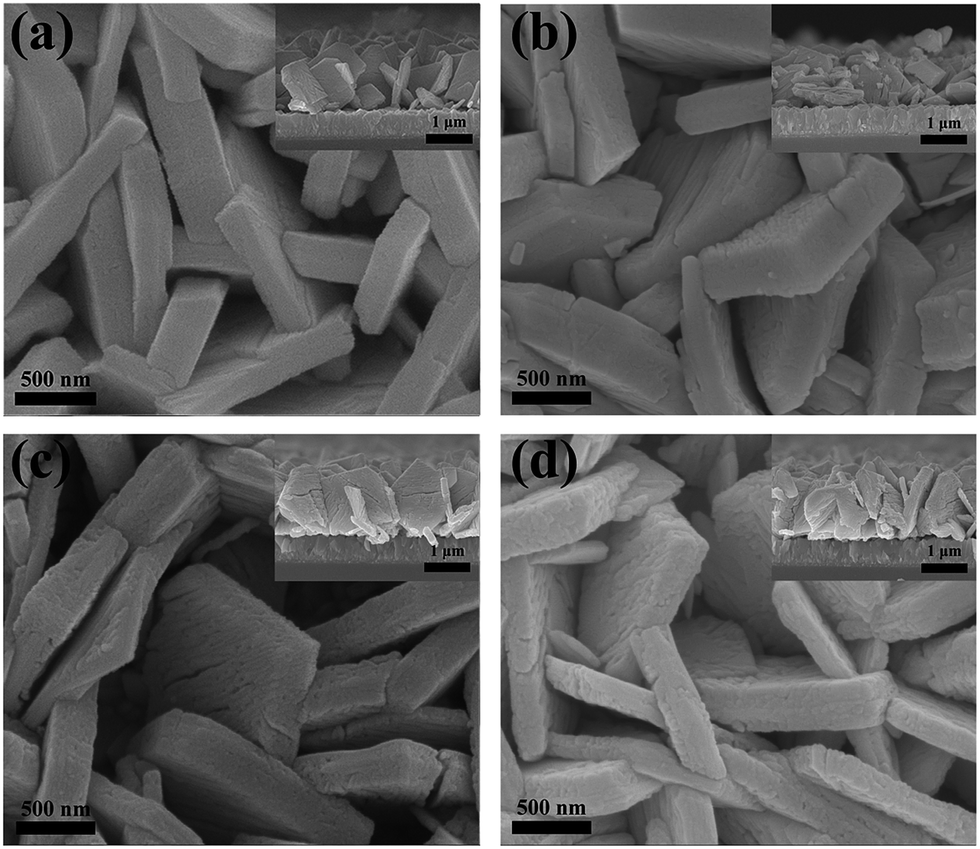 | ||
| Fig. 2 SEM images of WO3 films (a), WO3/C3N4-1.14% (b), 2.13% (c), 3.05% (d). | ||
To observe the crystal structures and binding mode of g-C3N4 nanoparticles and WO3 platelets, TEM image of g-C3N4/WO3 heterojunction film is presented in Fig. 3. As can be seen in Fig. 3a, the base material WO3 has plate-like structures. We can easily recognize the g-C3N4 nanoparticle from the HRTEM image (Fig. 3b), which was well attached to the WO3 platelet. The lattice d-spacing of 3.749 Å, corresponding to the (020) plane was examined by Fourier transform for the WO3 platelet, and lattice d-spacing of 3.274 Å (002) for the g-C3N4 nanoparticle.
 | ||
| Fig. 3 TEM image (a) and HRTEM image (b) of g-C3N4/WO3 films. | ||
The surface chemical composition of g-C3N4/WO3 heterojunction film was detected by XPS. The overall XPS survey spectrum of the sample is shown in Fig. 4a, which shows the existence of all the elements of g-C3N4 and WO3. It is observed that W 4f7/2 and W 4f5/2 peaks are located at 35.3 eV and 37.4 eV, respectively (Fig. 4b), which is consistent with the W6+.52 The O 1s main peak in Fig. 4c was dissociated into two peaks: W–O (530.1 eV) and surface hydroxide –OH (531.7 eV).31 The C 1s peaks (Fig. 4d) at 284.8 eV and 288.2 eV are ascribed to C–C and NC–N2 coordination.53 The asymmetrical N 1s peaks (Fig. 4e) can be deconvoluted into three characteristic peaks: triazine rings (C–N–C, 399.4 eV), tertiary nitrogen (N–C3, 400.1 eV) and amino functions (N–H, 401.6 eV).54 Simultaneously, the potentials for top of the valence band (VB) for the samples were estimated (Fig. 4f). This result indicates that the potential for the top of VB for g-C3N4 was 0.8 eV more negative than that for WO3. Based on this and optical absorption spectra, the energy level structure can be obtained.
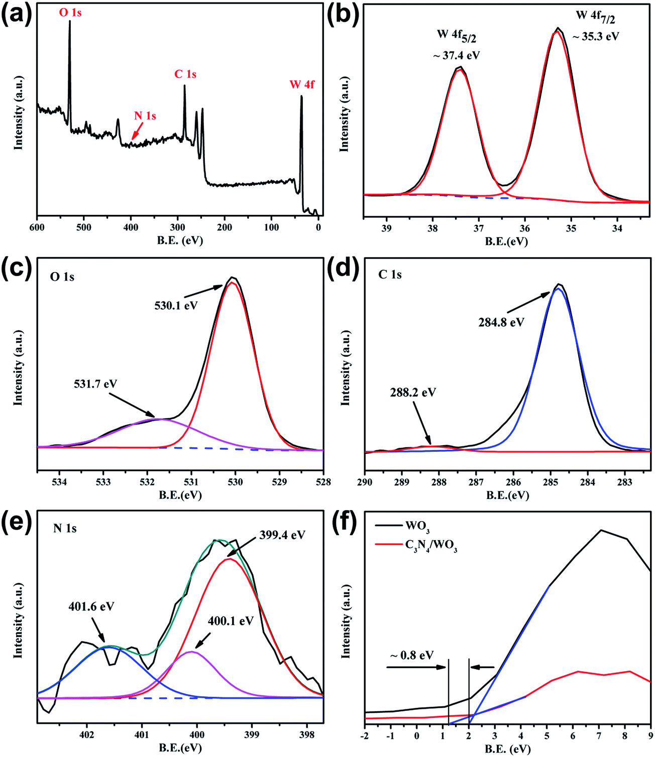 | ||
| Fig. 4 XPS spectra of the g-C3N4/WO3 heterojunction films: (a) the survey scan; (b) W 4f; (c) O 1s; (d) C 1s; (e) N 1s; (f) VB-XPS spectra. | ||
Fig. 5a depicts the UV-vis diffuse reflectance spectra of the pure WO3 and g-C3N4/WO3 films. For all samples, the optical absorption edge is estimated to be around 450 nm, which is consistent with the reported literature.45 The Tauc plot of band gap is shown in Fig. 5b. The band gap energy (Eg) can be calculated by the Tauc formula:55
| (αhν)1/2 = A(hν − Eg) | (1) |
 | ||
| Fig. 5 UV-vis absorption spectra (a) and the band gap determination (b) of WO3 and g-C3N4/WO3 heterojunction films. | ||
3.2 Photoelectrochemical study of as-prepared photoanodes
To investigate the PEC activity for water splitting, the g-C3N4/WO3 and WO3 films were used as photoanodes by linear sweep voltammetry (LSV) measurement under visible irradiation (Fig. 6). In Fig. 6a, the LSV was carried out with a potential range from 0.6 V to 2.0 V (vs. RHE) at a scan rate of 20 mV s−1. It can be seen that the g-C3N4/WO3 heterojunction films exhibited higher photocurrent density than the pure WO3 film under illumination. At +2.0 V (vs. RHE), the photocurrent density of g-C3N4/WO3 films with different content of g-C3N4 (1.14%, 2.13% and 3.05%) were 1.54, 2.10 and 1.48 mA cm−2, while the untreated WO3 film had a photocurrent of 0.78 mA cm−2. The g-C3N4/WO3 (2.13%) heterojunction film showed the best PEC performance, which exhibited an almost 3-fold higher photocurrent density than the pure WO3 film. While the weight of urea increases to 10 g, the photocurrent density decreased. This probably because excess g-C3N4 may result in the electrons in CB of WO3 combine with the holes in VB of g-C3N4 at the interface of the composite.45 | ||
| Fig. 6 Photocurrent densities (a) and IPCE (b) of the photoelectrodes. | ||
To quantitatively assess the PEC activity at different wavelength, the incident-photon-to-current-efficiencies (IPCE) measurements of the photoelectrodes were performed at a bias voltage of 1.6 V (vs. RHE) in 0.2 M Na2SO4 solution, and the results are shown in Fig. 6b. IPCE can be calculated by IPCE = (1240I)/(λJlight),58 where I (mA cm−2), λ (nm) and Jlight (mW cm−2) are the photocurrent density, the wavelength and power density of incident light, separately. All the photoelectrodes presented strong photo responses at 300–460 nm. Compared to the pure WO3 film, the g-C3N4/WO3 heterojunction film exhibited larger IPCE values. The formation of heterojunction resulted in enhanced efficiencies over the whole photo response region. The highest IPCE value of WO3 film at ∼350 nm increased from 25.1% to 53.1% after depositing g-C3N4 nanoparticles, which is in accordance with the photocurrent results.
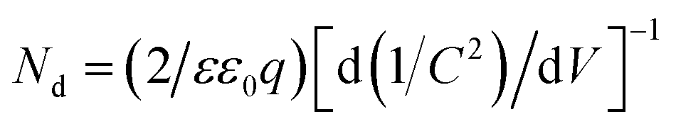
To elucidate the strong correlation between formation of heterojunction and the enhanced PEC performance, Mott–Schottky analysis under the visible light irradiation with a frequency of 1 kHz in a 0.2 M Na2SO4 aqueous electrolyte was carried out. The results were presented in Fig. 7. All the photoelectrodes exhibited an n-type characteristic with the positive slop, which reflected that electrons served as the majority carriers. The carrier density (Nd/cm−3) can be calculated based on the Mott–Schottky equation:59
 | (2) |
 | ||
| Fig. 7 Mott–Schottky plots of (a) WO3 and (b) g-C3N4/WO3 films. | ||
The flat band potential (Vfb) at semiconductor/electrolyte interface can also be estimated by the Mott–Schottky equation:63
| 1/C2 = (2/εε0qNd)[(V − Vfb) − kT/q] | (3) |
By extrapolating the X-intercepts of the liner region in Mott–Schottky plots, Vfb of pure WO3 plate film was found to be ∼0.67 V vs. RHE, while that of g-C3N4/WO3 heterojunction film shifted to the negative direction from 0.67 V to 0.52 V (1.14%), 0.43 V (2.13%) and 0.35 V (3.05%) vs. RHE. The typical n-type g-C3N4 has the values of Vfb around −1.13 V.64 The negative shifts of the flat band were prominent, which indicated fewer barrier for oxygen evolution, larger accumulation of electrons and better separation of photo-generated carriers in the heterojunction film.65
To investigate the kinetics of the electrochemical reaction process at the electrode surface, the capacitance and resistance of the electrode materials which indicated the separation efficiency between the photo-generated electrons and holes,66 electrochemical impedance spectroscopy (EIS) of the as-prepared electrodes were measured under the visible light illumination at an AC frequency varying from 10 kHz to 100 mHz with a 1.4 V (vs. RHE) bias. Fig. 8a shows the typical Nyquist curve of the g-C3N4/WO3 heterojunction film and pure WO3 film. The Nyquist plot collected for WO3 and g-C3N4/WO3 films, exhibits one semicircle, suggesting that the faradaic charge transfer is the limiting step for the electrochemical reaction process in the electrode surface.59 The apparent difference can be observed that the EIS Nyquist curve of g-C3N4/WO3 film has a smaller circular radius than that of the pure WO3 film. According to the equivalent circuit which consists of solution resistance (R1), charge transfer resistance (R2) in parallel to the constant phase element (CPE1),67 the resistance R1, R2 were calculated to be 58.6 Ω, 236.8 Ω for the g-C3N4/WO3 film (2.13%), and 43.6 Ω, 1028.1 Ω for the WO3 film, respectively. This suggests that g-C3N4/WO3 film owns lower charge transfer resistance than WO3 film, corresponding to the better PEC performance. Meanwhile, Bode plots are transformed by the Z-view software as presented in Fig. 8b. The frequency peak (fp) of g-C3N4/WO3 film shifts from 67.7 Hz to 13.8 Hz compared with pure WO3. From this, the lifetime of electrons for recombination with a time constant (τ) is calculated by the formula:68
| τ = 1/(2πfp) | (4) |
 | ||
| Fig. 8 EIS Nyquist plots (a) and Bode plots (b) of WO3 and g-C3N4/WO3 films. | ||
On the basis of the energy level structure reported in previous literature,44,45,69 the schematic diagram for the g-C3N4/WO3 heterojunction film which illustrates the photo-generated carriers transport process is shown in Fig. 9. The CB and VB positions of WO3 are about +0.74 and +3.4 V, respectively,70 while those of g-C3N4 are about −1.13 and +1.57 V, respectively.71 Under the artificial solar light irradiation, both g-C3N4 and WO3 would be excited and produce the photo-induced electrons and holes, simultaneously. In the g-C3N4/WO3 heterojunction film, the photo-generated electrons of g-C3N4 can easily migrate to the CB of WO3, and then transfer to the back-contacted FTO substrate through a direct pathway provided by the platelets. Similarly, the photo-generated holes of WO3 will transfer to the VB of g-C3N4 and take part in the oxygen evolution reaction. Thus, the photo-generated electron–hole pairs will be separated at the interface of the heterojunction and the long-lived charge carriers will be induced, resulting in enhancing PEC performance.
 | ||
| Fig. 9 Depiction of the charge separation process in the g-C3N4/WO3 films. | ||
4. Conclusions
In summary, the g-C3N4 nanoparticles on the surface of two-dimensional (2D) WO3 plates arrays were successfully in situ synthesized through a simple dipping-annealing method. The g-C3N4/WO3 (2.13 at%) heterojunction film possessed the best PEC performance, and exhibited a 3-fold higher photocurrent density with respect to the pure WO3 film under visible light irradiation. After introducing g-C3N4 nanoparticles, the g-C3N4/WO3 heterojunction film owned larger carriers density, longer electrons' lifetime and more negative flat band potential. The enhanced PEC performance of the heterojunction film is mainly attributed to the effective separation of photo-generated electron–hole pairs at the interface and the improvement of electrons' lifetime. Besides that, the 2D ordered structures are favorable for highly efficient and directional transport of electrons. The in situ formation of heterojunction facilitates the interfacial contact of two components, and optimizes the charge separation. The study demonstrates that in situ formation of heterojunction structure and low-dimensional morphological structure are expected to provide a promising electrode for photoelectric catalysis.Acknowledgements
This study was supported by the National Nature Science Foundation of China (51304253) and Hunan Provincial Innovation Foundation (CX2015B266).References
- T. Hisatomi, J. Kubota and K. Domen, Chem. Soc. Rev., 2014, 43, 7520–7535 RSC.
- A. Fujishima and K. Honda, Nature, 1972, 238, 37–38 CrossRef CAS PubMed.
- H. L. Zhu, D. M. Chen, D. Yue, Z. H. Wang and H. Ding, J. Nanopart. Res., 2014, 16, 1–10 CAS.
- Y. V. Kolen'Ko, A. Garshev, B. Churagulov, S. Boujday, P. Portes and C. Colbeau, J. Photochem. Photobiol., A, 2005, 172, 19–26 CrossRef PubMed.
- W. Smith, A. Wolcott, R. C. Fitzmorris, J. Z. Zhang and Y. Zhao, J. Mater. Chem., 2011, 21, 10792–10800 RSC.
- M. Yagi, S. Maruyama, K. Sone, K. Nagai and T. Norimatsu, J. Solid State Chem., 2008, 181, 175–182 CrossRef CAS PubMed.
- D. D. Qin, C. L. Tao, S. A. Friesen, T. H. Wang, O. K. Varghese, N. Z. Bao, Z. Y. Yang, T. E. Mallouk and C. A. Grimes, Chem. Commun., 2012, 48, 729–731 RSC.
- A. Memar, W. R. W. Daud, S. Hosseini, E. Eftekhari and L. J. Minggu, Sol. Energy, 2010, 84, 1538–1544 CrossRef CAS PubMed.
- Z. Jiao, J. Wang, L. Ke, X. W. Sun and H. V. Demir, ACS Appl. Mater. Interfaces, 2011, 3, 229–236 CAS.
- M. Butler, R. Nasby and R. K. Quinn, Solid State Commun., 1976, 19, 1011–1014 CrossRef CAS.
- F. Amano, D. Li and B. Ohtani, Chem. Commun., 2010, 46, 2769–2771 RSC.
- G. F. Cai, X. L. Wang, D. Zhou, J. H. Zhang, Q. Q. Xiong, C. D. Gu and J. P. Tu, RSC Adv., 2013, 3, 6896–6905 RSC.
- W. Mu, X. Xie, X. Li, R. Zhang, Q. Yu, K. Lv, H. Wei and Y. Jian, RSC Adv., 2014, 4, 36064–36070 RSC.
- W. Mu, X. Xie, R. Zhang, X. Li, K. Lv, Q. Yu, H. Wei and Y. Jian, RSC Adv., 2014, 4, 59740–59746 RSC.
- W. Li, J. Li, X. Wang and Q. Chen, Appl. Surf. Sci., 2012, 263, 157–162 CrossRef CAS PubMed.
- M. Takeuchi, Y. Shimizu, H. Yamagawa, T. Nakamuro and M. Anpo, Appl. Catal., B, 2011, 110, 1–5 CrossRef CAS PubMed.
- Y. Liu, Y. Li, W. Li, S. Han and C. Liu, Appl. Surf. Sci., 2012, 258, 5038–5045 CrossRef CAS PubMed.
- M. K. Aminian and M. Hakimi, Catal. Sci. Technol., 2014, 4, 657–664 Search PubMed.
- H. Qi, J. Wolfe, D. Wang, H. J. Fan, D. Fichou and Z. Chen, Nanoscale, 2014, 6, 13457–13462 RSC.
- J. Y. Zheng, G. Song, J. Hong, T. K. Van, A. U. Pawar, D. Y. Kim, C. W. Kim, Z. Haider and Y. S. Kang, Cryst. Growth Des., 2014, 14, 6057–6066 CAS.
- J. R. Navarro, A. Mayence, J. Andrade, F. Lerouge, F. Chaput, P. Oleynikov, L. Bergstrom, S. Parola and A. Pawlicka, Langmuir, 2014, 30, 10487–10492 CrossRef CAS PubMed.
- C. C. Huang, Y. H. Su, S. L. Tu, T. W. Yung, Y. F. Ke and T. H. Chen, Mater. Res. Innovations, 2014, 18, 72–75 Search PubMed.
- F. Amano, D. Li and B. Ohtani, J. Electrochem. Soc., 2011, 158, K42–K46 CrossRef CAS PubMed.
- F. Amano, M. Tian, G. Wu, B. Ohtani and A. Chen, ACS Appl. Mater. Interfaces, 2011, 3, 4047–4052 CAS.
- J. Su, X. Feng, J. D. Sloppy, L. Guo and C. A. Grimes, Nano Lett., 2011, 11, 203–208 CrossRef CAS PubMed.
- M. Law, L. E. Greene, J. C. Johnson, R. Saykally and P. Yang, Nat. Mater., 2005, 4, 455–459 CrossRef CAS PubMed.
- K. H. Ng, H. A. Kadir, L. J. Minggu and M. B. Kassim, Mater. Sci. Forum, 2013, 756, 219–224 CrossRef.
- C. X. Kronawitter, L. Vayssieres, S. H. Shen, L. J. Guo, D. A. Wheeler, J. Z. Zhang, B. R. Antoun and S. S. Mao, Energy Environ. Sci., 2011, 4, 3889–3899 CAS.
- S. A. K. Leghari, S. Sajjad and J. Zhang, RSC Adv., 2013, 3, 15354–15361 RSC.
- C. W. Lai and S. Sreekantan, Int. J. Hydrogen Energy, 2013, 38, 2156–2166 CrossRef CAS PubMed.
- M. M. Mohamed, S. A. Ahmed and K. S. Khairou, Appl. Catal., B, 2014, 150, 63–73 CrossRef PubMed.
- W. Luo, T. Yu, Y. Wang, Z. Li, J. Ye and Z. Zou, J. Phys. D: Appl. Phys., 2007, 40, 1091–1096 CrossRef CAS.
- L. Yin, D. Chen, M. Feng, L. Ge, D. Yang, Z. Song, B. Fan, R. Zhang and G. Shao, RSC Adv., 2015, 5, 328–337 RSC.
- X. Wang, K. Maeda, A. Thomas, K. Takanabe, G. Xin, J. M. Carlsson, K. Domen and M. Antonietti, Nat. Mater., 2009, 8, 76–80 CrossRef CAS PubMed.
- Z. Zhao, Y. Sun and F. Dong, Nanoscale, 2015, 7, 15–37 RSC.
- M. Groenewolt and M. Antonietti, Adv. Mater., 2005, 17, 1789–1792 CrossRef CAS PubMed.
- S. C. Yan, Z. S. Li and Z. G. Zou, Langmuir, 2009, 25, 10397–10401 CrossRef CAS PubMed.
- Y. Yuan, W. Xu, L. Yin, S. Cao, Y. Liao, Y. Tng and C. Xue, Int. J. Hydrogen Energy, 2013, 38, 13159–13163 CrossRef CAS PubMed.
- F. Dong, Y. Sun, L. Wu, M. Fu and Z. Wu, Catal. Sci. Technol., 2012, 2, 1332–1335 CAS.
- Y. He, J. Cai, T. Li, Y. Wu, Y. Yi, M. Luo and L. Zhao, Ind. Eng. Chem. Res., 2012, 51, 14729–14737 CrossRef CAS.
- Y. Wang, X. Bai, C. Pan, J. He and Y. Zhu, J. Mater. Chem., 2012, 22, 11568 RSC.
- M. Ou, Q. Zhong and S. Zhang, J. Sol-Gel Sci. Technol., 2014, 72, 443–454 CrossRef CAS.
- H. Xing, H. Ma, Y. Fu, M. Xue, X. Zhang, X. Dong and X. Zhang, Mater. Technol., 2015, 30, 122–127 CrossRef CAS PubMed.
- Y. Zang, L. Li, Y. Zuo, H. Lin, G. Li and X. Guan, RSC Adv., 2013, 3, 13646 RSC.
- H. Katsumata, Y. Tachi, T. Suzuki and S. Kaneco, RSC Adv., 2014, 4, 21405 RSC.
- Y. Bai, H. Yu, Z. Li, R. Amal, G. Q. Lu and L. Wang, Adv. Mater., 2012, 24, 5850–5856 CrossRef CAS PubMed.
- M. Fang, H. Jia, W. He, Y. Lei, L. Zhang and Z. Zheng, Phys. Chem. Chem. Phys., 2015, 17, 13531–13538 RSC.
- Y. Peng, Q. Chen, D. Wang, H. Zhou and A. Xu, CrystEngComm, 2014, 17, 569–576 RSC.
- J. Yang, W. Li, J. Li, D. Sun and Q. Chen, J. Mater. Chem., 2012, 22, 17744 RSC.
- Y. Hou, F. Zuo, A. Dagg and P. Feng, Nano Lett., 2012, 12, 6464–6473 CrossRef CAS PubMed.
- L. Ge, C. C. Han and J. Liu, Appl. Catal., B, 2011, 108, 100–107 CrossRef PubMed.
- S. Han, J. Li, X. Chen, Y. Huang, C. Liu, Y. Yang and W. Li, Int. J. Hydrogen Energy, 2012, 37, 16810–16816 CrossRef CAS PubMed.
- S. Wang, C. Li, T. Wang, P. Zhang, A. Li and J. Gong, J. Mater. Chem. A, 2014, 2, 2885–2890 CAS.
- H. Katsumata, T. Sakai, T. Suzuki and S. Kaneco, Ind. Eng. Chem. Res., 2014, 53, 8018–8025 CrossRef CAS.
- A. Subrahmanyam and A. Karuppasamy, Sol. Energy Mater. Sol. Cells, 2007, 91, 266–274 CrossRef CAS PubMed.
- W. Z. Li, J. Li, X. Wang, J. Ma and Q. Y. Chen, Int. J. Hydrogen Energy, 2010, 35, 13137–13145 CrossRef CAS PubMed.
- W. Z. Li, J. Li, X. Wang, J. Ma and Q. Y. Chen, Appl. Surf. Sci., 2010, 256, 7077–7082 CrossRef CAS PubMed.
- Y. Ling, G. Wang, D. A. Wheeler, J. Z. Zhang and Y. Li, Nano Lett., 2011, 11, 2119–2125 CrossRef CAS PubMed.
- G. Wang, Y. Ling, D. A. Wheeler, K. E. George, K. Horsley, C. Heske, J. Z. Zhang and Y. Li, Nano Lett., 2011, 11, 3503–3509 CrossRef CAS PubMed.
- E. Salje and K. Viswanathan, Acta Crystallogr., Sect. A: Cryst. Phys., Diffr., Theor. Gen. Crystallogr., 1975, 31, 356–359 CrossRef.
- L. Su, L. Zhang, J. Fang, M. Xu and Z. Lu, Sol. Energy Mater. Sol. Cells, 1999, 58, 133–140 CrossRef CAS.
- R. Sivakumar, A. Moses Ezhil Raj, B. Subramanian, M. Jayachandran, D. Trivedi and C. Sanjeeviraja, Mater. Res. Bull., 2004, 39, 1479–1489 CrossRef CAS PubMed.
- N. T. Hahn and C. B. Mullins, Chem. Mater., 2010, 22, 6474–6482 CrossRef CAS.
- L. Huang, Y. Li, H. Xu, Y. Xu, J. Xia, K. Wang, H. Li and X. Cheng, RSC Adv., 2013, 3, 22269–22279 RSC.
- J. Zhang, J. H. Bang, C. Tang and P. V. Kamat, ACS Nano, 2009, 4, 387–395 CrossRef PubMed.
- W. D. Zhang, L. C. Jiang and J. S. Ye, J. Phys. Chem. C, 2009, 113, 16247–16253 CAS.
- S. K. Pilli, T. G. Deutsch, T. E. Furtak, L. D. Brown, J. A. Turner and A. M. Herring, Phys. Chem. Chem. Phys., 2013, 15, 3273–3278 RSC.
- M. Zhou, J. Bao, Y. Xu, J. Zhang, J. Xie, M. Guan, C. Wang, L. Wen, Y. Lei and Y. Xie, ACS Nano, 2014, 8, 7088–7098 CrossRef CAS PubMed.
- A. T. Doan, X. D. N. Thi, P. H. Nguyen, V. N. N. Thi, S. J. Kim and V. Vo, Bull. Korean Chem. Soc., 2014, 35, 1794–1798 CrossRef CAS.
- M. Miyauchi, Phys. Chem. Chem. Phys., 2008, 10, 6258–6265 RSC.
- S. Yan, S. Lv, Z. Li and Z. Zou, Dalton Trans., 2010, 39, 1488–1491 RSC.
Footnote |
| † Electronic supplementary information (ESI) available. See DOI: 10.1039/c5ra11464k |
| This journal is © The Royal Society of Chemistry 2015 |