DOI:
10.1039/C5RA10972H
(Paper)
RSC Adv., 2015,
5, 71519-71528
Synthesis and properties of novel diisopropyl-functionalized polyglycolide–PEG copolymers†
Received
9th June 2015
, Accepted 11th August 2015
First published on 11th August 2015
Abstract
Because of the importance of glycolide-based polymers in materials and medical applications, we have synthesized alternative novel thermosensitive biomaterials to be possible candidates instead of the well-known polymers. The synthesis of L-diisopropyl glycolide monomer was performed in two steps, novel PEG based poly(diisopropyl glycolide) diblock and triblock copolymers (MePEG–PDIPG, PDIPG–PEG–PDIPG) were synthesized with high conversions by ring opening polymerization. The molar mass distributions of the copolymers were very narrow, and the NMR measurements confirmed that no racemization had occurred in the polymer synthesis even at 180 °C. Gel–sol experiments were conducted after each component’s length in MePEG–PDIPG and PDIPG–PEG–PDIPG was adjusted with care during the synthesis. The polymer showed a sol property at 42–45 °C, which is suitable for injection and a gel property at body temperature. After loading paclitaxel into gels effectively, the anticancer drug showed prolonged release (60 days). SEM analysis confirmed that the gels turned into a more porous structure due to drug release.
1. Introduction
Drug delivery can be simply expressed as the delivery of various types of drugs to humans or animals using proper formulations. Drug delivery systems have now become a multi-billion dollar industry with the versatile application of many interdisciplinary sciences.1,2 Also, it will undoubtedly continue to be one of the important research areas in the future. Thermosensitive biodegradable polymers for use as drug delivery systems have been one of the most preferred polymers in the biomedical field in the last decade because they can easily be subjected to a physical change with external heat due to their thermosensitivity, and because no effort is needed to remove them from the body due to their biodegradability.3–5 PEG based copolymers of poly(lactide), poly(glycolide), and poly(lactide-co-glycolide) (PLGA) are one of the most preferred thermosensitive biodegradable polymers in the biomedical field.6–9 In contrast, there have been a few reports describing the use of symmetric glycolic acid derivatives as monomers or comonomers for the preparation of the glycolide family of polymers.10–17 Novel versatile biomaterials are needed day after day for the growing pharmaceutical and biomedical industry. In particular, poly(diisopropyl glycolide) may be considered as a highly promising material for those industries in the future due to its enhanced properties. There are only a few studies related to the homopolymer of diisopropyl glycolide in the literature.18–22 The synthesis of the poly(diisopropyl glycolide) homopolymer was performed, and the conformational aspects were evaluated by means of optical rotatory dispersion (ORD) and circular dichroism (CD).18,19 Also, homopolymerization of poly(diisopropyl glycolide) was examined by varying the catalyst, temperature, and time, and the optical purity of the polymers was investigated.20,21 The polymerization kinetics and spectroscopic and thermal properties of poly(diisopropyl glycolide) were also studied.22 In the present work, novel PEG-based diblock and triblock copolymers of poly(diisopropyl glycolide) were synthesized in order to give a thermosensitive property to these PDIPG homopolymers for applications in drug delivery systems. Then, the gel–sol transition temperature was adjusted by changing the length of each component. Aqueous solutions of these copolymers were sols at around 42–45 °C, and then they were loaded with bioactive molecules. After fast cooling to body temperature, the loaded copolymer formed a gel that could serve as a sustained-release matrix for medicines. Homogenous drug loading and the sustained release of paclitaxel over 60 days were observed successfully.
2. Experimental section
Materials
L-Valine (98%), sodium nitrite (NaNO2), p-toluene sulfonic acid monohydrate (PTSA) (98.5%), diethyl ether (99.5%), toluene (99.7%), dichloromethane (99%), and methanol (99.9%) were purchased from Sigma-Aldrich (Germany) and used without further purification except when mentioned specifically. MePEG-2000 homopolymer (Fluka), PEG-2000 homopolymer (Sigma), and tin(II) 2-ethylhexanoate (Sn(Oct)2) (Aldrich, 95%) were used in the syntheses of the di- and triblock copolymers. The paclitaxel anticancer drug (Alfa Aesar 99.5%) was used as received. The acetonitrile (Sigma-Aldrich, 99.9%) mobile phase was filtered out with a filtration system prior to use.
Characterization
1H- and 13C-NMR experiments were carried out with a Bruker Avance DPX 400 and a Bruker Avance III 500 MHz for the structural analysis and for the determination of the number average molecular weight of the copolymers. The ATR-FTIR spectra of samples were recorded using an ATR Bruker-Tensor 27 spectrometer between 600 and 4000 cm−1. The elemental analysis was carried out on a Costech Elemental Combustion System (ECS 4010) elemental analyzer. Analysis of the copolymers was carried out with gel permeation chromatography (GPC) at 30 °C on a Shimadzu prominence GPC system equipped with a RID-10A refractive index detector, a LC-20AD solvent delivery unit, a CTO-10AS column oven and a set of two columns, PSS SDV 5 μL 1000 Å and PSS SDV 5 μL 50 Å. THF (HPLC grade) was used as the mobile phase at 1.0 mL min−1. The sample concentration was adjusted to 2 mg mL−1, and the injection volume was set at 50 μL. The calibration curve was made with seven polystyrene standards having an average molecular weight range of 162 to 34
300 Da. The HPLC spectra were recorded on a 1260 Infinity Agilent with a UV detector. Data was analyzed with the Chem Station software program. A ZORBAX SB-C18 4.6 × 150 mm, 3.5 μm HPLC column was used for the analysis of the paclitaxel anticancer drug. A scanning electron microscope (Jeol 6060) was used in the microstructural investigations of the copolymer gels. The samples collected were frozen in liquid nitrogen and dried by freeze-drying (Labconco). Thermogravimetric analyses (TGA) were performed on a Perkin Elmer TGA 4000 under nitrogen atmosphere between 20 and 600 °C at a heating rate of 40 °C min−1. Differential scanning calorimeter (DSC) analyses were performed using a Mettler Toledo DSC Star System or a Perkin Elmer DSC 4000 under nitrogen atmosphere between −65 and 220 °C at a heating rate of 10 °C min−1 (or 40 °C min−1) with a double run for the determination of the melting point and glass transition temperatures of the copolymers.
Synthesis of L-2-hydroxy-3-methylbutanoic acid (2)
L-2-Hydroxy-3-methylbutanoic acid was prepared with a modified protocol.23 A solution of sodium nitrite (28.8 g, 0.42 mol) in water (200 mL) was added dropwise into a solution of L-valine (12 g, 0.1 mol) in 1.0 M H2SO4 (200 mL) over 2 h in an ice-bath, and the reaction mixture was stirred overnight at 0 °C. The progress of the reaction was followed with thin layer chromatography (TLC, silica gel, distilled water). Then, the TLC plate was treated with a ninhydrin solution that only stained L-valine. The reaction mixture was saturated with NaCl, extracted with diethyl ether (6 × 60 mL), and the combined organic extracts were dried over Na2SO4. After removing the ether under reduced pressure, the residue was recrystallized from toluene to afford pure L-2-hydroxy-3-methylbutanoic acid as a white solid (50%). 1H-NMR (400 MHz, CDCl3) δ: 0.92 (3H, d), 1.04 (3H, d), 2.09–2.2 (1H, m), 4.15 (1H, d), 6.2–8.6 (2H, b). 13C-NMR (100 MHz, CDCl3) δ: 16.1, 18.9, 32.1, 75, 179.2. ATR-FTIR (νmax/cm−1): 3413 (OH), 2972, 2935, 2880 (CH), 1702 (C
O).
Synthesis of L-3,6-diisopropyl-1,4-dioxane-2,5-dione (L-DIPG) (3)
L-3,6-Diisopropyl-1,4-dioxane-2,5-dione was synthesized by modifying the method in the literature.10 A mixture of L-2-hydroxy-3-methylbutanoic acid (25 g, 0.21 mol) and p-toluene sulfonic acid monohydrate (0.5 g, 2.5 mmol) in toluene (200 mL) was refluxed for five days in order to remove water with Dean–Stark apparatus. After the mixture was cooled, toluene was removed by rotary evaporation until a small volume of solvent remained. The toluene solution was left to crystallize at 4 °C for 3–4 hours and the resulting crystals were separated by decantation. A second crystallization was performed in order to eliminate impurities. A small volume of toluene was added to the residue. Then, it was heated at 55 °C to dissolve the crystals. The solution was kept at 4 °C for a while. The resulting crystals were collected by cold filtration and dried under vacuum to give 8.5 g (40%) of L-3,6-diisopropyl-1,4-dioxane-2,5-dione. Elemental analysis for C10H16O4 (200.23 g mol−1): Calc. C 59.98, H 8.05%; found: C 59.97, H 7.75%. 1H-NMR (400 MHz, CDCl3) δ: 1.05 (6H, d), 1.15 (6H, d), 2.44–2.55 (2H, m), 4.73 (2H, d). 13C-NMR (100 MHz, CDCl3) δ: 16, 18.7, 29.6, 79.8, 166.6. ATR-FTIR (νmax/cm−1): 2969, 2939, 2878 (CH), 1748 (C
O).
Synthesis of MePEG–PDIPG diblock copolymers
The MePEG–PDIPG diblock copolymers were synthesized in the melt via ring opening polymerization. 240 mg of MePEG-2000 (0.12 mmol), 400 mg of L-DIPG (2 mmol), and 20 mg of Sn(Oct)2 (0.05 mmol) were added to the polymerization tube. The reaction was stirred at 120 °C for 3 hours under a nitrogen atmosphere. Purification of copolymer 9 can be performed with the following method: synthesized diblock copolymer 9 was dissolved in the minimum volume needed of dichloromethane (4 mL) before it was precipitated with an excess volume of cold methanol (40 mL). Then the product 9 was dried under vacuum overnight. In a similar way, various molecular weights of the diblock copolymers (6, 7, 8, 10) could be obtained by changing the feed ratio of L-DIPG but keeping a constant molar amount of MePEG. 1H-NMR (400 MHz, CDCl3) δ: 1.05–1.09 (6H, t), 2.15–2.59 (1H, m), 3.66 (4H, s), 4.99 (1H, d). 13C-NMR (100 MHz, CDCl3) δ: 17, 18.6, 30.3, 70.6, 77, 168.8. ATR-FTIR (νmax/cm−1): 2970, 2935, 2877 (CH), 1753 (CO).
Synthesis of PDIPG–PEG–PDIPG triblock copolymers
In order to perform the synthesis of the PDIPG–PEG–PDIPG triblock copolymers, the PEG homopolymer with hydroxyl groups at both ends was used instead of the MePEG homopolymer which has a methoxy group on one end and a hydroxyl group on the other end. A series of triblock copolymers were obtained by changing the molar amount of L-DIPG but keeping a constant molar amount of PEG.
Determination of the thermosensitivity properties of the copolymers
The thermosensitivity of the copolymers was determined by inverting a vial at different temperatures using a controlled water bath.4 Firstly, various amounts of the copolymers were mixed with distilled water to prepare a series of suspensions in 1.5 mL vials. All of the suspensions were vortexed to get a homogenous mixture if possible. After the homogenous mixtures were kept at 4 °C for 30 min, they were immersed in a temperature controlled water bath. The gel–sol transition temperatures of the copolymers were examined between 4 and 80 °C with 2 °C increments. The vials were kept in water baths for 2 min at each temperature before inverting. The critical gel–sol transition temperature was determined as the temperature at which the gel turned into a sol form immediately after inverting the tube.
Preparation of phosphate buffer solution (PBS)
2 g of NaCl, 0.05 g of KCl, 0.36 g of Na2HPO4, and 0.06 g of KH2PO4 were dissolved in distilled water. Then, the pH was adjusted to 7.4 with dilute HCl, and the volume of the mixture was brought to 250 mL with distilled water.
Paclitaxel loading into copolymer gel
The anticancer drug paclitaxel was used to determine the release behavior of the synthesized copolymers. Paclitaxel was loaded into the produced copolymer gels effectively at 1.0%. One of the drug loaded copolymer gels was prepared with the following method: 2 mg paclitaxel, 200 mg PDIPG–PEG–PDIPG 12 triblock copolymer, and 300 μL distilled water were added to the vial, and the sample was vortexed for 2 min at room temperature to obtain a drug loaded gel. Other drug-loaded gels were prepared with a similar method.
Drug release studies
1 mL of 2.0% Tween 80 in PBS at pH = 7.4 was added to the upper side of the drug loaded gels for the drug release experiments. These samples were inserted into an incubator, and they were shaken at a constant speed of 200 rpm at 37 °C. At different time intervals, 1 mL of the release medium was taken from the vials and replaced with 1 mL of fresh medium. The amount of paclitaxel released into the medium was measured using HPLC with a UV detector.
3. Result and discussion
General aspects of the polyglycolide family polymers
The most widely investigated polymers in the controlled release drug delivery of therapeutic agents are aliphatic polyesters such as poly(D,L-lactide) (PLA) and poly(D,L-lactide-coglycolide) (PLGA) because of their predictable degradation kinetics, biocompatibility, ease of fabrication and regulatory approval by the Food and Drug Administration (FDA). These two polymers are relatively hydrophobic polyesters, they are not stable in a damp medium and they biodegrade into non-toxic by-products (lactic acid, glycolic acid, CO2, and H2O). Substituents in the glycolide monomer drastically change its physiochemical properties. When the hydrogen substituent (glycolide) is replaced with a methyl group (lactide) in the cyclic dimer monomer, the bio-applications are considerably affected due to the change in hydrophobicity, crystallinity, biodegradation, and the mechanical and thermal properties. Here we especially examined the physicochemical properties when one hydrogen substituent was changed with an isopropyl group in the cyclic dimer. It is very important and practical in terms of an industrial perspective to synthesize the PEG based poly(L-diisopropyl glycolide) polymer in bulk from valine, which is a basic and cheap amino acid. More hydrophobic polymers due to the isopropyl group may increase the hydrophobic drug loading for application in various drug delivery systems. On the other hand, it is also very convenient to prepare the thermosensitive PDIPG copolymer with hydrophilic PEG to control the hydrophobic/hydrophilic balance. Thus, the solution of the PDIPG–PEG block copolymer was a liquid at around 42–45 °C for the injection and a gel at body temperature for the release of the bioactive drug into its surroundings. These controlled release biocompatible hydrogel systems provide a special advantage in terms of local drug delivery; e.g., around a solid tumor site. These novel biodegradable injectable controlled release systems could be good candidates for the treatment of solid brain tumors or solid tumors which are just under the skin. They are good candidates for the treatment of brain tumors because the blood barrier limits drug release or causes excess drug release.4 Also, side effects can be greatly diminished with the local therapy of drug loaded gels like PDIPG–PEG due to there being no systematic circulation of the drug in the body.4
Monomer synthesis
L-2-Hydroxy-3-methylbutanoic acid 2 was synthesized from L-2-amino-3-methylbutanoic acid 1 by using sodium nitrite in the presence of sulfuric acid for 24 hours at 0 °C.23 Conversion of the free amine to an alcohol group in compound 2 was easily confirmed by the appearance of a new –OH peak at 3413 cm−1 in the ATR-FTIR spectrum. Then, the synthesis of L-3,6-diisopropyl-1,4-dioxane-2,5-dione 3 was carried out from the condensation of compound 2 at reflux temperature for 5 days in the presence of p-toluenesulfonic acid monohydrate in toluene (Scheme 1).10 It was found that time was an important factor in the synthesis of the L-diisopropyl glycolide monomer. The GPC and FTIR analyses confirmed that there was still L-2-hydroxy-3-methylbutanoic acid 2, the starting material, after 24 hours while the monomer started to turn into the oligomeric species after a longer reaction time, especially after more than 5 days. The formation of the L-diisopropyl glycolide monomer was proven by the shifting of the carbonyl stretching of the carboxylic acid group in compound 2 from 1702 cm−1 to 1748 cm−1 in the cyclic monomer. Also, the complete disappearance of the OH peak at 3413 cm−1 in the ATR-FTIR spectrum proved the synthesis of the ring-closed compound. Similar results were observed by proton and carbon NMR, as well. The CH proton in compound 2 shifted from 4.15 ppm to 4.73 ppm in the monomer. Ester formation was also observed in 13C-NMR by the shift from 179.2 ppm (acid carbonyl group) to 166.6 ppm (ester carbonyl group). Here, we presented the detailed synthesis and characterization analysis of the L-DIPG monomer, which differs from the literature.18–22 All spectra for the characterizations of compound 2 and 3 can be found in the ESI† section.
 |
| | Scheme 1 Synthesis of L-3,6-diisopropyl-1,4-dioxane-2,5-dione. | |
Polymer synthesis
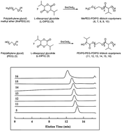
MePEG–PDIPG diblock and PDIPG–PEG–PDIPG triblock copolymers were obtained from the ring-opening polymerization of the diisopropyl glycolide monomer with poly(ethylene glycol) methyl ether (MePEG-2000) or poly(ethylene glycol) (PEG-2000) under a nitrogen atmosphere (Scheme 2). All of the polymerization reactions were performed in bulk at 120 °C under melt conditions except for copolymers 10 and 16. Both of these copolymers needed higher temperature (180 °C) to melt due to the high molar amount of hydrophobic component (Table 1). Stannous octoate (Sn(Oct)2) was selected as the catalyst due to the almost complete conversions which could be obtained and low toxicity when compared to other heavy metal salts.24,25 The molecular weight of MePEG was purposely selected as 2000 g mol−1 for the synthesis of all of the copolymers because higher molecular weight PEG (above ∼10k) is inappropriate for filtration through the membrane of a human kidney due to the large hydrodynamic radius of PEG in the aqueous phase.3
 |
| | Scheme 2 Synthesis of the MePEG–PDIPG and PDIPG–PEG–PDIPG diblock and triblock copolymers. | |
Table 1 Conditions and characterization of the diblock and triblock copolymers
| Conditions for synthesis of copolymers |
Characterization of diblock and triblock copolymers |
| ID |
Copolymer |
PEG (mmol) |
L-DIPG (mmol) |
Sn(Oct)2 (mmol) |
Time (hour) |
Temperature (°C) |
Mwa (g mol−1) |
Mna (g mol−1) |
Mnb (g mol−1) |
Theoretical |
Mw/Mna |
% Conv.b |
RUb of DIPG |
L-DIPG/Sn(Oct)2 |
| Determined by GPC. Determined by the 1H-NMR spectrum (the conversion was calculated using the signal from the CH (δ 4.7 ppm) of the unreacted monomer, and the CH (δ 4.9 ppm) of the polymer), RU: repeating unit. |
| 6 |
MePEG–PDIPG |
0.12 |
0.25 |
0.05 |
3 |
120 |
3050 |
2930 |
2670 |
2420 |
1.04 |
97.3 |
7 |
5 |
| 7 |
0.75 |
3 |
120 |
3880 |
3670 |
3120 |
3250 |
1.06 |
97.8 |
11 |
15 |
| 8 |
1.5 |
3 |
120 |
4740 |
4380 |
4380 |
4500 |
1.08 |
99.9 |
24 |
30 |
| 9 |
2 |
3 |
120 |
5100 |
4730 |
5060 |
5330 |
1.08 |
99.5 |
30 |
40 |
| 10 |
10 |
8 |
180 |
18 900 |
14 430 |
16 370 |
18 670 |
1.31 |
95.6 |
144 |
200 |
| 11 |
PDIPG–PEG–PDIPG |
0.12 |
0.6 |
0.05 |
3 |
120 |
3960 |
3800 |
3310 |
3000 |
1.04 |
96.5 |
13 |
12 |
| 12 |
0.8 |
3 |
120 |
4300 |
4100 |
3470 |
3330 |
1.05 |
97.4 |
15 |
16 |
| 13 |
1 |
3 |
120 |
4690 |
4450 |
3710 |
3670 |
1.05 |
97.7 |
17 |
20 |
| 14 |
1.5 |
3 |
120 |
5470 |
5190 |
4650 |
4500 |
1.05 |
99.6 |
27 |
30 |
| 15 |
3 |
3 |
120 |
7190 |
6800 |
6760 |
7000 |
1.06 |
94.1 |
48 |
60 |
| 16 |
10 |
8 |
180 |
19 390 |
16 000 |
15 800 |
18 670 |
1.21 |
97 |
138 |
200 |
Also, the hydrophobic PDIPG length was adjusted by changing the molar amount of diisopropyl glycolide (0.25–10.0 mmol) but keeping a constant molar amount of the hydrophilic MePEG (0.12 mmol), as seen in Table 1. Also, the [DIPG]/[cat] ratio was kept between 5 and 200 for the synthesis of the di- and triblock copolymers. The number average molecular weight of the copolymers was determined from the ratio of the CH protons of monomer and the CH2 protons of MePEG or PEG in 1H-NMR. The number average molecular weights estimated by 1H-NMR were close to the ones determined by GPC.
Polymer characterization
GPC was performed to evaluate the molar mass distributions of the various copolymers. Fig. 1 shows the GPC curves of the PEG-2000 and PEG-2000-initiated triblock copolymers 11 to 16. The peak of PEG-2000 5 appeared at an elution time of 14.5 min, with a polydispersity index of 1.03. On the GPC curves of the copolymers, the peak corresponding to PEG was not detected, indicating that the copolymers were effectively synthesized with no residual PEG homopolymer. Very high (>95%) conversions were observed even before the purification steps in many syntheses. The molar mass distributions of many of the diblock and triblock copolymers were very narrow, with polydispersity indices in the 1.04 to 1.08 range except for copolymers 10 (1.31) and 16 (1.21). Moreover, the elution time shifted to 13.9 min for copolymer 11, 13.7 min for copolymer 13, and 13.3 min for copolymer 15, with corresponding Mn values of 3800, 4450, and 6800 g mol−1, respectively. Similar behavior was observed for other diblock copolymers. The molecular weight of the copolymers was purposely kept between 3 kDa and 7 kDa for the gel–sol experiments. Copolymers 10 and 16 were synthesized to show that higher molar mass copolymers could be obtained for any other applications if desired.
 |
| | Fig. 1 GPC curves of the PDIPG–PEG–PDIPG triblock copolymers. | |
The structure of the MePEG–PDIPG diblock copolymers was analyzed with 1H- and 13C-NMR (Fig. 2). For the 1H-NMR spectrum of MePEG–PDIPG 9, the resonances at 4.99 ppm (CH) (f), in the 2.48–2.26 ppm range (CH) (b), and in the 1.18–1.01 ppm range (CH3) (a) belong to the PDIPG blocks. The methylene protons (d) in the CH2 group of MePEG were recorded at around 3.66 ppm, and the slight singlet peak at 3.41 ppm can be attributed to the methoxy protons (–O–CH3) (c) at the end of the MePEG blocks. The α-methylene protons of the PDIPG-connecting EO units (PDIPG–COO–CH2–CH2–) (e), which confirm copolymer formation, appear as a multiplet in the 4.38–4.23 ppm range, together with the CH protons of the hydroxylated diisopropyl end units (Fig. 2a). Fig. 2b shows the 13C-NMR spectrum of copolymer MePEG45/PDIPG30 9 in CDCl3. The resonances of the carbon atoms in the copolymer were assigned to the –CH2–CH2 of the EO units (d) at 70.6 ppm in the PEG blocks, –(CH3)CH(CH3) (a), –(CH3)CH(CH3) (b), –CH (c), –CH (e), and –C
O (f) at 17, 18.6, 30.3, 77, and 168.8 ppm in the PDIPG block units, respectively. A single sharp peak at 168.8 ppm confirmed the absence of racemization. Also no racemization was noticed for all of the other polymer batches including copolymer 10 synthesized at 180 °C. Also, MePEG–PDIPG copolymer 9 was analyzed with ATR-FTIR spectroscopy. The C–H stretching in compound 9 was assigned to the peaks at 2970, 2935, and 2877 cm−1 while the peak of the carbonyl group was observed at 1753 cm−1 in the ATR-FTIR spectrum as seen in Fig. 2c. Similar results were obtained when the other diblock copolymers were characterized by 1H-, 13C-NMR, and ATR-FTIR (ESI†). The only exception was that the 1H- and 13C-NMR peaks of the end-repeating units of PDIPG were observed to be more visible when the length of PDIPG drastically decreased (for example Fig. 27 or 31 in the ESI†). The additional peak at 174.6 ppm in the 13C-NMR spectra was the carbonyl group of the hydroxylated end unit, which was more apparent if a couple of repeating units existed in the copolymer (low molecular weight copolymers). On the other hand, as expected, when a higher molar amount of the DIPG monomer was used, the end units and the neighboring units to the end were not so visible in the NMR spectrum due to the existence of so many inner repeating units (Fig. 30 or 36 in ESI†). There was only the main repeating unit for the PDIPG block units in the NMR spectrum (i.e. the carbonyl peak at 168.8 ppm). This phenomenon matched well with the literature.27 The general features of the NMR and ATR-FTIR spectra of the triblock copolymers were similar with those spectra of the diblock copolymers (ESI,† Fig. 7–12, 19–24 and 31–36).
 |
| | Fig. 2 1H-NMR (a), 13C-NMR (b), and ATR-FTIR (c) spectra of MePEG–PDIPG 9. | |
Gel–sol transition properties
All of the PEG based poly(diisopropyl glycolide) diblock and triblock copolymers (i.e. 7, 8, 11, 12, and 13) in various concentrations were tested to determine the gel–sol transition temperature. Our aim was especially to search for appropriate copolymers that exhibited a sol behavior at around 42–45 °C, which was suitable for injection, and then a gel with subsequent rapid cooling to body temperature. The gel–sol transition temperature was managed by the changing of the concentration and the biodegradable block content. Diblock copolymer 8 at 25% conc. and triblock copolymer 12 at 40% conc. showed gel behavior at about 37 °C and sol behavior at 42–45 °C (Fig. 3a and b). The upper left region of each curve represented the sol phase and the opposite region (lower right) the gel phase. Also, it was observed that the gel–sol transition occurred at lower concentrations with increasing molecular weight of the hydrophobic PDIPG block (Fig. 3b). The diblock copolymers showed a steeper slope than the triblock copolymers. The gel–sol transition of the diblock copolymers was more sensitive to the change of concentration. Therefore, the range of the gel to sol transition temperatures of the diblock copolymers was broad in a narrow concentration range. It was confirmed that a very high diisopropyl glycolide content relative to the PEG moiety in the triblock chain did not provide any homogenous suspensions even at 5% concentration (i.e. copolymer 15). Therefore, each component’s length in the MePEG–PDIPG and PDIPG–PEG–PDIPG copolymers was adjusted with great care during the syntheses. The molecular weight of the copolymers was purposely kept within a specific range for the gel–sol experiments.
 |
| | Fig. 3 Image of the gel and sol behavior of the copolymer (a), gel–sol curves of the PDIPG–MePEG 7, 8 and PDIPG–PEG–PDIPG 11, 12 copolymers (b). | |
Release studies
The release behavior of the paclitaxel anticancer drug from the MePEG–PDIPG 7, 8 and PDIPG–PEG–PDIPG 11, 12 copolymers was examined over 60 days (Fig. 4). At the end of the first day, there was an initial burst release of about 25% and 6% for diblock copolymers 7 and 8, respectively. It was about 12% and 8% for triblock copolymers 11 and 12, respectively. These results confirmed that the higher hydrophobic length of PDIPG caused a lower burst release possibly due to the good hydrophobic–hydrophobic interactions between the PDIPG block and paclitaxel. After the initial burst release, the drug was slowly released by diffusion over the next 59 days due to the very low solubility of paclitaxel under in vitro conditions. As seen in Fig. 4, 57%, 52%, 54%, and 31% of paclitaxel was released from the MePEG–PDIPG 7, MPEG–PDIPG 8, PDIPG–PEG–PDIPG 11, and PDIPG–PEG–PDIPG 12 copolymer gels by the end of the 60 days. The different release behavior of paclitaxel from the gels is associated with the hydrophobic PDIPG block length. For example, when the triblock copolymers 11 and 12 were compared with each other, the increase in the hydrophobic chain in copolymer 12 caused slow drug release (31 vs. 54) due to reasons mentioned above. A similar behavior but less distinct was also observed in the diblock copolymers, as well.
 |
| | Fig. 4 Paclitaxel release curves from the diblock and triblock gels. Each point of the plot is the result of an average of at least three independent reproductions. | |
The SEM micrographs of the free drug (Fig. 5a) and the drug loaded diblock copolymer 8 gel were recorded before (day 0, Fig. 5b) and after releasing paclitaxel in PBS/Tween 80 (2%) at 37 °C for 1 day (Fig. 5c) and 14 days (Fig. 5d). No significant differences were observed in the surface morphology in the first 24 h of drug release in PBS/Tween 80 (2%) (Fig. 5b vs. c). But, larger pore sizes and rougher surfaces were observed after the drug release from the gel continued in PBS/Tween 80 (2%) for 2 weeks (Fig. 5d). Therefore, SEM analysis confirmed that the gels turned into a more porous structure due to the drug release.
 |
| | Fig. 5 SEM analyses of the drug (a), the drug-loaded gel before the release study (day 0) (b), the drug-loaded gel after 1 day (c), and after 14 days (d). | |
We also performed a degradation test using GPC to confirm if the formation of the more porous structure resulted from the hydrolytic degradation of the polymers in the buffer media in addition to the drug release. GPC showed that the diblock 8 and triblock 12 copolymer degraded into oligomeric species (15%, Mn: 1300 and 500 g mol−1 for diblock 8) and (26%, Mn: 1000 g mol−1, for triblock 12) at 37 °C in PBS media at the end of two weeks (Fig. 6). In conclusion, GPC showed the degradation of copolymer after two weeks. These newly formed oligomeric species were more soluble in the buffer media, which helped the formation of a more porous structure with the removal of the oligomers when the buffer media was replaced with fresh buffer media.
 |
| | Fig. 6 The GPC curves of copolymer 8 and 12 after and before degradation. The diblock copolymer 8 after 2 weeks of degradation (a) and before degradation (b); the triblock copolymer 12 after 2 weeks of degradation (c) and before degradation (d). | |
Thermal properties
The determination of the decomposition characteristics of the copolymers was performed by thermogravimetric analysis (TGA). Also, the TGA analyses of the MePEG and PEG homopolymers were recorded for comparison, as seen in Fig. 7. The thermal degradation profile of MePEG45–PDIPG144 10 (under nitrogen flow) displayed two decomposition stages. The first one was due to the decomposition of PDIPG with 88% weight loss at 337.1 °C. The second stage, appearing at higher temperatures, was due to MePEG decomposition with 10.15% weight loss at 415.6 °C (pure MePEG: 415.1 °C). After heating to 600 °C, the char yield of compound 10 was found to be 1.85%. Similar behavior was also seen for PDIPG69–PEG45–PDIPG69 16. The thermal degradation profile of compound 16 under nitrogen flow showed two decomposition stages, as presented in Fig. 7. The decomposition for 87% weight loss was at 328.2 °C and for 11.8% weight loss it was at 417.6 °C and these were closely related to the PDIPG and PEG parts (pure PEG: 420.5 °C), respectively. After heating to 600 °C, the char yield of compound 10 was found to be 1.2%. It was also confirmed from TGA curves that the weight percentage of each block was compatible with the weight percentage of components in the crude feed.
 |
| | Fig. 7 The TGA curves of MePEG-2000 4, MePEG45–PDIPG144 10, PEG-2000 5, PDIPG69–PEG45–PDIPG69 16. | |
The DSC thermograms of the different copolymers revealed that the higher block length of PDIPG in the copolymers caused a higher melting peak for PDIPG and a lower melting peak for MePEG/PEG (Fig. 8). For example, the melting temperatures of MePEG and PEG-2000 were around 60 °C26 and 50 °C,27 respectively. The Tm values of MePEG were found to be 48.2 and 44.8 °C for diblock copolymers 7 and 8, respectively, and the Tm values of PEG were 36 and 30 °C for triblock copolymers 11 and 14, respectively. Therefore, the presence of the PDIPG blocks attached to the PEG blocks reduced the melting temperature of the corresponding PEG. Similar behavior was observed in polylactide–PEG copolymers in the literature.26 This situation proved that the crystallization of each component was remarkably affected by the presence of the other component.
 |
| | Fig. 8 The DSC thermograms of the diblock and triblock copolymers. All of the samples were recorded in the second run. | |
In the cases of diblock copolymer 10 and triblock copolymer 16, which had a higher number of repeating units of PDIPG, the Tm values of MePEG and PEG were not exactly detected due to the low content of PEG. In contrast, the copolymer of MePEG45–PDIPG24 8 exhibited two small melting peaks at 121 and 136 °C, and the copolymer of MePEG45–PDIPG144 10 exhibited a double melting peak at 181 and 190 °C. It was clearly understood that a higher PDIPG length increased the melting point of PDIPG in the copolymers. The Tm value of MePEG45–PDIPG11 7 was not detected because of the short length of PDIPG. The presence of two different crystal structures or two different thicknesses of crystal lamellae with the same type of crystal structure or the simultaneous melting–reorganization/recrystallization–remelting of the lamellae originally formed during the crystallization process could be the reasons for the formation of the double melting peaks in the DSC heating profiles of the PDIPG component in the copolymer.26 In addition, the glass transition temperatures of the copolymers of MePEG45–PDIPG144 10 and PDIPG69–PEG45–PDIPG69 16, were found to be −14 °C and −3 °C, respectively (data not shown).
4. Conclusion
In this work; the synthesis and characterization of novel biomaterials, which could be an alternative to the commonly studied polyester copolymers such as PEG based poly(lactide), poly(lactide-co-glycolide) and poly(ε-caprolactone) in the literature, were performed. Then, the use of these biomaterials in controlled drug delivery systems was studied. Their characterization was performed by thermal (DSC, TGA), spectroscopic (NMR, ATR-FTIR), chromatographic (GPC), and microscopic (SEM) methods. In the second stage, the thermosensitive properties, by considering their physical changes, of those copolymers were studied in detail. When the molecular weight of hydrophobic biodegradable block (PDIPG) increased, the gel to sol transition occurred at lower concentrations. The results confirmed the relationship between the gelation properties and the copolymer structure, as well as presenting more information for these copolymers in drug delivery applications. The paclitaxel loaded gels of the MePEG–PDIPG diblock and PDIPG–PEG–PDIPG triblock copolymers described here would be ideal for future use as an effective treatment for localized solid tumors.
Acknowledgements
This study was granted by TUBITAK, Turkey, with project number 112T865.
References
- K. E. Uhrich, S. M. Cannizzaro, R. S. Langer and K. M. Shakesheff, Chem. Rev., 1999, 99, 3181–3198 CrossRef CAS PubMed.
- Y. Zhang, H. F. Chan and K. W. Leong, Adv. Drug Delivery Rev., 2013, 65, 104–120 CrossRef CAS PubMed.
- B. Jeong, Y. H. Bae, D. S. Lee and S. W. Kim, Nature, 1997, 388, 860–862 CrossRef CAS PubMed.
- O. Mert, G. Esendağlı, L. A. Doğan and A. S. Demir, RSC Adv., 2012, 2, 176–185 RSC.
- O. Mert, E. Doganci, H. Y. Erbil and A. S. Demir, Langmuir, 2008, 24, 749–757 CrossRef CAS PubMed.
- J. Liu, Y. Jiang, Y. Cui, C. Xu, X. Ji and Y. Luan, Int. J. Pharm., 2014, 473, 560–571 CrossRef CAS PubMed.
- K. Jelonek, S. Li, X. Wu, J. Kasperczyk and A. Marcinkowski, Int. J. Pharm., 2015, 485, 357–364 CrossRef CAS PubMed.
- L. Yu, T. Ci, S. Zhou, W. Zeng and J. Ding, Biomater. Sci., 2013, 1, 411–420 RSC.
- W. L. Chen, Y. F. Peng, S. K. Chiang and M. H. Huang, Int. J. Nanomed., 2015, 10, 2815–2822 CAS.
- M. Yin and G. L. Baker, Macromolecules, 1999, 32, 7711–7718 CrossRef CAS.
- X. Jiang, E. B. Vogel, M. R. Smith III and G. L. Baker, Macromolecules, 2008, 41, 1937–1944 CrossRef CAS.
- X. Jiang, E. B. Vogel, M. R. Smith III and G. L. Baker, J. Polym. Sci., Part A: Polym. Chem., 2007, 45, 5227–5236 CrossRef CAS PubMed.
- G. L. Baker, E. B. Vogel and M. R. Smith III, Polym. Rev., 2008, 48, 64–84 CrossRef CAS PubMed.
- T. Trimaille, R. Gurny and M. Möller, J. Biomed. Mater. Res., Part A, 2007, 80, 55–65 CrossRef PubMed.
- T. Trimaille, K. Mondon, R. Gurny and M. Möller, Int. J. Pharm., 2006, 319, 147–154 CrossRef CAS PubMed.
- Q. Zhang, H. Ren and G. L. Baker, Polym. Chem., 2015, 6, 1275–1285 RSC.
- Q. Zhang, H. Ren and G. L. Baker, Beilstein J. Org. Chem., 2014, 10, 1365–1371 CrossRef PubMed.
- Y. Iwakura, K. Iwata, S. Matsuo and A. Tohara, Makromol. Chem., 1969, 122, 275–280 CrossRef CAS PubMed.
- S. Matsuo, A. Tohara, Y. Iwakura and K. Iwata, Makromol. Chem., 1973, 168, 241–249 CrossRef CAS PubMed.
- Y. Iwakura, K. Iwata, S. Matsuo and A. Tohara, Makromol. Chem., 1971, 146, 21–32 CrossRef CAS PubMed.
- N. Cohen-Arazi, A. J. Domb and J. Katzhendler, Macromol. Biosci., 2013, 13, 1689–1699 CrossRef CAS PubMed.
- F. Jing, M. R. Smith III and G. L. Baker, Macromolecules, 2007, 40, 9304–9312 CrossRef CAS.
- J. Müller, S. C. Feifel, T. Schmiederer, R. Zocher and R. D. Süssmuth, ChemBioChem, 2009, 10, 323–328 CrossRef PubMed.
- H. R. Kricheldorf, M. Berl and N. Scharnagl, Macromolecules, 1988, 21, 286–293 CrossRef CAS.
- M. Spinu, C. Jackson, M. Y. Keating and K. H. J. Gardner, J. Macromol. Sci., Part A: Pure Appl. Chem., 1996, 33, 1497–1530 CrossRef PubMed.
- C. Huang, S. Tsai and C. Chen, J. Polym. Sci., Part B: Polym. Phys., 2006, 44, 2438–2448 CrossRef CAS PubMed.
- I. Rashkov, N. Manolova, S. M. Li, J. L. Espartero and M. Vert, Macromolecules, 1996, 29, 50–56 CrossRef CAS.
Footnote |
| † Electronic supplementary information (ESI) available. See DOI: 10.1039/c5ra10972h |
|
| This journal is © The Royal Society of Chemistry 2015 |
Click here to see how this site uses Cookies. View our privacy policy here.