M. T. H. Aunkora,
I. M. Mahbubul*a,
R. Saidurb and
H. S. C. Metselaar*a
aDepartment of Mechanical Engineering, Faculty of Engineering, University of Malaya, 50603 Kuala Lumpur, Malaysia. E-mail: immahbubul@gmail.com; h.metselaar@um.edu.my; Fax: +603 7967 5317; Tel: +603 7967 7611
bCenter of Research Excellence in Renewable Energy (CoRE-RE), King Fahd University of Petroleum and Minerals (KFUPM), Dhahran 31261, Saudi Arabia
First published on 12th August 2015
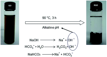
A one-step, novel, easy, fast, facile, economic, and environmental friendly route to reduce graphene oxide (GO) is studied and explained in this study. The household baking soda (sodium bicarbonate/NaHCO3) was applied here as a reducing agent. In aqueous solution, NaHCO3 hydrolyses into hydroxide ion and leads to deoxygenate GO sheets. The confirmation of oxygen reduction was checked with UV-visible spectroscopy, X-ray diffraction, Fourier transform infrared spectrometry and Raman spectroscopy. Thermal behavior of graphene was analyzed by thermogravimetry analysis and differential scanning calorimetry. Again, atomic force microscopy, scanning electron microscopy, field emission scanning electron microscopy and transmission electron microscopy were used for morphological study. Energy dispersive X-ray spectrometer was applied to analyze the elemental composition. The results of the above experiments demonstrate that the house-hold baking soda is able to produce functional graphene, which can be a suitable replacement of hydrazine, sodium borohydrate or other toxic reducing agents. This method is high yielding and safe to use in biomaterial applications.
000 cm2 v−1 s−1),3 high thermal conductivity (∼5000 W m−1 K−1),4 high optical transmittance (∼97.7%).5 These unique behavior of graphene opened a new window for its wide range of application at different branches of technology such as Li ion battery,6 catalyst engineering,7 biosensor,8 solar cells,9 photovoltaic devices,10 p–n junction materials,11 super capacitor.12 However, large quantities of graphene are required to fulfil the huge demand of aforementioned applications.
Inexpensive scale-up production of graphene is one of the major challenges of this scientific field. Several approaches have designed to synthesis graphene from pristine graphite such as: epitaxial growth,13 vacuum thermal annealing,14 micromechanical cleavage,15 chemical vapor deposition,16 thermal exfoliation,17 liquid phase exfoliation,18 chemical based detonation,19 carbon nano tube unzipping.20 These methods can produce crystalline graphene, however, they are not suitable for large scale production.21
The most potential procedure for mass production of graphene could be the chemical oxidation–reduction reaction. It is considered as an easier, scalable, fast, dynamic, facile, and economic approach compared to other methods. This method involves the production of graphite oxide from graphite flakes by chemical oxidation, subsequent exfoliation of graphite oxide to graphene oxide (GO) in aqueous solution and finally reduction of GO to functional graphene sheet. Recently, different reducing agents are using to convert GO to graphene sheet such as: hydrazine,22 sodium borohydrate,23 hydroquinone.24 Unfortunately, all of these reducers are poisonous, corrosive and hazardous to human and environment, which hinder their application in the several field of biomaterials, biosensors or biomedical engineering. Furthermore, the existing oxygen molecules on the sheet (after reduction) and additional functional groups of reducer molecules could arise π–π stacking, resulting sheet resistant. In order to address the above problems, different green and ecofriendly reducers have chosen by the researchers to produce high quality graphene such as: metal powder,25 amino acid,26 plant extract,27 organic acid,28 metal oxide,29 cellulose,30 biological agent.31 However, the green reducers used in nontoxic synthesis have some remarkable limitations like possibility of product contamination (metal powders), repeated downstream processing (amino acid, plant extract), complex and controlled experimental conditions (biological agent), lengthy reduction time.
Therefore, a novel and green route of reduction is needed to be explored that would be free from the above limitations. In this work, we reported a new approach to the direct synthesis of graphene from GO. We used household baking soda as a reducing agent containing NaHCO3. There are some significant reasons for the selection of NaHCO3. Firstly, it is economically cheap, environmentally safe, and readily available in everywhere. Secondly, it is mild alkaline in nature and able to reduce the reaction time.32 It is important to mention that the alkaline pH has a great impact over the graphene sheet.33 In an investigation, Navarro et al.34 claimed that acidic pH can cause higher degrees of defect or disorder in the resulting graphene. It can reduce the size of the sheet and arise irreversible agglomeration. These types of sheets are not suitable for electronic devices due to their lower electron conductivity. Additionally, lower pH is responsible for giving rise to other graphitic nano forms such as multi-walled carbon nanotubes, fullerenes, and nano-onions. In contrast, alkaline condition lessens the defects of the derived sheet and produces bigger graphitic domain. It can promote colloidal stability of the GO sheet. It also increases the negative charge of the sheet resulting magnetic repulsion in-between individual layer. Thus, it can prevent irreversible aggregation and re-establish new sp2 bonding network upon reduction. It is free from the introduction of further functional groups (which are not suitable for the practical application of graphene). This study highlighted the possible mechanism of deoxygenation in the presence of NaHCO3. We hope that this research will shed a light in the field of green synthesis for deoxygenation of graphene oxide.
![]() | ||
| Fig. 1 (a) GO synthesised by modified hummer method, (b) magnetic repulsion of GO at room temperature, (c) baking soda reduced graphene oxide (SRGO), and (d) SRGO after drying. | ||
UV-vis absorption spectroscopy is commonly used to insure the reduction of oxygen-containing groups and such results of this study are reported in Fig. 3. The UV-vis absorption spectrum of GO displays an absorption peak at 231 nm, which represents the π–π* transition of C
C bonds. The spectrum also shows a weak shoulder at around 300 nm attributed to the n–π* transition of carboxyl groups (COOH). Upon complete reduction the C
C plasmon peak red shifted to ∼270 nm, which reveals that the π-electron has increased in SRGO. It is also consistent with the ordering of structure through possible rearrangement of atoms. This result strongly evaluates the findings of previous related works, as mentioned in Table 1. On the other hand, the shoulder related to COOH group of GO was absent in the spectrum of SRGO. It also supports the omission of oxygen after reduction.
C bonds) of GO and RGO at different reduction methods reported in previous literatures
| Name of reducer | π–π* transition of GO | π–π* transition of RGO | References |
|---|---|---|---|
| L-Cysteine | 230 nm | 270 nm | 26a |
| Aqueous phytoextract | 232 nm | 270.9 nm | 27a |
| Benzylamine | 230 nm | 260 nm | 35 |
| Dopamine | 230 nm | 271 nm | 36 |
| Gallic acid | 230 nm | 273 nm | 28b |
| Glucose amine | 231 nm | 273 nm | 27c |
| Glycine | 230 nm | 267 nm | 37 |
| Humanin | 227 nm | 265 nm | 38 |
| Iron powder | 230 nm | Not mentioned | 25b |
| L-Ascorbic acid | 230 nm | 264 nm | 28c |
| Metal salt | 229 nm | 259 nm | 39 |
| Nascent hydrogen | 232 nm | 266 nm | 40 |
| Natural cellulose | 230 nm | 269 nm | 30 |
| L-Aspartic acid | 250 nm | 269 nm | 26b |
| Oligothiophene | Not mentioned | Between 243–247 nm | 41 |
| Reducing sugar | 230 nm | 261 nm | 42 |
| Sodium borohydrate | 230 nm | 260 nm | 23 |
| Solvothermal | 229 nm | 269 nm | 43 |
| Tannin | 230 nm | 275 nm | 27c |
| Tea solution | 228 nm | 271 nm | 44 |
| Yeast | 230 nm | 263 nm | 31c |
| Zn powder | 230 nm | 272 nm | 45 |
Fig. 4 shows the XRD profile of pristine graphite, GO, and SRGO. In pristine graphite, a strong and sharp 002 diffraction peak appeared at 2θ = 26.49° corresponding to a d-spacing of 3.35 Å (Fig. 4a). After the treatment of oxidizing agents, this characteristic peak (002) has been shifted to a lower angle in GO.28a In the current study, it was recorded 2θ = 10.43° with 8.47 Å d-spacing (Fig. 4b). The increment of d-spacing from 3.35 Å to 8.47 Å confirms the introduction of oxygen functional groups in between the graphene sheet. It also indicates the formation of new sp3 bonding responsible for structural defects or atomic scale roughness.26a The XRD pattern of SRGO displays two peaks at 24.68° (d-spacing = 3.36 Å) and 26.52° (d-spacing = 3.60 Å) (Fig. 4c). The weak or even invisible peak at 24.68° is related to the fully exfoliated graphene sheet.46 However, other weak peak at 26.52° reveals that the van der Waals forces cause restacking of sheet due to the elimination of oxygen with less hydrophilicity.47 In fact, the aggregation or stacking of graphene layer can inevitably occur, if it is reduced directly.22
Raman spectroscopy is an important technique to characterize GO before and after reduction and such results of this study is mentioned in Fig. 5. Typically, the Raman spectra of GO and RGO display two fundamental peak within the range of 1500–1700 cm−1 termed as D peak and G peak. The characteristic of D peak is the consequence of breathing mode of six atom rings. It is caused by the defects or disorders present over the plane. And the G peak is the result of first order scattering of E2g phonons. It is caused by the sp2 bonded carbon which constructs graphene. In this study, a sharp and intense G peak was observed at 1581 cm−1 caused by the lattice structure of graphite. It also displayed a weak D band at 1353 cm−1 (disordered band) caused by the graphitic edges. After the treatment of oxidising agents, the Raman spectra of GO displayed the D peak and G peak at 1350 and 1614 cm−1, respectively. Here the G band is broaden and blue-shifted to higher wave number indicating size degradation of the in-plane sp2 domains.48 While the GO is being reduced with baking soda, these two peaks are shifted to a lower wave length of 1347 and 1596 cm−1, respectively. Significantly, the shifting of G peak to the lower wave number is important upon reduction. In SRGO the G peak is sharper than the GO. This phenomenon reveals the “self-healing” mechanism followed by the elimination of oxygen.48a,49 On the other hand, the intensity ratio of D peak and G peak is applied to measure the average size of sp2 domain as well as the degree of disorder. According to the Tuinstra–Koenig relation, the ID/IG ratio is inversely proportional to the crystalline size of sp2 domain.50 In GO the ID/IG ratio was found to be 1.47. After reduction, it was decreased to 1.37. This achievement also suggests that the green reduction of GO with baking soda is able to repair disorders and restore new π-conjugated structure.25b On the other hand, the spectrum of graphene could display another weak peak, which is resonance-enhanced by a two phonon scattering process. It is termed as 2D (G′) peak.51 This peak indicates the increment of new layer in SRGO.52 However, the main application of this peak is the determination of mono, bi or multiple layer graphene. In case of single layer graphene, it is generally observed at 2679 cm−1.27a If the graphene is multiple layer (2–4), it will be shifted towards a higher wave number by 19 cm−1.53 In this work, the 2D peak position was observed at 2695 cm−1, which shifted to 16 cm−1 higher wave number. This result confirms the formation of multiple layer graphene (SRGO).
The FTIR spectroscopy of this study is plotted in Fig. 6. The FTIR spectra of pristine graphite contains only two characteristic peaks at 1610 and 3450 cm−1 attributed to aromatic C
C bonding (the structural vibration) and adsorbed water molecules (the vibration of O–H stretching), respectively.54 In GO, the appearance of different intense band at around 1706, 1386, 1219, and 1052 cm−1 are ascribed to the carboxyl group (–C
O), OH deformation, epoxy group (C–O–C), and alkoxy group (C–O stretching). And the broad band at approximately 3395 cm−1 is responsible for OH stretching.55 All these peaks confirm the penetration of the above oxygen moieties over the graphene sheet. This phenomenon also indicates the destruction of sp2 character and generation of defects in the sheet owing to extensive oxidation. Secondly, the peak for the aromatic C
C stretching of GO sample was observed at 1625.97 cm−1. Notably, among the aforementioned oxygen groups, the hydroxyl and epoxy groups are the dominant locate at the basal plane while the carboxyl groups are found at the edge of the sheet, as shown in Fig. 7. The binding of these numerous oxygen containing groups make GO hydrophilic in nature. However, upon reduction with baking soda the carboxyl peak (1706 cm−1) was not observed in the SRGO, which indicates its successful elimination.28c,56 Secondly, the alkoxy peak (1052 cm−1) and epoxy peak (1220 cm−1) were almost disappeared.56 And the hydroxyl stretching peak (3395 cm−1) became weaker than the GO.32 It is noteworthy that the intensity of the peak related to sp2 hybridized C
C was increased to 1570 cm−1 for SRGO. Significantly, the appearance of two novel peak at 2918 and 2850 cm−1 related to the symmetric and asymmetric stretching vibration of –CH2 (methylene group) point out the successful retention of new sp2 network.57 This achievement not only confirms the removal of oxygen moieties but also promulgates the successful production of functional graphene sheet. It also reveals that the use of household baking soda could be a potential replacement of toxic hydrazine or sodium borohydrate.
The thermal stability of GO and SRGO was investigated by TGA and is shown in Fig. 8. In this experiment, the samples were heated under nitrogen atmosphere with a rate of 10 °C min−1 from 25–700 °C. GO is thermally unstable and begins to lose weight just after heating.58 The curve of GO displays three significant level of mass loss. The first level showed about 17% mass loss at around 100 °C. It is owing to the evaporation of absorbed water from the sheet. It demonstrates that around 17% of water molecules could be trapped between the sheets. It is also considered as the higher amount of oxygen quantity.54,58 The second level occurred at around 220 °C with 46% mass loss. It is attributed to the thermal decomposition of labile oxygen moieties. Finally, the third level displayed about 60% mass losses at around 600 °C. It is related to the pyrolysis of existing moieties as well as burning of carbon plane. However, the residual weight of GO sample is about 36% at 700 °C. In contrast, the SRGO sample exhibits about 11, 16, and 33 wt% mass loss at around 100, 220, and 600 °C, respectively. All these mass losses of SRGO are lower than the GO. In addition, the residual weight of SRGO was found ∼63% at 700 °C, which is almost twice than the GO. These aforementioned results suggest that most of the oxygen holding groups are vanished upon reduction. It also increases the van der Waals interaction between the sheets leading to better re-graphitization.59 Therefore, the as prepared SRGO possesses higher thermal stability compare to the GO precursor.40,60
DSC is another useful technique to study the thermal behavior of graphene. The heat flow curve by DSC is shown in Fig. 9. In GO, a strong exothermal peak was centred at around 200 °C. It is ascribed to the vanishing of labile oxygen. It also shows an endothermal peak at around 170 °C attributed to the burning of skeletal carbon. On the other hand, after heating the SRGO sample up to 450 °C, the SRGO curve shows neither sudden weight loss nor obvious exothermal peak. It displays an endothermal peak at around 175 °C, which is sharper than the GO. This result is consistent with the result of Ji and co-workers.61 It also reveals that the SRGO possesses higher thermal stability than GO.
AFM is a powerful tool to study the morphology of graphene and carbon related materials. Fig. 10 shows the typical tapping mode AFM image of GO and SRGO. However, it is well established that the van der Waals thickness of a pristine graphite is about 0.34 nm that is automatically flat.22 After oxidation, the newly synthesised GO become thicker due to the attachment of covalently bonded oxygen. This insertion of oxygen prolapses the sp2 hybridized carbons over and under the original plane of graphene. In this work, the average thickness of GO sheet is obtained about 1.20 nm, which corresponds to the earlier literature.62 In contrast, the average thickness of as-formed SRGO fragment is about 2.5 nm (Fig. 10b). This increment of thickness is ascribed to the tendency of graphene layers to restack in the absence of additional stabilizing molecules.27b
![]() | ||
| Fig. 10 Tapping mode AFM measurement of (a) GO and (b) SRGO, the samples are deposited on to SiO2/Si substrate. | ||
To investigate surface morphology, the SEM image of graphite, GO, and prepared SRGO were obtained. Fig. 11a shows the SEM image of natural graphite powder. Morphologically, it exhibits flaky appearance for the strong sp2 carbon to carbon bonding in the plane. Nevertheless, at higher concentration, the GO surface shows soft-carpet like morphology as seen in Fig. 11b. It may be the attachment of residual water molecules, carboxyl and hydroxyl groups to carbon basal plane.63 Furthermore, the higher magnification as portrayed in Fig. 11c clearly expresses the wrinkled and coarse feature of GO. After reduction, the SRGO materials show aggregated structure where individual sheets of different size are closely associated with each other (Fig. 11d). Here one single sheet is looked like a disordered solid (Fig. 11e). However, it is obvious that the edge of GO is quite distinguishable than that of SRGO. Significantly, the edges of the GO sheets look crumpled due to extensive oxidation (Fig. 11c), which is later magnified and discussed by FESEM study. By contrast, the edges of SRGO were sharp (Fig. 11e and f) that supports the investigation of Gurunathan and co-workers.38 This morphological difference between the folded edge of GO and sharp edge of SRGO demonstrate that the household baking soda can play an important role to convert the GO into functional graphene.
Fig. 12a shows the FESEM image of GO. The thin, crystal, and shrivelling morphology of GO were observed where the sheets were slightly joined with each other. Generally, it is termed as random orientation aiding to form agglomerate. In this work, most of the GO sheets are exfoliated into mono or multiple layer graphene, with some of the sheets being overlapped. Fig. 12b and c show the higher magnification FE-SEM of GO. From these images, it can be demonstrate that the boarder of sheets are partially folded, which significantly reduces the total surface energy of the sheets.43 On the other hand, the heaping of individual sheets through different self-assembly techniques develops the surface morphology of SRGO film. As a result, the sheets take crumpled or sea wave like texture as seen in Fig. 12d. Similar trend like this also observed by other researchers.45,64 This morphological difference assures the change of microstructure after reduction. Recently, it is claimed that the average lateral size of giant GO sheets is around 18.4 μm, although the relative range of size (wide) distributes between one to several micrometre. In this work, the lateral size (wide) of GO and SRGO sheets were 7 and 10 μm that is similar to the work of green graphene synthesis using humanin.38
Energy dispersive X-ray spectrometer is applied to analyze the elemental composition as well as the purity of the materials (as displayed in Fig. 13). In pristine graphite (can be seen in Fig. 13a), the atomic weight percentage of carbon was found to be 98%. The rest 2% of oxygen is related to the atmospheric or aerial oxygen binding over the sheet surface with weak van der Waals interactions.65 In GO (can be seen in Fig. 13b), the atomic weight percentage of carbon and oxygen were found to be 58.4% and 38.9%, respectively.66 The enhancement of atomic oxygen in GO reveals the successful oxidation of graphite. Noticeably, the elimination of impurities other than trace amounts of S (2.7 atomic weight percentage) confirms that the GO precursor prepared by modified hummer method is pure. In contrast, the atomic weight percentage of carbon is increased to 86.6% in SRGO (refer to Fig. 13c).67 This result suggests that a considerable amount of the oxygen functionalities were removed upon deoxygenation. Here, the remaining oxygen is mainly the absorbed water or oxygen molecules. And the trace amounts of sodium are the supplement of baking soda (NaHCO3).
TEM is another useful tool to study the nano structure of graphene. The TEM image of both GO and SRGO at lower magnification range is portrayed in Fig. 14. The Fig. 14a shows the amorphous GO sheets before reduction. Morphologically, the GO nano sheets show transparent, stable, uniform and silk like appearance under high energy electron beam, as in previously reported work.68 In this stage the inter layer coherence is not demolished. For this reason, the GO nano sheets trend to entangle with each other in a random manner and produce multiple layer agglomerates. However, this scrolling and corrugation feature is the intrinsic nature of graphene.69 Because the 2D graphene membrane become thermodynamically stable by crumpling via bucking or bending.70 Notably, the scrolling and corrugation are found not only at the edge but also in the middle of the sheets. Thus, the GO nano sheets become folded or coiled. Contrarily, after subsequent washing the TEM image of SRGO exhibits a wrinkle paper-like structure, as shown in Fig. 14b. It is attributed to the abundance of carbon to carbon chemical bonding either in a single layer or several layers.71
GO is electrically insulant because the bounded oxygen moieties, which increase the resistance against electron flow.72 In this study, we measured the resistance of GO and SRGO pellet by using four point probe method. The resistance of GO pellet was 4.45 × 10⁶ Ω while the SRGO was as low as 3.36 × 101 Ω. It reveals that the resistance of SRGO reduces about 105 times in comparison to that of GO. This achievement also reconfirms the washing of oxygen groups from the sheet.
| This journal is © The Royal Society of Chemistry 2015 |